Revision 4
#62834
Store at -20C
877-616-CELL (2355)
877-678-TECH (8324)
3 Trask Lane | Danvers | Massachusetts | 01923 | USA
For Research Use Only. Not for Use in Diagnostic Procedures.
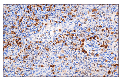
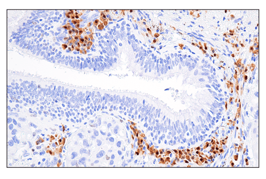
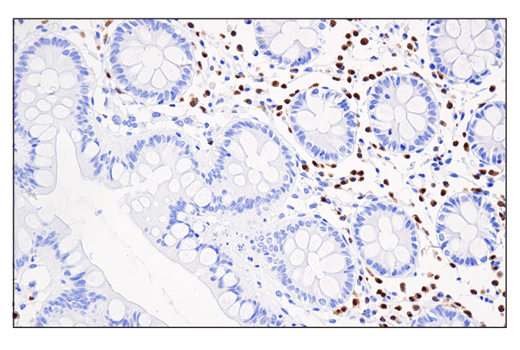
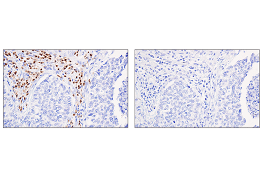
Applications:
W, IP, IHC-Bond, IHC-P, FC-FP
Reactivity:
H M
Sensitivity:
Endogenous
MW (kDa):
51
Source/Isotype:
Rabbit IgG
UniProt ID:
#Q15306
Entrez-Gene Id:
3662
Product Usage Information
| Application | Dilution |
|---|---|
| Western Blotting | 1:1000 |
| Immunoprecipitation | 1:100 |
| IHC Leica Bond | 1:50 - 1:200 |
| Immunohistochemistry (Paraffin) | 1:50 - 1:200 |
| Flow Cytometry (Fixed/Permeabilized) | 1:200 - 1:800 |
Storage
For a carrier free (BSA and azide free) version of this product see product #56471.
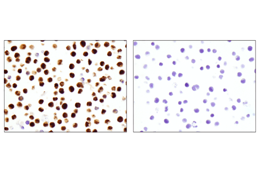
Specificity/Sensitivity
Source / Purification
Background
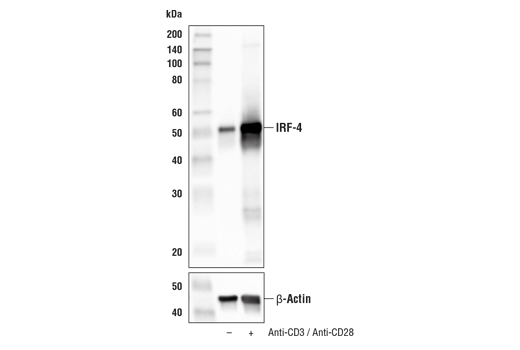
IRF-4 was independently cloned by three groups and demonstrated to have roles in different contexts of lymphoid regulation (3-5). First, IRF-4 (Pip) was found to associate with PU.1, a hematopoietic specific member of the ETS family, and to regulate the expression of B-cell specific genes (3). Second, it was characterized as a lymphoid-specific member of the IRF family (LSIRF) able to bind to ISRE (4). Third, it was identified in activated T cells as a factor that binds to the promoter of the interleukin-5 gene (ICSAT), and it was shown to repress gene activation induced by IFN (5). IRF-4 is expressed in all stages of B cell development and in mature T cells, and is inducible in primary lymphocytes by antigen mimetic stimuli such as concavalin A, CD3 crosslinking, anti-IgM and PMA treatment (4,5). Mice deficient in IRF-4 show normal distribution of B and T lymphocytes at 4 to 5 weeks, but later develop progressive generalized lymphadenopathy, suggesting a role for IRF-4 in the function and homeostasis of mature B- and T-lymphocytes (6).
Background References
- Taniguchi, T. et al. (2001) Annu Rev Immunol 19, 623-55.
- Honda, K. and Taniguchi, T. (2006) Nat Rev Immunol 6, 644-58.
- Eisenbeis, C.F. et al. (1995) Genes Dev 9, 1377-87.
- Matsuyama, T. et al. (1995) Nucleic Acids Res 23, 2127-36.
- Yamagata, T. et al. (1996) Mol Cell Biol 16, 1283-94.
- Mittrücker, H.W. et al. (1997) Science 275, 540-3.
Species Reactivity
Species reactivity is determined by testing in at least one approved application (e.g., western blot).
Western Blot Buffer
IMPORTANT: For western blots, incubate membrane with diluted primary antibody in 5% w/v BSA, 1X TBS, 0.1% Tween® 20 at 4°C with gentle shaking, overnight.
Applications Key
W: Western Blotting IP: Immunoprecipitation IHC-Bond: IHC Leica Bond IHC-P: Immunohistochemistry (Paraffin) FC-FP: Flow Cytometry (Fixed/Permeabilized)
Cross-Reactivity Key
H: Human M: Mouse
Trademarks and Patents
Cell Signaling Technology is a trademark of Cell Signaling Technology, Inc.
Alexa Fluor is a registered trademark of Life Technologies Corporation.
KARPAS cell line source: Dr. Abraham Karpas at the University of Cambridge.
All other trademarks are the property of their respective owners. Visit cellsignal.com/trademarks for more information.
Limited Uses
Except as otherwise expressly agreed in a writing signed by a legally authorized representative of CST, the following terms apply to Products provided by CST, its affiliates or its distributors. Any Customer's terms and conditions that are in addition to, or different from, those contained herein, unless separately accepted in writing by a legally authorized representative of CST, are rejected and are of no force or effect.
Products are labeled with For Research Use Only or a similar labeling statement and have not been approved, cleared, or licensed by the FDA or other regulatory foreign or domestic entity, for any purpose. Customer shall not use any Product for any diagnostic or therapeutic purpose, or otherwise in any manner that conflicts with its labeling statement. Products sold or licensed by CST are provided for Customer as the end-user and solely for research and development uses. Any use of Product for diagnostic, prophylactic or therapeutic purposes, or any purchase of Product for resale (alone or as a component) or other commercial purpose, requires a separate license from CST. Customer shall (a) not sell, license, loan, donate or otherwise transfer or make available any Product to any third party, whether alone or in combination with other materials, or use the Products to manufacture any commercial products, (b) not copy, modify, reverse engineer, decompile, disassemble or otherwise attempt to discover the underlying structure or technology of the Products, or use the Products for the purpose of developing any products or services that would compete with CST products or services, (c) not alter or remove from the Products any trademarks, trade names, logos, patent or copyright notices or markings, (d) use the Products solely in accordance with CST Product Terms of Sale and any applicable documentation, and (e) comply with any license, terms of service or similar agreement with respect to any third party products or services used by Customer in connection with the Products.
Revision 4
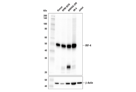
(D6A8) Rabbit mAb #8457 (lower). As expected, IRF-4 protein is not expressed in Jurkat cells. KARPAS-299 cell line source: Dr. Abraham Karpas at the University of Cambridge.
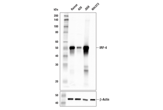
(D6A8) Rabbit mAb #8457 (lower). As expected, IRF-4 protein is not expressed in NIH/3T3 cells.
Revision 4
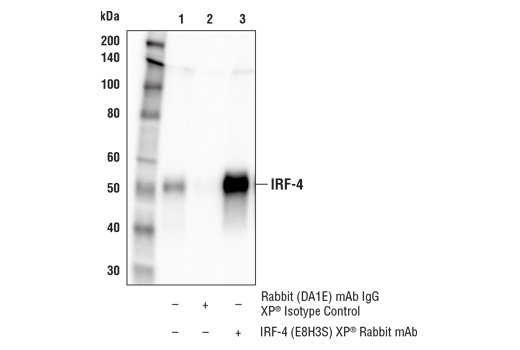
antibody to avoid cross-reactivity with IgG.

Revision 4
Revision 4
Revision 4