| Cat. # | Size | Qty. | Price |
|---|---|---|---|
| 40415T | 1 Kit (9 x 20 microliters) |
|
| Product Includes | Quantity | Applications | Reactivity | MW(kDa) | Isotype |
|---|---|---|---|---|---|
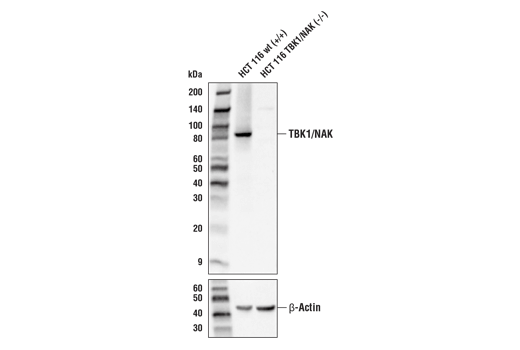
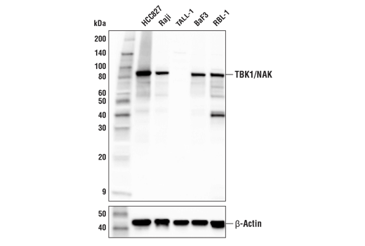
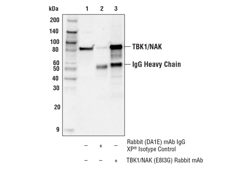
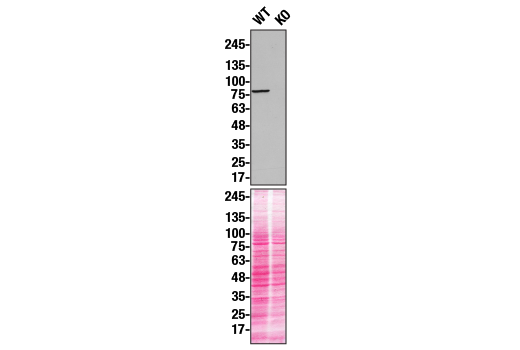
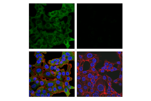
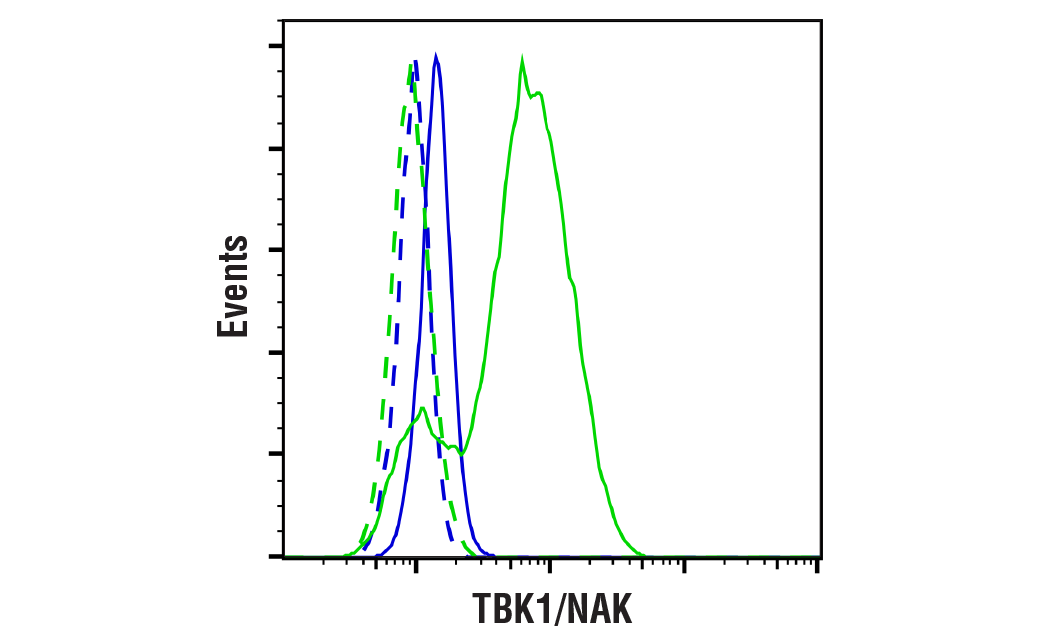
| TBK1/NAK (E8I3G) Rabbit mAb 38066 | 20 µl |
|
H M R | 84 | Rabbit IgG |
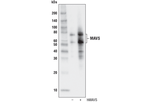
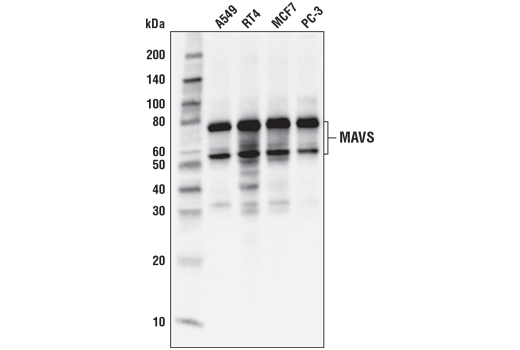
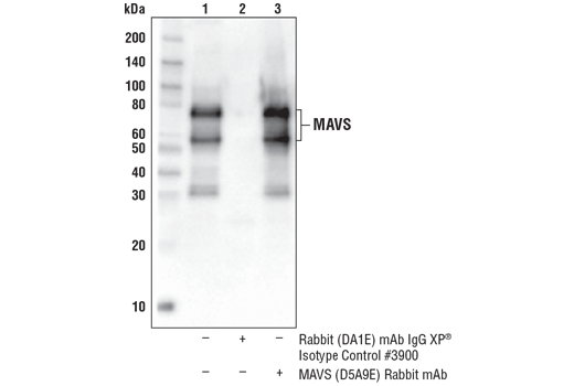
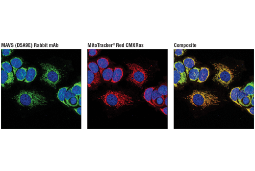
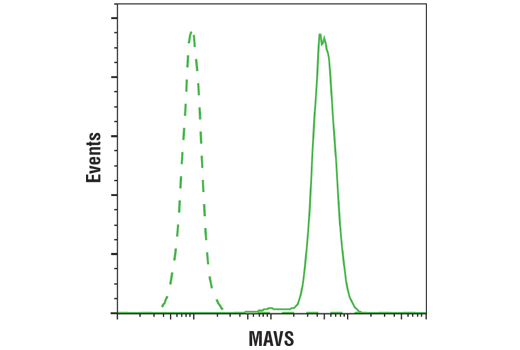
| MAVS (D5A9E) Rabbit mAb 24930 | 20 µl |
|
H | 75, 52 | Rabbit IgG |
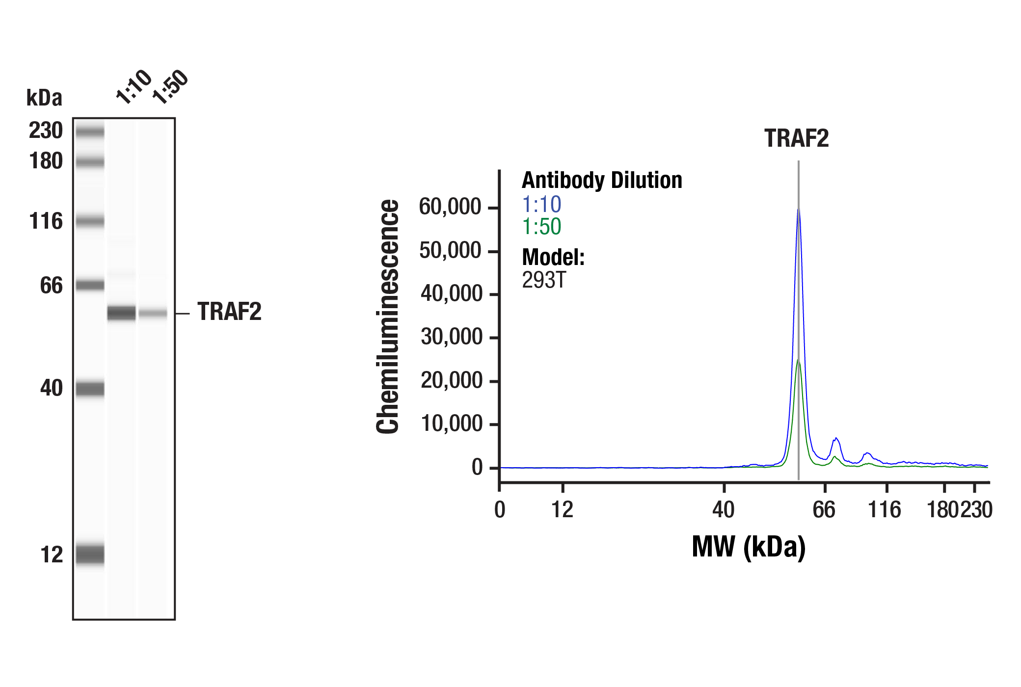
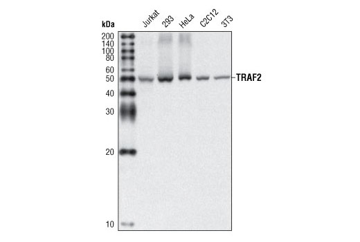
| TRAF2 (C192) Antibody 4724 | 20 µl |
|
H M Mk | 53 | Rabbit |
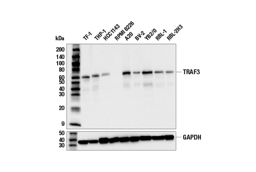
| TRAF3 (E8H3B) Rabbit mAb 33640 | 20 µl |
|
H M R | 62 | Rabbit IgG |
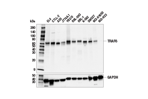
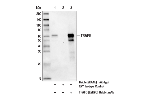
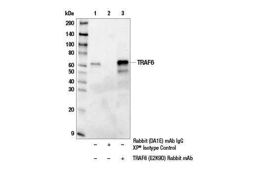
| TRAF6 (E2K9D) Rabbit mAb 67591 | 20 µl |
|
H M R | 60 | Rabbit IgG |
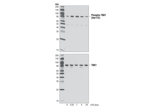
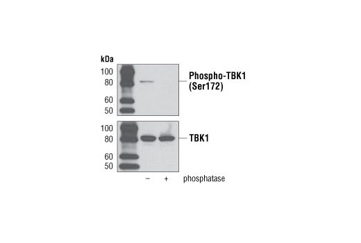
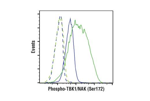
| Phospho-TBK1/NAK (Ser172) (D52C2) XP® Rabbit mAb 5483 | 20 µl |
|
H M | 84 | Rabbit IgG |
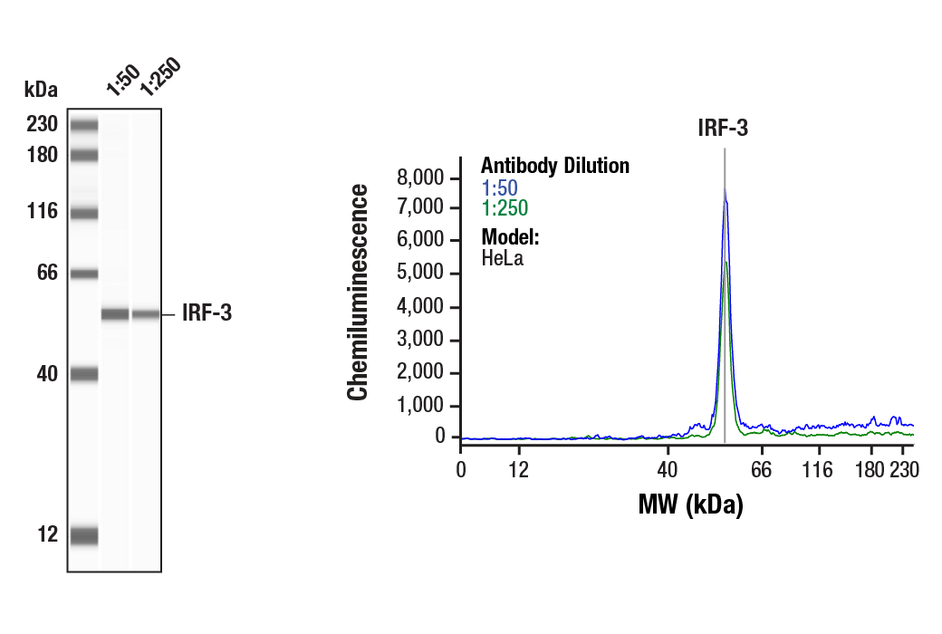
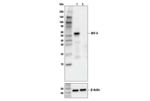
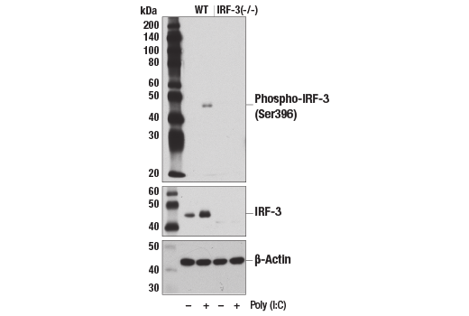
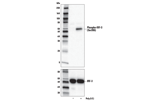
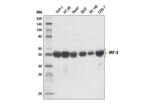
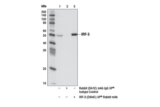
| IRF-3 (D6I4C) XP® Rabbit mAb 11904 | 20 µl |
|
H Mk | 50-55 | Rabbit IgG |
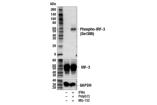
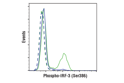
| Phospho-IRF-3 (Ser386) (E7J8G) XP® Rabbit mAb 37829 | 20 µl |
|
H | 50-55 | Rabbit IgG |
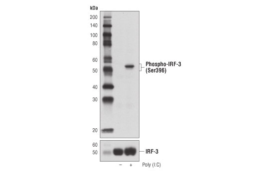
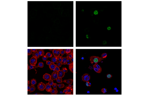
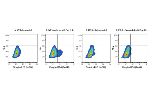
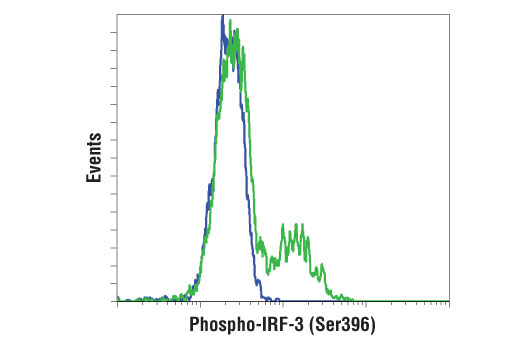
| Phospho-IRF-3 (Ser396) (D6O1M) Rabbit mAb 29047 | 20 µl |
|
H M R | 45-55 | Rabbit IgG |
| Anti-rabbit IgG, HRP-linked Antibody 7074 | 100 µl |
|
Goat |
Product Information
Polyclonal antibodies are produced by immunizing animals with a synthetic peptide corresponding to residues surrounding Cys192 of human TRAF2. Antibodies are purified by protein A and peptide affinity chromatography. Monoclonal antibodies are produced by immunizing animals with recombinant human IRF-3 protein and recombinant protein specific to a central region of mouse TRAF6, or synthetic phosphopeptides corresponding to residues surrounding Ser172 of human TBK1, Ser386 of human IRF-3, and Ser396 of human IRF-3 protein, or synthetic peptides corresponding to residues surrounding Leu202 of human MAVS and Ala327 of human TRAF3, or near the carboxy terminus of human TBK1/NAK protein.
Recognition of conserved molecular structures of viruses by host pattern-recognition receptors (PRRs) initiates innate antiviral immune responses. Several families of PRRs have been demonstrated to sense different microbial components (1,2). The RIG-I-like receptor (RLR) family, including RIG-I, MDA5, and LGP2, are the primary PRRs to recognize viral RNAs (3,4). Upon binding to viral RNA, these receptors undergo conformation change, leading to their interaction with mitochondrial antiviral signaling protein (MAVS). MAVS subsequently forms large prion-like polymers and serves as a platform to recruit multiple components, including TRAF proteins and TBK1, to form the so-called MAVS signalosome. MAVS signalosome, in turn, activates the IRF-3 and NF-kB pathways, leading to the production of type I IFNs and pro-inflammatory cytokines (5-9).
Explore pathways related to this product.
STRING - Known and Predicted Protein-Protein Interactions.
Except as otherwise expressly agreed in a writing signed by a legally authorized representative of CST, the following terms apply to Products provided by CST, its affiliates or its distributors. Any Customer's terms and conditions that are in addition to, or different from, those contained herein, unless separately accepted in writing by a legally authorized representative of CST, are rejected and are of no force or effect.
Products are labeled with For Research Use Only or a similar labeling statement and have not been approved, cleared, or licensed by the FDA or other regulatory foreign or domestic entity, for any purpose. Customer shall not use any Product for any diagnostic or therapeutic purpose, or otherwise in any manner that conflicts with its labeling statement. Products sold or licensed by CST are provided for Customer as the end-user and solely for research and development uses. Any use of Product for diagnostic, prophylactic or therapeutic purposes, or any purchase of Product for resale (alone or as a component) or other commercial purpose, requires a separate license from CST. Customer shall (a) not sell, license, loan, donate or otherwise transfer or make available any Product to any third party, whether alone or in combination with other materials, or use the Products to manufacture any commercial products, (b) not copy, modify, reverse engineer, decompile, disassemble or otherwise attempt to discover the underlying structure or technology of the Products, or use the Products for the purpose of developing any products or services that would compete with CST products or services, (c) not alter or remove from the Products any trademarks, trade names, logos, patent or copyright notices or markings, (d) use the Products solely in accordance with CST Product Terms of Sale and any applicable documentation, and (e) comply with any license, terms of service or similar agreement with respect to any third party products or services used by Customer in connection with the Products.