Western Blotting Protocol
For western blots, incubate membrane with diluted primary antibody in 5% w/v nonfat dry milk, 1X TBS, 0.1% Tween® 20 at 4°C with gentle shaking, overnight.
NOTE: Please refer to primary antibody product webpage for recommended antibody dilution.
A. Solutions and Reagents
From sample preparation to detection, the reagents you need for your Western Blot are now in one convenient kit: #12957 Western Blotting Application Solutions Kit
NOTE: Prepare solutions with reverse osmosis deionized (RODI) or equivalent grade water.
- 20X Phosphate Buffered Saline (PBS): (#9808) To prepare 1 L 1X PBS: add 50 ml 20X PBS to 950 ml dH2O, mix.
- 10X Tris Buffered Saline (TBS): (#12498) To prepare 1 L 1X TBS: add 100 ml 10X to 900 ml dH2O, mix.
- 1X SDS Sample Buffer: Blue Loading Pack (#7722) or Red Loading Pack (#7723) Prepare fresh 3X reducing loading buffer by adding 1/10 volume 30X DTT to 1 volume of 3X SDS loading buffer. Dilute to 1X with dH2O.
- 10X Tris-Glycine SDS Running Buffer: (#4050) To prepare 1 L 1X running buffer: add 100 ml 10X running buffer to 900 ml dH2O, mix.
- 10X Tris-Glycine Transfer Buffer: (#12539) To prepare 1 L 1X Transfer Buffer: add 100 ml 10X Transfer Buffer to 200 ml methanol + 700 ml dH2O, mix.
- 10X Tris Buffered Saline with Tween® 20 (TBST): (#9997) To prepare 1 L 1X TBST: add 100 ml 10X TBST to 900 ml dH2O, mix.
- Nonfat Dry Milk: (#9999).
- Blocking Buffer: 1X TBST with 5% w/v nonfat dry milk; for 150 ml, add 7.5 g nonfat dry milk to 150 ml 1X TBST and mix well.
- Wash Buffer: (#9997) 1X TBST.
- Primary Antibody Dilution Buffer: 1X TBST with 5% nonfat dry milk; for 20 ml, add 1.0 g nonfat dry milk to 20 ml 1X TBST and mix well.
- Biotinylated Protein Ladder Detection Pack: (#7727).
- Blue Prestained Protein Marker, Broad Range (11-250 kDa): (#59329).
- Blotting Membrane and Paper: (#12369) This protocol has been optimized for nitrocellulose membranes. Pore size 0.2 µm is generally recommended.
- Secondary Antibody Conjugated to HRP: Anti-mouse IgG, HRP-linked Antibody (#7076).
- Detection Reagent: SignalFire™ ECL Reagent (#6883).
B. Protein Blotting
A general protocol for sample preparation.
- Treat cells by adding fresh media containing regulator for desired time.
- Aspirate media from cultures; wash cells with 1X PBS; aspirate.
- Lyse cells by adding 1X SDS sample buffer (100 µl per well of 6-well plate or 500 µl for a 10 cm diameter plate). Immediately scrape the cells off the plate and transfer the extract to a microcentrifuge tube. Keep on ice.
- Sonicate for 10–15 sec to complete cell lysis and shear DNA (to reduce sample viscosity).
- Heat a 20 µl sample to 95–100°C for 5 min; cool on ice.
- Microcentrifuge for 5 min.
Load 20 µl onto SDS-PAGE gel (10 cm x 10 cm).
NOTE: Loading of prestained molecular weight markers (#59329, 10 µl/lane) to verify electrotransfer and biotinylated protein ladder (#7727, 10 µl/lane) to determine molecular weights are recommended.
- Electrotransfer to nitrocellulose membrane (#12369).
C. Membrane Blocking and Antibody Incubations
NOTE: Volumes are for 10 cm x 10 cm (100 cm2) of membrane; for different sized membranes, adjust volumes accordingly.
I. Membrane Blocking
- (Optional) After transfer, wash nitrocellulose membrane with 25 ml TBS for 5 min at room temperature.
- Incubate membrane in 25 ml of blocking buffer for 1 hr at room temperature.
- Wash three times for 5 min each with 15 ml of TBST.
II. Primary Antibody Incubation
- Incubate membrane and primary antibody (at the appropriate dilution and diluent as recommended in the product webpage) in 10 ml primary antibody dilution buffer with gentle agitation overnight at 4°C.
- Wash three times for 5 min each with 15 ml of TBST.
- Incubate membrane with Anti-mouse IgG, HRP-linked Antibody (#7076 at 1:2000) and Anti-biotin, HRP-linked Antibody (#7075 at 1:1000–1:3000) to detect biotinylated protein markers in 10 ml of blocking buffer with gentle agitation for 1 hr at room temperature.
- Wash three times for 5 min each with 15 ml of TBST.
- Proceed with detection (Section D).
D. Detection of Proteins
Directions for Use:
- Wash membrane-bound HRP (antibody conjugate) three times for 5 minutes in TBST.
- Prepare 1X SignalFire™ ECL Reagent (#6883) by diluting one part 2X Reagent A and one part 2X Reagent B (e.g. for 10 ml, add 5 ml Reagent A and 5 ml Reagent B). Mix well.
- Incubate substrate with membrane for 1 minute, remove excess solution (membrane remains wet), wrap in plastic and expose to X-ray film.
* Avoid repeated exposure to skin.
posted June 2005
revised June 2020
Protocol Id: 19
Immunohistochemistry (Paraffin)
A. Solutions and Reagents
- Xylene
- Ethanol, anhydrous denatured, histological grade (100% and 95%)
- Deionized water (dH2O)
- Hematoxylin (optional)
- Wash Buffer:
- 1X Tris Buffered Saline with Tween® 20 (TBST): To prepare 1L 1X TBST add 100 ml 10X Tris Buffered Saline with Tween® 20 (#9997) to 900 ml dH20, mix.
- Antibody Diluent TBST/5% Normal Goat Serum: to 5 mL 1X TBST, add 250 µl Normal Goat Serum #5425).
- 1X Citrate Unmasking Solution: To prepare 250 mL of 1X citrate unmasking solution, dilute 25 ml of SignalStain® Citrate Unmasking Solution (10X) (#14746) with 225 mL of dH2O.
- 3% Hydrogen Peroxide: To prepare, add 10 ml 30% H2O2 to 90 ml dH2O.
- Blocking Solution: TBST/5% Normal Goat Serum or 1X Animal-Free Blocking Solution.
- TBST/5% Normal Goat Serum: to 5 ml 1X TBST, add 250 µl Normal Goat Serum (#5425).
- 1X Animal-Free Blocking Solution: to 4 mL of dH2O add 1 ml of Animal-Free Blocking Solution (5X) (#15019).
- Detection System: VECTASTAIN® Elite ABC, including biotinylated secondary antibody (Vector Laboratories).
- Substrate: Vector® NovaRED™ (Vector Laboratories).
- Hematoxylin: Hematoxylin (#14166).
- Mounting Medium: SignalStain® Mounting Medium (#14177).
B. Deparaffinization/Rehydration
NOTE: Do not allow slides to dry at any time during this procedure.
- Deparaffinize/hydrate sections:
- Incubate sections in three washes of xylene for 5 minutes each.
- Incubate sections in two washes of 100% ethanol for 10 minutes each.
- Incubate sections in two washes of 95% ethanol for 10 minutes each.
- Wash sections twice in dH2O for 5 minutes each.
C. Antigen Unmasking
For Citrate: Heat slides in a microwave submersed in 1X citrate unmasking solution until boiling is initiated; follow with 10 min at a sub-boiling temperature (95°-98°C). Cool slides on bench top for 30 min.
D. Staining
- Wash sections in dH2O three times for 5 minutes each.
- Incubate sections in 3% hydrogen peroxide for 10 minutes.
- Wash sections in dH2O twice for 5 minutes each.
- Wash sections in wash buffer for 5 minutes.
- Block each section with 100-400 µl of preferred blocking solution for 1 hour at room temperature.
- Remove blocking solution and add 100-400 µl primary antibody diluted in recommended antibody diluent to each section. Incubate overnight at 4°C.
- Prepare ABC solution per manufacturer's recommendations.
- Remove primary antibody and wash section three times with wash buffer for 5 minutes each.
- Add 100-400 µl biotinylated secondary antibody, diluted in TBST per manufacturer’s recommendation, to each section. Incubate 30 minutes at room temperature.
- Remove secondary antibody solution and wash sections three times with wash buffer for 5 minutes each.
- Cover sections with 100-400 µl pre-mixed ABC solution as needed and incubate in a humidified chamber for 30 min at room temperature.
- Wash section three times with wash buffer for 5 min each.
- Prepare Vector® NovaRED™ per manufacturer's recommendations.
- Apply 100-400 µl substrate to each section and monitor closely. 1-10 minutes generally provides an acceptable staining intensity.
- If desired, counterstain sections with hematoxylin (#14166).
- Wash sections in dH2O two times for 5 minutes each.
- Dehydrate sections:
- Incubate sections in 95% ethanol two times for 10 seconds each.
- Repeat in 100% ethanol, incubating sections two times for 10 seconds each.
- Repeat in xylene, incubating sections two times for 10 seconds each.
- Mount sections with coverslips and mounting medium (#14177).
posted June 2005
revised March 2016
Protocol Id: 295
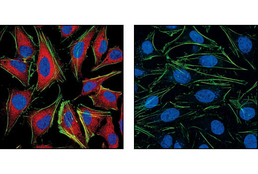
Immunofluorescence (Immunocytochemistry)
A. Solutions and Reagents
Achieve higher quality immunofluorescent images using the efficient and cost-effective, pre-made reagents in our #12727 Immunofluorescence Application Solutions Kit
NOTE: Prepare solutions with reverse osmosis deionized (RODI) or equivalent grade water.
- 20X Phosphate Buffered Saline (PBS): (9808) To prepare 1L 1X PBS: add 50 ml 20X PBS to 950 ml dH2O, mix. Adjust pH to 8.0.
- Formaldehyde: 16%, methanol free, Polysciences, Inc. (cat# 18814), use fresh and store opened vials at 4°C in dark, dilute in 1X PBS for use.
- Blocking Buffer: (1X PBS / 5% normal serum / 0.3% Triton™ X-100): To prepare 10 ml, add 0.5 ml normal serum from the same species as the secondary antibody (e.g., Normal Goat Serum (#5425) to 9 ml 1X PBS) and mix well. While stirring, add 30 µl Triton™ X-100.
- Antibody Dilution Buffer: (1X PBS / 1% BSA / 0.3% Triton™ X-100): To prepare 10 ml, add 30 µl Triton™ X-100 to 10 ml 1X PBS. Mix well then add 0.1 g BSA (#9998), mix.
Recommended Fluorochrome-conjugated Anti-Mouse secondary antibodies:
- Prolong® Gold AntiFade Reagent (#9071), Prolong® Gold AntiFade Reagent with DAPI (#8961).
B. Specimen Preparation - Cultured Cell Lines (IF-IC)
NOTE: Cells should be grown, treated, fixed and stained directly in multi-well plates, chamber slides or on coverslips.
-
Aspirate liquid, then cover cells to a depth of 2–3 mm with 4% formaldehyde diluted in 1X PBS.
NOTE: Formaldehyde is toxic, use only in a fume hood.
- Allow cells to fix for 15 min at room temperature.
- Aspirate fixative, rinse three times in 1X PBS for 5 min each.
- Proceed with Immunostaining (Section C).
C. Immunostaining
NOTE: All subsequent incubations should be carried out at room temperature unless otherwise noted in a humid light-tight box or covered dish/plate to prevent drying and fluorochrome fading.
- Block specimen in Blocking Buffer for 60 min.
- While blocking, prepare primary antibody by diluting as indicated on product webpage in Antibody Dilution Buffer.
- Aspirate blocking solution, apply diluted primary antibody.
- Incubate overnight at 4°C.
- Rinse three times in 1X PBS for 5 min each.
- Incubate specimen in fluorochrome-conjugated secondary antibody diluted in Antibody Dilution Buffer for 1–2 hr at room temperature in the dark.
- Rinse three times in 1X PBS for 5 min each.
- Coverslip slides with Prolong® Gold Antifade Reagent (#9071) or Prolong® Gold Antifade Reagent with DAPI (#8961).
- For best results, allow mountant to cure overnight at room temperature. For long-term storage, store slides flat at 4°C protected from light.
posted November 2006
revised November 2013
Protocol Id: 148
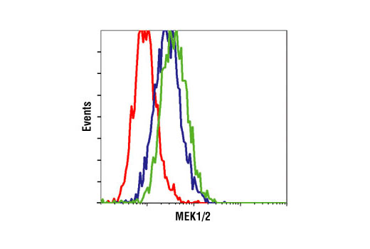
Flow Cytometry, Methanol Permeabilization Protocol for Mouse
Antibodies
A. Solutions and Reagents
All reagents required for this protocol may be efficiently purchased together in our Intracellular Flow
Cytometry Kit (Methanol) #13593, or
individually using the catalog numbers listed below.
NOTE: Prepare solutions with reverse osmosis deionized (RODI) or equivalent grade water.
- 1X Phosphate Buffered Saline (PBS): To prepare 1 L 1X PBS: add 100 ml 10X PBS (#12528) to 900 ml water mix.
- 4% Formaldehyde, Methanol-Free (#47746)
- 100% Methanol (#13604): Chill before use
- Antibody Dilution Buffer: Purchase ready-to-use Flow Cytometry Antibody Dilution Buffer (#13616), or prepare a 0.5% BSA PBS buffer by
dissolving 0.5 g Bovine Serum Albumin (BSA) (#9998) in 100 ml 1X PBS. Store at 4°C.
- Recommended Anti-Mouse secondary antibodies::
- Anti-Mouse IgG (H+L), F(ab')2 Fragment (Alexa Fluor® 488 Conjugate) #4408
- Anti-Mouse IgG (H+L), F(ab')2 Fragment (Alexa Fluor® 594 Conjugate) #8890
- Anti-Mouse IgG (H+L), F(ab')2 Fragment (Alexa Fluor® 647 Conjugate) #4410
- Anti-Mouse IgG (H+L), F(ab')2 Fragment (PE Conjugate) #8887
NOTE: When including fluorescent cellular dyes in your experiment (including viability dyes, DNA dyes, etc.), please refer to the dye product page for the recommended protocol. Visit www.cellsignal.com for a full listing of cellular dyes validated for use in flow cytometry.
B. Fixation
NOTE: Adherent cells or tissue should be dissociated and in single-cell suspension prior to fixation.
NOTE: Optimal centrifugation conditions will vary depending upon cell type and reagent volume. Generally, 150-300g for 1-5 minutes will be sufficient to pellet the cells.
NOTE: If using whole blood, lyse red blood cells and wash by centrifugation prior to fixation.
NOTE: Antibodies targeting CD markers or other extracellular proteins may be added prior to fixation if the epitope is disrupted by formaldehyde and/or methanol. The antibodies will remain bound to the target of interest during the fixation and permeabilization process. However, note that some fluorophores (including PE and APC) are damaged by methanol and thus should not be added prior to permeabilization. Conduct a small-scale experiment if you are unsure.
- Pellet cells by centrifugation and remove supernatant.
- Resuspend cells in approximately 100 µl 4% formaldehyde per 1 million cells. Mix well to dissociate pellet and prevent cross-linking of individual cells.
- Fix for 15 min at room temperature (20-25°C).
- Wash by centrifugation with excess 1X PBS. Discard supernatant in appropriate waste container. Resuspend cells in 0.5-1 ml 1X PBS. Proceed to Permeabilization step.
- Alternatively, cells may be stored overnight at 4°C in 1X PBS.
C. Permeabilization
- Permeabilize cells by adding ice-cold 100% methanol slowly to pre-chilled cells, while gently vortexing, to a final concentration of 90% methanol.
- Permeabilize for a minimum of 10 min on ice.
- Proceed with immunostaining (Section D) or store cells at -20°C in 90% methanol.
D. Immunostaining
NOTE: Count cells using a hemocytometer or alternative method.
- Aliquot desired number of cells into tubes or wells. (Generally, 5x10⁵ to 1x10⁶ cells per assay.)
- Wash cells by centrifugation in excess 1X PBS to remove methanol. Discard supernatant in appropriate
waste container. Repeat if necessary.
- Resuspend cells in 100 µl of diluted primary antibody, prepared in Antibody Dilution Buffer at a recommended dilution or as determined via titration.
- Incubate for 1 hr at room temperature.
- Wash by centrifugation in Antibody Dilution Buffer or 1X PBS. Discard supernatant. Repeat.
- Resuspend cells in 100 µl of diluted fluorochrome-conjugated secondary antibody (prepared in Antibody Dilution Buffer at the recommended dilution).
- Incubate for 30 min at room temperature. Protect from light.
- Wash by centrifugation in Antibody Dilution Buffer or 1X PBS. Discard supernatant. Repeat.
- Resuspend cells in 200-500 µl of 1X PBS and analyze on flow cytometer.
posted June 2005
revised June 2020
Protocol Id: 406