Revision 1
#36422
Store at -20C
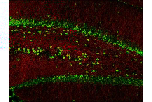
Microglia LPS-Related Module Antibody Sampler Kit
1 Kit
(9 x 20 microliters)
877-616-CELL (2355)
877-678-TECH (8324)
3 Trask Lane | Danvers | Massachusetts | 01923 | USA
For Research Use Only. Not for Use in Diagnostic Procedures.
| Product Includes | Product # | Quantity | Mol. Wt | Isotype/Source |
|---|---|---|---|---|
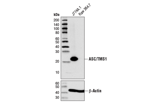
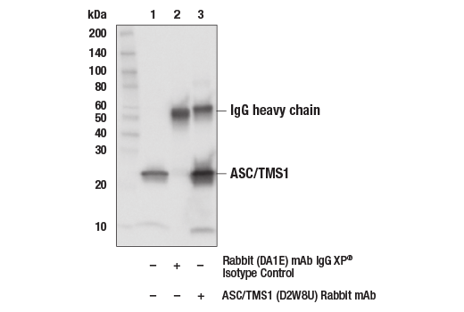
| ASC/TMS1 (D2W8U) Rabbit Monoclonal Antibody | 67824 | 20 µl | 22 kDa | Rabbit IgG |
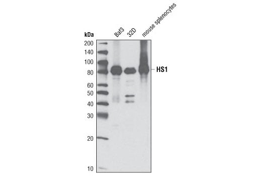
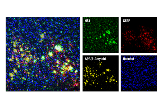
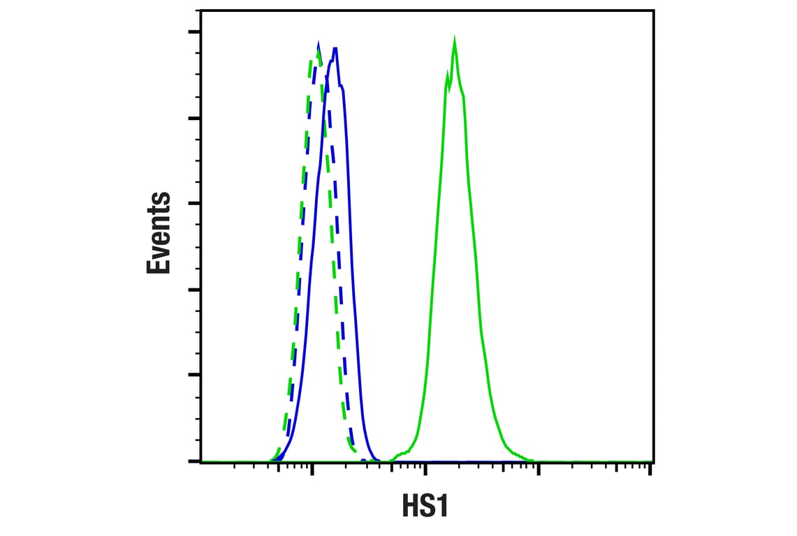
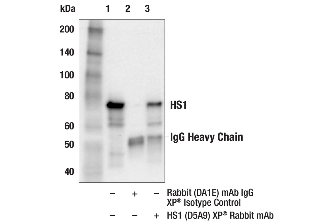
| HS1 (D5A9) Rabbit Monoclonal Antibody | 3892 | 20 µl | 80 kDa | Rabbit IgG |
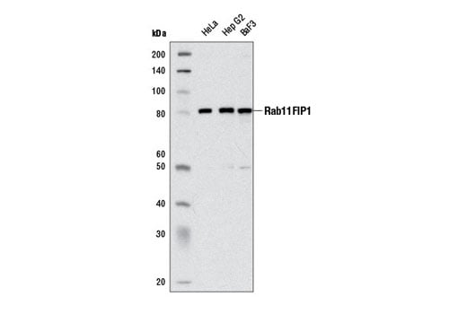
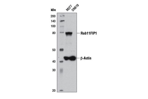
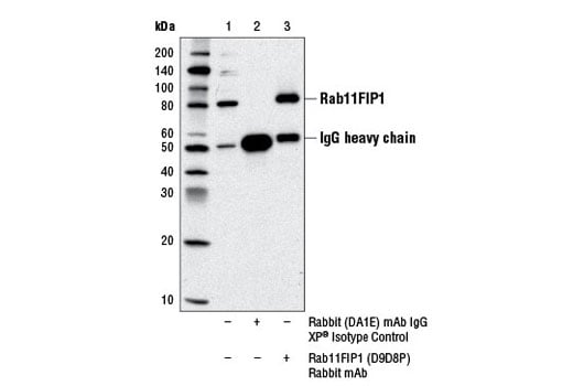
| Rab11FIP1 (D9D8P) Rabbit Monoclonal Antibody | 12849 | 20 µl | 85 kDa | Rabbit IgG |
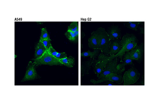
| Integrin alpha4 (D2E1) Rabbit Monoclonal Antibody | 8440 | 20 µl | 70, 140, 150, kDa | Rabbit IgG |
| IQGAP1 (D8K4X) Rabbit Monoclonal Antibody | 20648 | 20 µl | 195 kDa | Rabbit IgG |
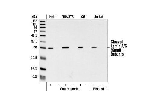
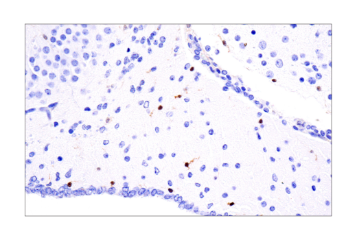
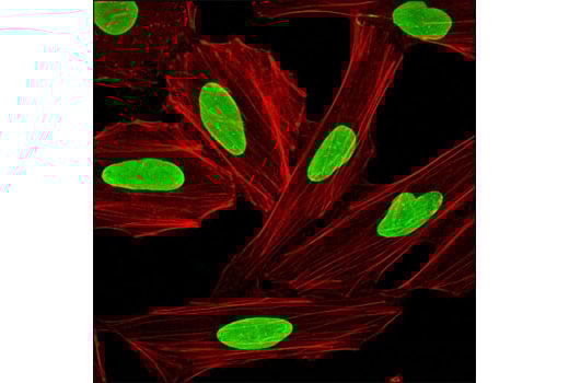
| Cleaved Lamin A (Small Subunit) (30H5) Mouse Monoclonal Antibody | 2036 | 20 µl | 28 kDa | Mouse IgG1 |
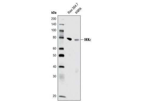
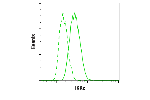
| IKK epsilon (D61F9) Rabbit Monoclonal Antibody | 3416 | 20 µl | 80 kDa | Rabbit IgG |
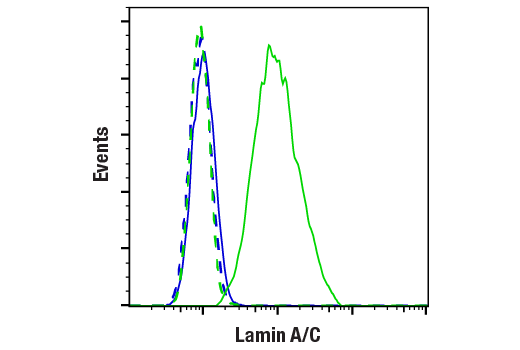
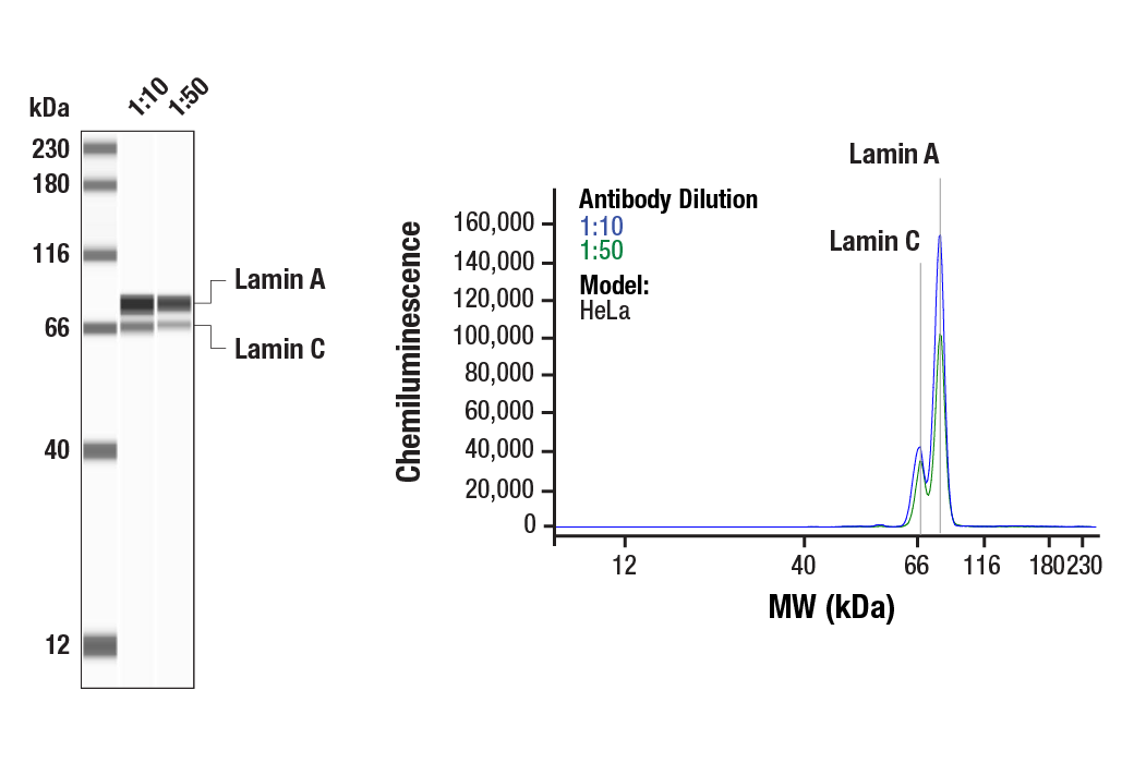
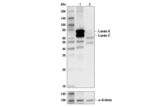
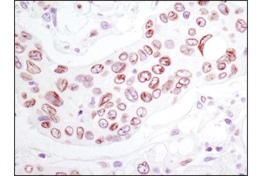
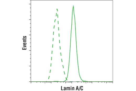
| Lamin A/C (4C11) Mouse Monoclonal Antibody | 4777 | 20 µl | 74 (Lamin A), 63 (Lamin C) kDa | Mouse IgG2a |
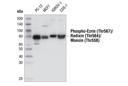
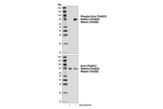
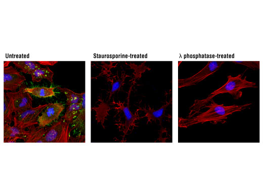
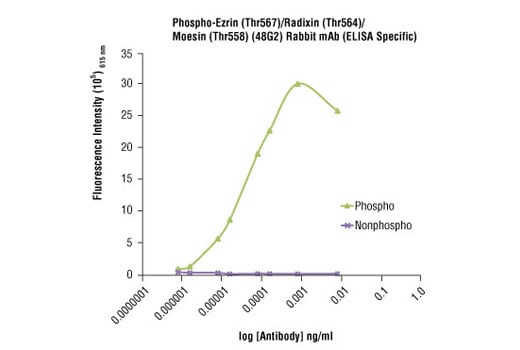
| Phospho-Ezrin (Thr567)/Radixin (Thr564)/Moesin (Thr558) (48G2) Rabbit Monoclonal Antibody | 3726 | 20 µl | 75 Moesin. 80 Ezrin, Radixin. kDa | Rabbit IgG |
| Anti-rabbit IgG, HRP-linked Antibody | 7074 | 100 µl | Goat |
Please visit cellsignal.com for individual component applications, species cross-reactivity, dilutions, protocols, and additional product information.
Description
Storage
Background
The Rab11-family interacting proteins (Rab11-FIPs) facilitate Rab11-dependent vesicle recycling through interaction with the conserved carboxyl terminal Rab11 binding domain (5,6). Rab11FIP1 has been shown to play a role in endocytic sorting and trafficking of EGFR and integrin subunits (6). Integrins are α/β heterodimeric cell surface receptors that mediate cell adhesion and migration and regulate cell growth and survival. Two significant α4 integrins, α4β1 and α4β7, interact with VCAM-1, fibronectin, and MAdCAM-1 at cell adhesions and have been shown to play an important role in cell trafficking during inflammatory processes (7-9). Lamins are nuclear membrane structural components important for maintaining normal cell functions. Lamin A/C is cleaved by caspase-6 and serves as a marker for caspase-6 activation. The cleavage of lamins results in nuclear dysregulation and cell death (10,11). The ezrin, radixin, and moesin (ERM) proteins function as linkers between the plasma membrane and the actin cytoskeleton and are involved in cell adhesion, membrane ruffling, and microvilli formation (12). ERM proteins undergo intra or intermolecular interaction between their amino- and carboxy-terminal domains, existing as inactive cytosolic monomers or dimers (13). Phosphorylation at a carboxy-terminal threonine residue (Thr567 of ezrin, Thr564 of radixin, Thr558 of moesin) disrupts the amino- and carboxy-terminal association and may play a key role in regulating ERM protein conformation and function (14,15). IQGAPs are scaffolding proteins involved in mediating cytoskeletal function that contain multiple protein interaction domains (16). IQGAP1 is ubiquitously expressed and has been found to interact with APC (17) and the CLIP170 complex in response to small GTPases, promoting cell polarization and migration (18). IKKε is an IKK-related kinase that functions as part of the signal-stimulated noncanonical pathway of NF-kB activation (19). IKKε plays a role in the immune response and also impacts cell proliferation and transformation (20).
Background References
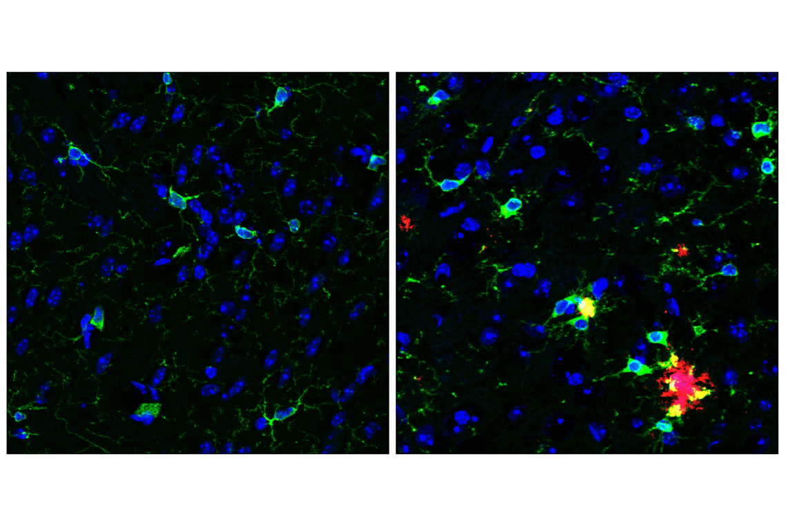
- Friedman, B.A. et al. (2018) Cell Rep 22, 832-47.
- Zhang, Y. et al. (2014) J Neurosci 34, 11929-47.
- Kitamura, D. et al. (1995) Biochem Biophys Res Commun 208, 1137-46.
- Srinivasula, S.M. et al. (2002) J Biol Chem 277, 21119-22.
- Hales, C.M. et al. (2001) J Biol Chem 276, 39067-75.
- Baetz, N.W. and Goldenring, J.R. (2013) Mol Biol Cell 24, 643-58.
- Hood, J.D. and Cheresh, D.A. (2002) Nat Rev Cancer 2, 91-100.
- Liu, S. et al. (2000) J Cell Sci 113 (Pt 20), 3563-71.
- Kummer, C. and Ginsberg, M.H. (2006) Biochem Pharmacol 72, 1460-8.
- Oberhammer, F.A. et al. (1994) J Cell Biol 126, 827-37.
- Rao, L. et al. (1996) J Cell Biol 135, 1441-55.
- Tsukita, S. and Yonemura, S. (1999) J Biol Chem 274, 34507-10.
- Mangeat, P. et al. (1999) Trends Cell Biol 9, 187-92.
- Matsui, T. et al. (1998) J Cell Biol 140, 647-57.
- Gautreau, A. et al. (2000) J Cell Biol 150, 193-203.
- Briggs, M.W. and Sacks, D.B. (2003) EMBO Rep 4, 571-4.
- Watanabe, T. et al. (2004) Dev Cell 7, 871-83.
- Fukata, M. et al. (2002) Cell 109, 873-85.
- Sun, S.C. et al. (2013) Trends Immunol 34, 282-9.
- Verhelst, K. et al. (2013) Biochem Pharmacol 85, 873-80.
Trademarks and Patents
Cell Signaling Technology is a trademark of Cell Signaling Technology, Inc.
All other trademarks are the property of their respective owners. Visit cellsignal.com/trademarks for more information.
Limited Uses
Except as otherwise expressly agreed in a writing signed by a legally authorized representative of CST, the following terms apply to Products provided by CST, its affiliates or its distributors. Any Customer's terms and conditions that are in addition to, or different from, those contained herein, unless separately accepted in writing by a legally authorized representative of CST, are rejected and are of no force or effect.
Products are labeled with For Research Use Only or a similar labeling statement and have not been approved, cleared, or licensed by the FDA or other regulatory foreign or domestic entity, for any purpose. Customer shall not use any Product for any diagnostic or therapeutic purpose, or otherwise in any manner that conflicts with its labeling statement. Products sold or licensed by CST are provided for Customer as the end-user and solely for research and development uses. Any use of Product for diagnostic, prophylactic or therapeutic purposes, or any purchase of Product for resale (alone or as a component) or other commercial purpose, requires a separate license from CST. Customer shall (a) not sell, license, loan, donate or otherwise transfer or make available any Product to any third party, whether alone or in combination with other materials, or use the Products to manufacture any commercial products, (b) not copy, modify, reverse engineer, decompile, disassemble or otherwise attempt to discover the underlying structure or technology of the Products, or use the Products for the purpose of developing any products or services that would compete with CST products or services, (c) not alter or remove from the Products any trademarks, trade names, logos, patent or copyright notices or markings, (d) use the Products solely in accordance with CST Product Terms of Sale and any applicable documentation, and (e) comply with any license, terms of service or similar agreement with respect to any third party products or services used by Customer in connection with the Products.
Revision 1

Revision 1
Revision 1

Revision 1
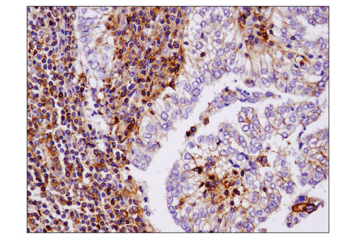
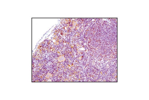
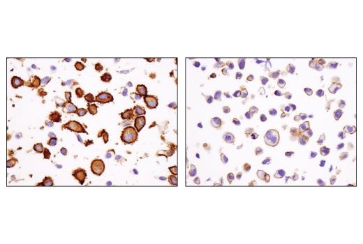
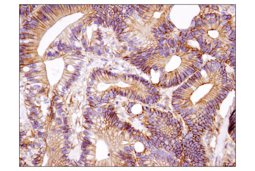
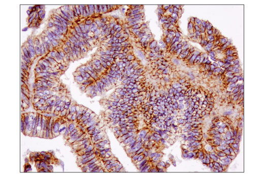
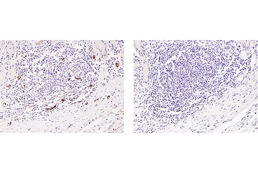
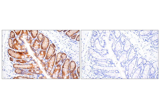
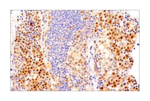
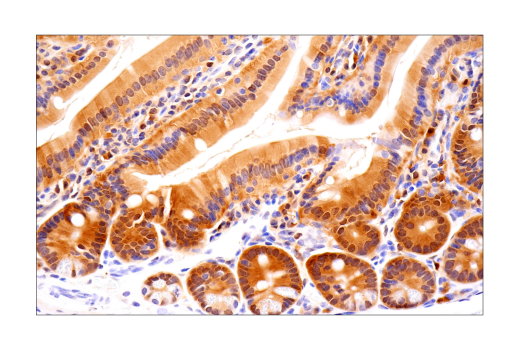
Immunohistochemical analysis of paraffin-embedded human infiltrating papillary carcinoma of the breast using IQGAP1 (D8K4X) XP® Rabbit mAb.
Revision 1
Revision 1

Revision 1
Revision 1


Revision 1
Revision 1
Revision 1
Revision 1
Revision 1

Revision 1
Revision 1
Revision 1
Revision 1
Revision 1