Revision 1
#49938
Store at -20C
Microglia Proliferation Module Antibody Sampler Kit
1 Kit
(7 x 20 microliters)
877-616-CELL (2355)
877-678-TECH (8324)
3 Trask Lane | Danvers | Massachusetts | 01923 | USA
For Research Use Only. Not for Use in Diagnostic Procedures.
| Product Includes | Product # | Quantity | Mol. Wt | Isotype/Source |
|---|---|---|---|---|
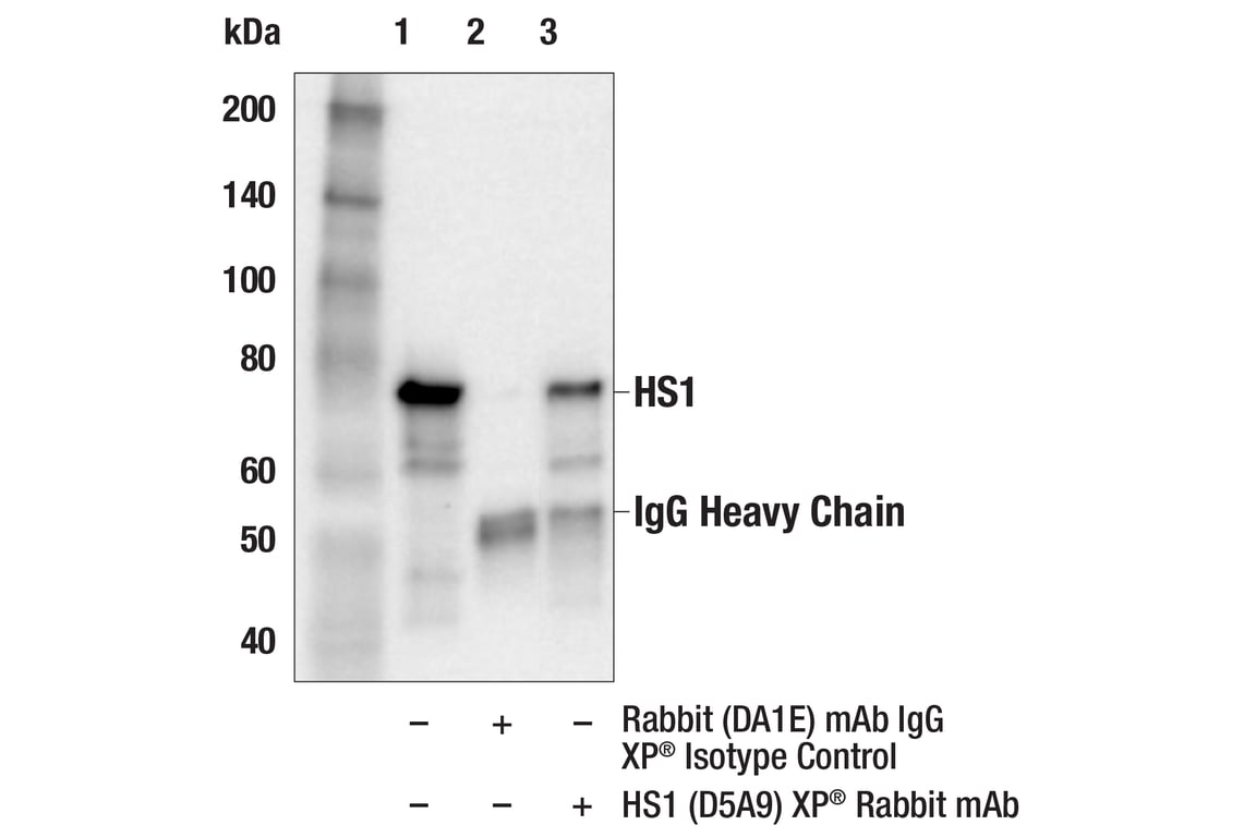
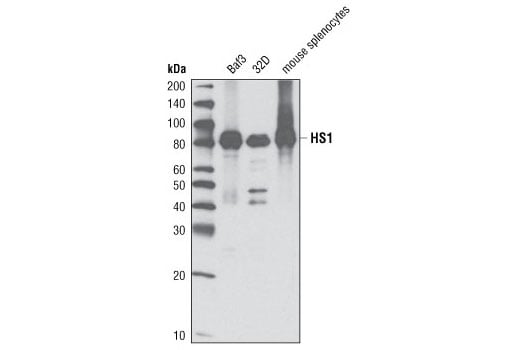
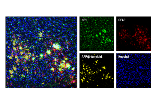
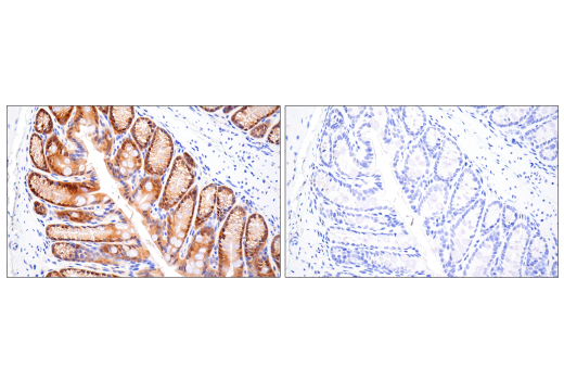
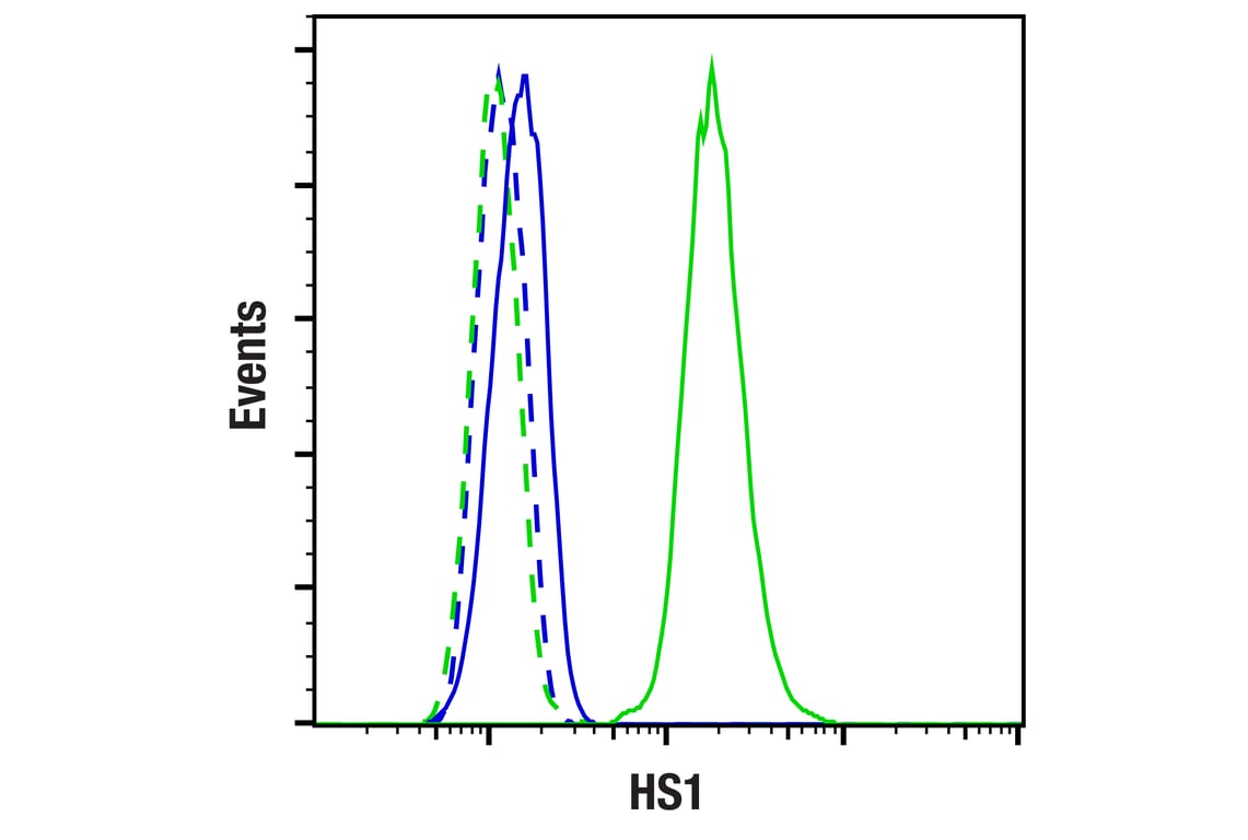
| HS1 (D5A9) Rabbit Monoclonal Antibody | 3892 | 20 µl | 80 kDa | Rabbit IgG |
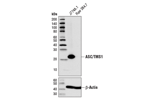
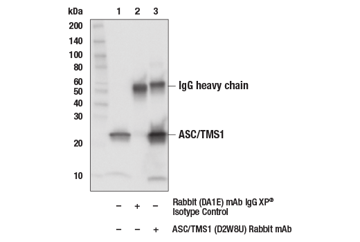
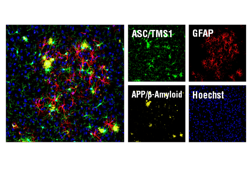
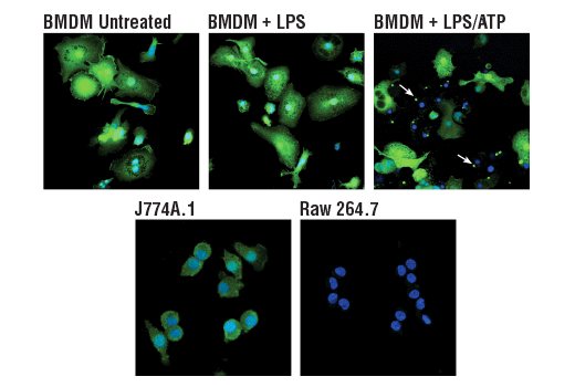
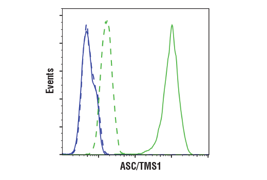
| ASC/TMS1 (D2W8U) Rabbit Monoclonal Antibody | 67824 | 20 µl | 22 kDa | Rabbit IgG |
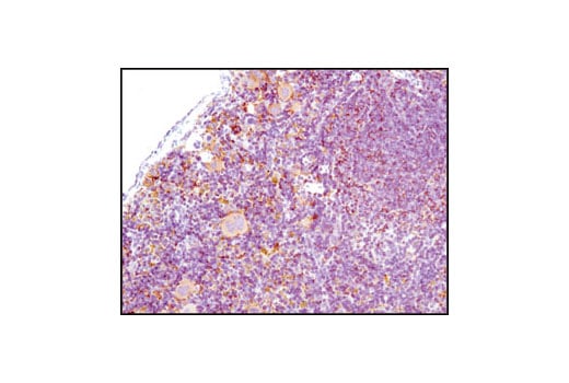
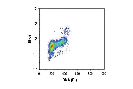
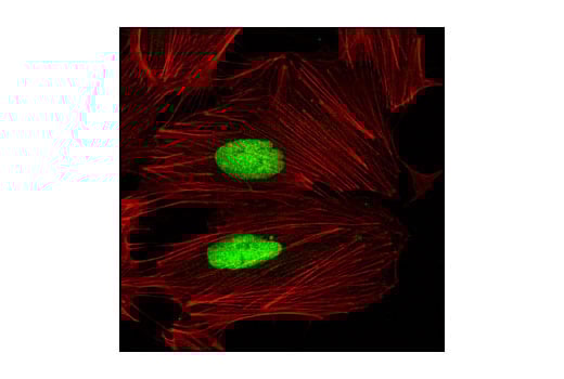
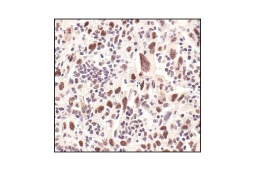
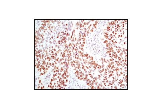
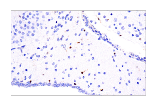
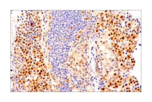
| Ki-67 (D3B5) Rabbit Monoclonal Antibody | 9129 | 20 µl | Rabbit IgG | |
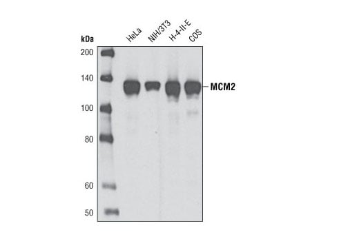
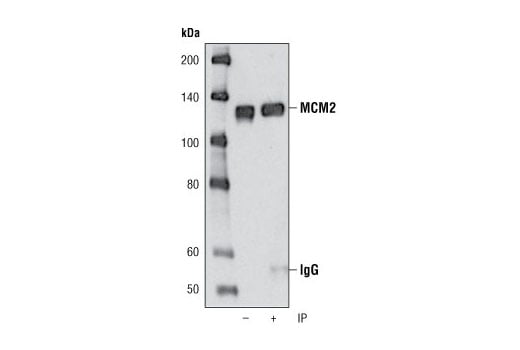
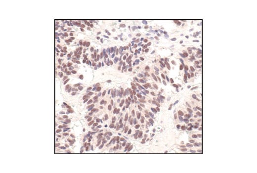
| MCM2 (D7G11) Rabbit Monoclonal Antibody | 3619 | 20 µl | 125 kDa | Rabbit IgG |
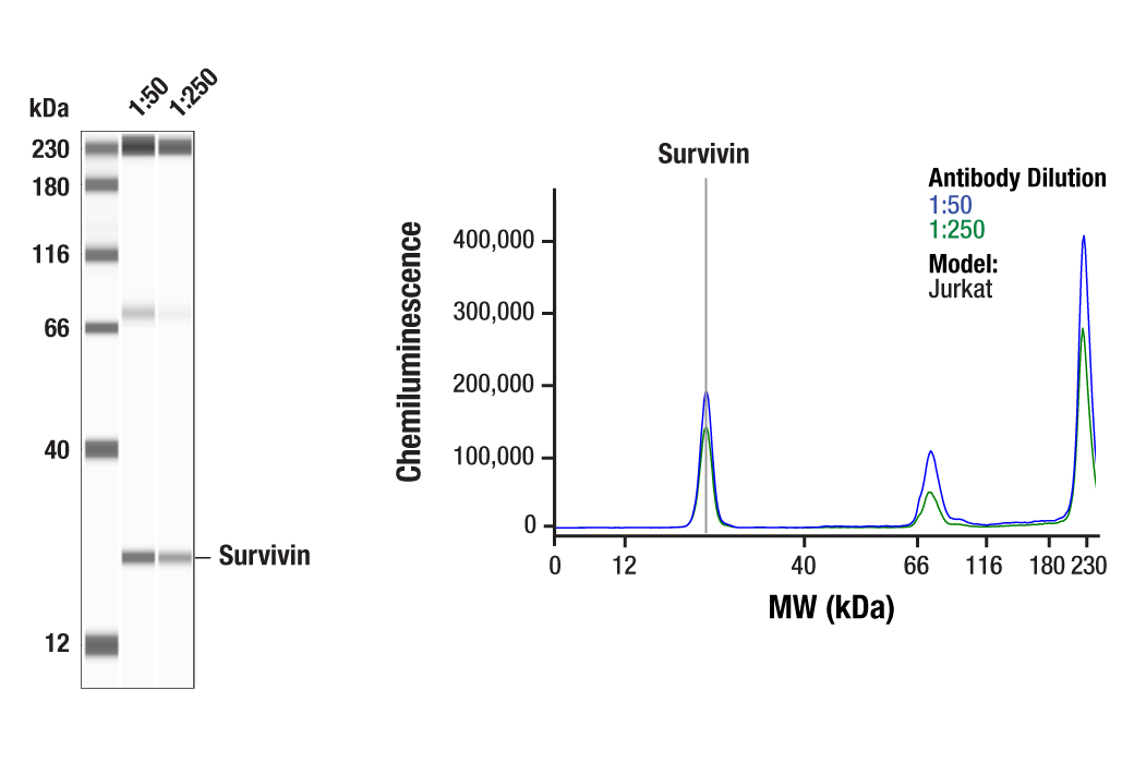
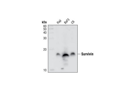
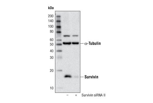
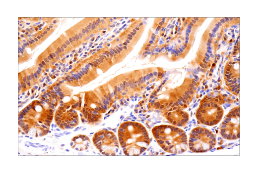
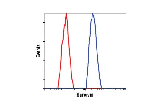
| Survivin (71G4B7) Rabbit Monoclonal Antibody | 2808 | 20 µl | 16 kDa | Rabbit IgG |
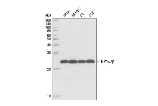
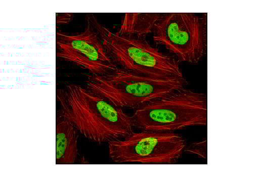
| HP1 alpha/beta (C7F11) Rabbit Monoclonal Antibody | 2623 | 20 µl | 25 kDa | Rabbit IgG |
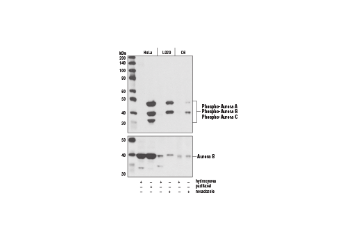
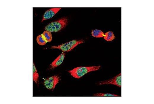
| Phospho-Aurora A (Thr288)/Aurora B (Thr232)/Aurora C (Thr198) (D13A11) Rabbit Monoclonal Antibody | 2914 | 20 µl | 35, 40, 48 kDa | Rabbit IgG |
| Anti-rabbit IgG, HRP-linked Antibody | 7074 | 100 µl | Goat |
Please visit cellsignal.com for individual component applications, species cross-reactivity, dilutions, protocols, and additional product information.
Description
Storage
Background
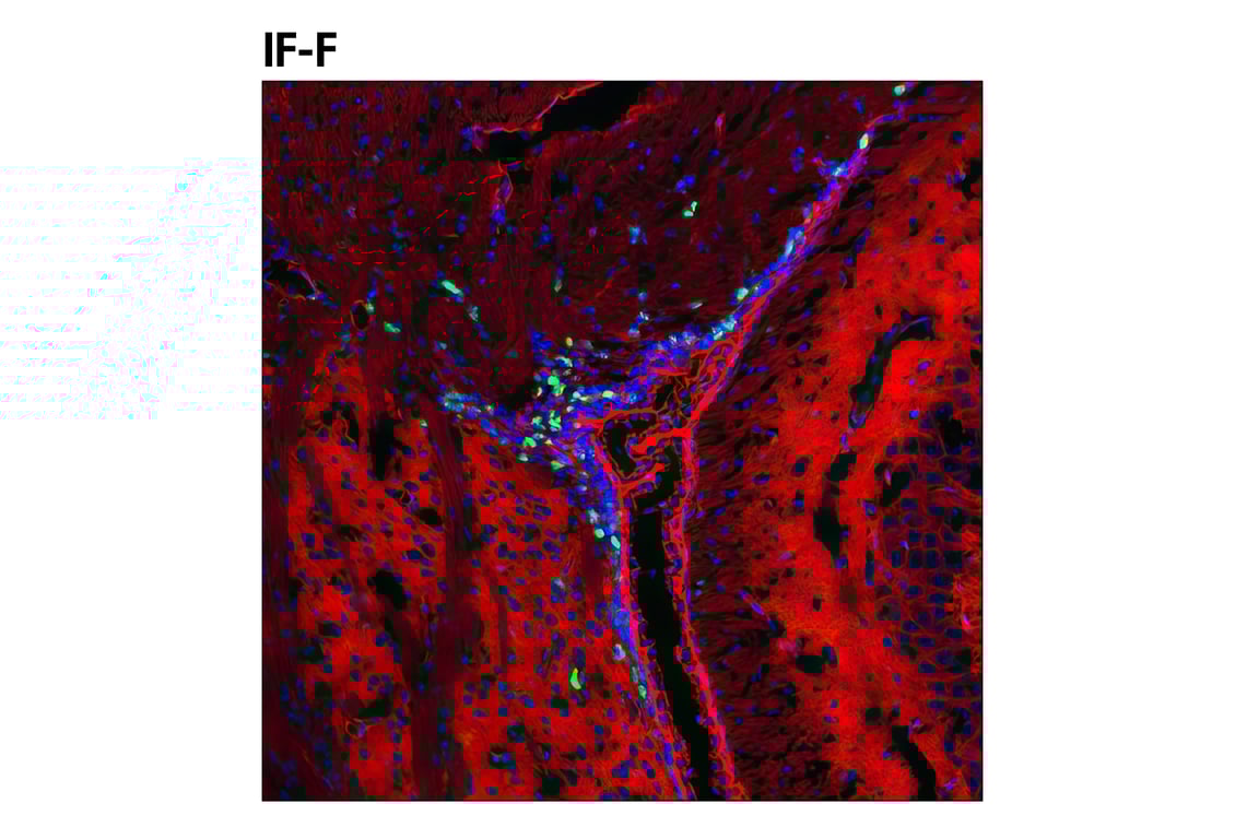
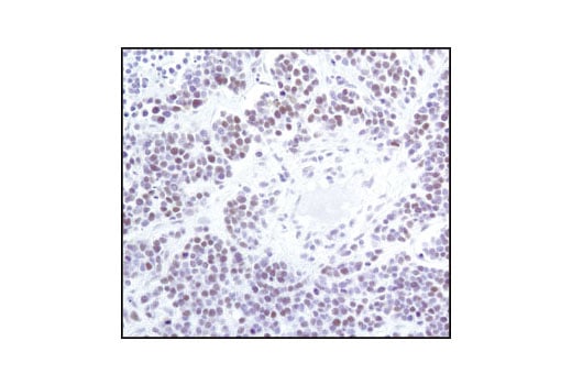
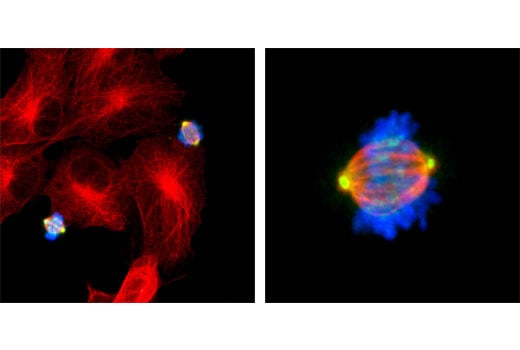
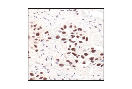
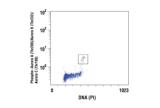
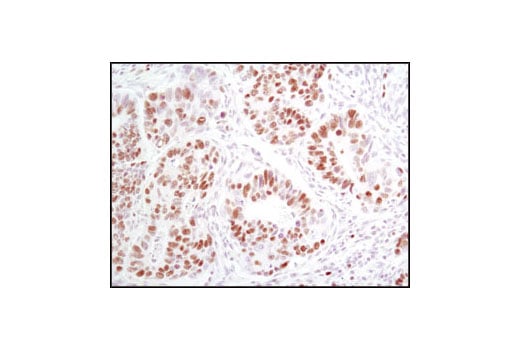
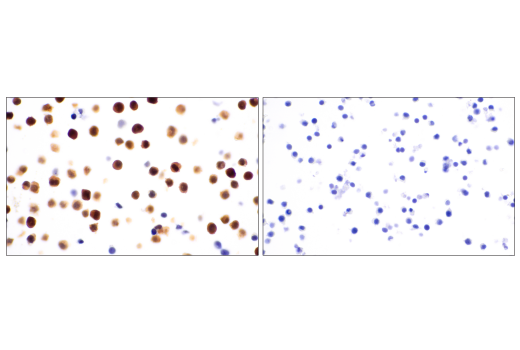
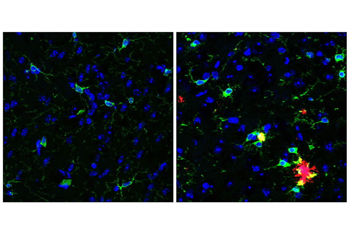
Ki-67 is a nuclear nonhistone protein (5) universally expressed among proliferating cells and absent in quiescent cells (6). Minichromosome maintenance protein 2 (MCM2) is a nuclear protein that plays a role in DNA replication and cell division (7) and is commonly used as a marker for cell proliferation, including brain tissue (8). Survivin binds and inhibits caspase-3, controlling the checkpoint in the G2/M-phase of the cell cycle by inhibiting apoptosis and promoting cell division (9). Aurora A, B, and C are a family of highly conserved serine/threonine kinases that regulate chromosomal alignment and segregation during mitosis and meiosis. Their activity requires autophosphorylation of a threonine within their kinase domain at site Thr288 of Aurora A, Thr232 of Aurora B, and Thr198 of Aurora C (10). Heterochromatin protein 1 (HP1) α and β are heterochromatic adaptor molecules involved in both gene silencing and higher order chromatin structure (11).
Background References
- Friedman, B.A. et al. (2018) Cell Rep 22, 832-47.
- Zhang, Y. et al. (2014) J Neurosci 34, 11929-47.
- Kitamura, D. et al. (1995) Biochem Biophys Res Commun 208, 1137-46.
- Srinivasula, S.M. et al. (2002) J Biol Chem 277, 21119-22.
- Gerdes, J. et al. (1983) Int J Cancer 31, 13-20.
- Weigel, M.T. and Dowsett, M. (2010) Endocr Relat Cancer 17, R245-62.
- Mincheva, A. et al. (1994) Cytogenet Cell Genet 65, 276-7.
- Doorn, K.J. et al. (2014) Neural Plast 2014, 959154.
- Li, F. et al. (1999) Nat Cell Biol 1, 461-6.
- Goldenson, B. and Crispino, J.D. (2015) Oncogene 34, 537-45.
- Maison, C. and Almouzni, G. (2004) Nat Rev Mol Cell Biol 5, 296-304.
Trademarks and Patents
Cell Signaling Technology is a trademark of Cell Signaling Technology, Inc.
All other trademarks are the property of their respective owners. Visit cellsignal.com/trademarks for more information.
Limited Uses
Except as otherwise expressly agreed in a writing signed by a legally authorized representative of CST, the following terms apply to Products provided by CST, its affiliates or its distributors. Any Customer's terms and conditions that are in addition to, or different from, those contained herein, unless separately accepted in writing by a legally authorized representative of CST, are rejected and are of no force or effect.
Products are labeled with For Research Use Only or a similar labeling statement and have not been approved, cleared, or licensed by the FDA or other regulatory foreign or domestic entity, for any purpose. Customer shall not use any Product for any diagnostic or therapeutic purpose, or otherwise in any manner that conflicts with its labeling statement. Products sold or licensed by CST are provided for Customer as the end-user and solely for research and development uses. Any use of Product for diagnostic, prophylactic or therapeutic purposes, or any purchase of Product for resale (alone or as a component) or other commercial purpose, requires a separate license from CST. Customer shall (a) not sell, license, loan, donate or otherwise transfer or make available any Product to any third party, whether alone or in combination with other materials, or use the Products to manufacture any commercial products, (b) not copy, modify, reverse engineer, decompile, disassemble or otherwise attempt to discover the underlying structure or technology of the Products, or use the Products for the purpose of developing any products or services that would compete with CST products or services, (c) not alter or remove from the Products any trademarks, trade names, logos, patent or copyright notices or markings, (d) use the Products solely in accordance with CST Product Terms of Sale and any applicable documentation, and (e) comply with any license, terms of service or similar agreement with respect to any third party products or services used by Customer in connection with the Products.
Revision 1
Revision 1
Revision 1

Revision 1
Revision 1

Revision 1
Revision 1

Revision 1
Revision 1
Revision 1
Revision 1
Revision 1


Revision 1
Revision 1
Revision 1
Revision 1