Revision 2
#43110
Store at -20C
Mitophagy Antibody Sampler Kit
1 Kit
(9 x 20 microliters)
877-616-CELL (2355)
877-678-TECH (8324)
3 Trask Lane | Danvers | Massachusetts | 01923 | USA
For Research Use Only. Not for Use in Diagnostic Procedures.
| Product Includes | Product # | Quantity | Mol. Wt | Isotype/Source |
|---|---|---|---|---|
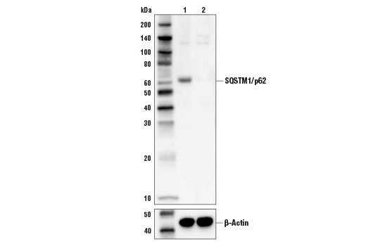
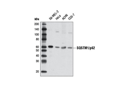
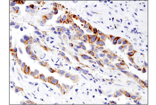
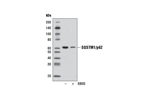
| SQSTM1/p62 (D5E2) Rabbit Monoclonal Antibody | 8025 | 20 µl | 62 kDa | Rabbit IgG |
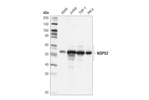
| NDP52 (D1E4A) Rabbit Monoclonal Antibody | 60732 | 20 µl | 52, 60 kDa | Rabbit IgG |
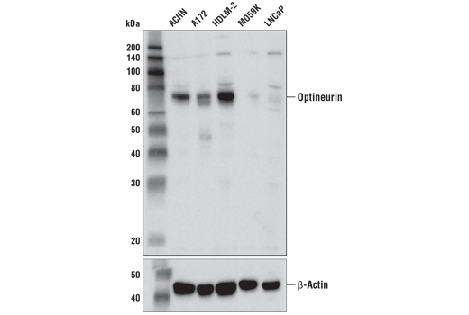
| Optineurin (D2L8S) Rabbit Monoclonal Antibody | 58981 | 20 µl | 75 kDa | Rabbit IgG |
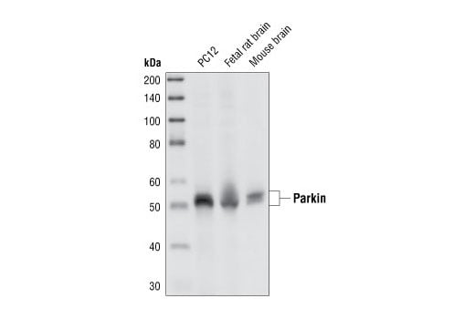
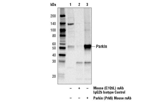
| Parkin (Prk8) Mouse Monoclonal Antibody | 4211 | 20 µl | 50 kDa | Mouse IgG2b |
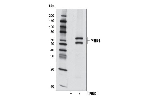
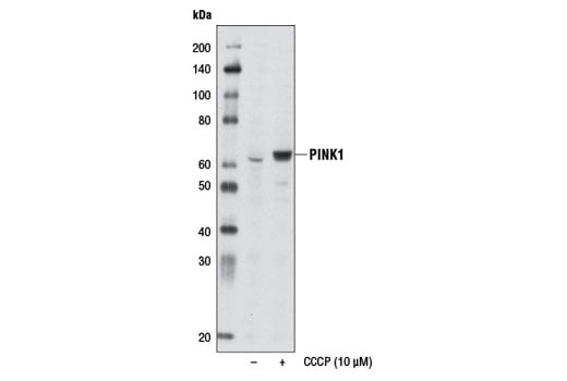
| PINK1 (D8G3) Rabbit Monoclonal Antibody | 6946 | 20 µl | 60, 50 kDa | Rabbit IgG |
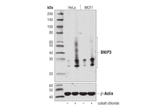
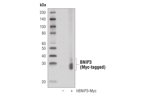
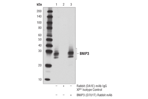
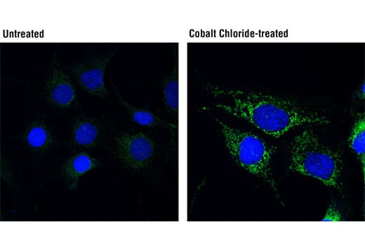
| BNIP3 (D7U1T) Rabbit Monoclonal Antibody | 44060 | 20 µl | 22-28, 50-55 kDa | Rabbit IgG |
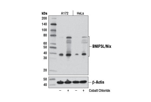
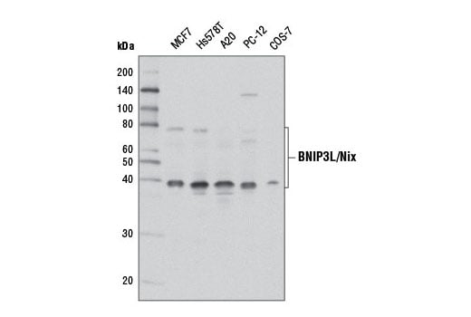
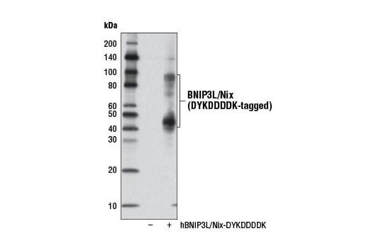
| BNIP3L/Nix (D4R4B) Rabbit Monoclonal Antibody | 12396 | 20 µl | 38, 76 kDa | Rabbit IgG |
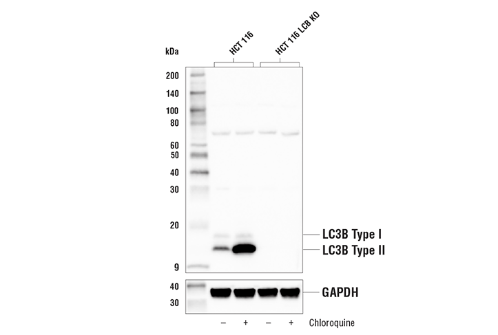
| LC3B (D11) Rabbit Monoclonal Antibody | 3868 | 20 µl | 14, 16 kDa | Rabbit IgG |
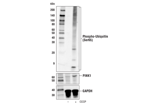
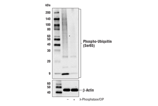
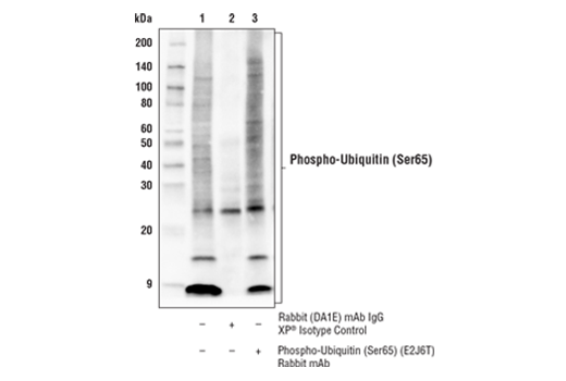
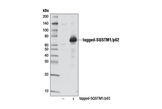
| Phospho-Ubiquitin (Ser65) (E2J6T) Rabbit Monoclonal Antibody | 62802 | 20 µl | Rabbit IgG | |
| Anti-rabbit IgG, HRP-linked Antibody | 7074 | 100 µl | Goat |
Please visit cellsignal.com for individual component applications, species cross-reactivity, dilutions, protocols, and additional product information.
Description
Storage
Background
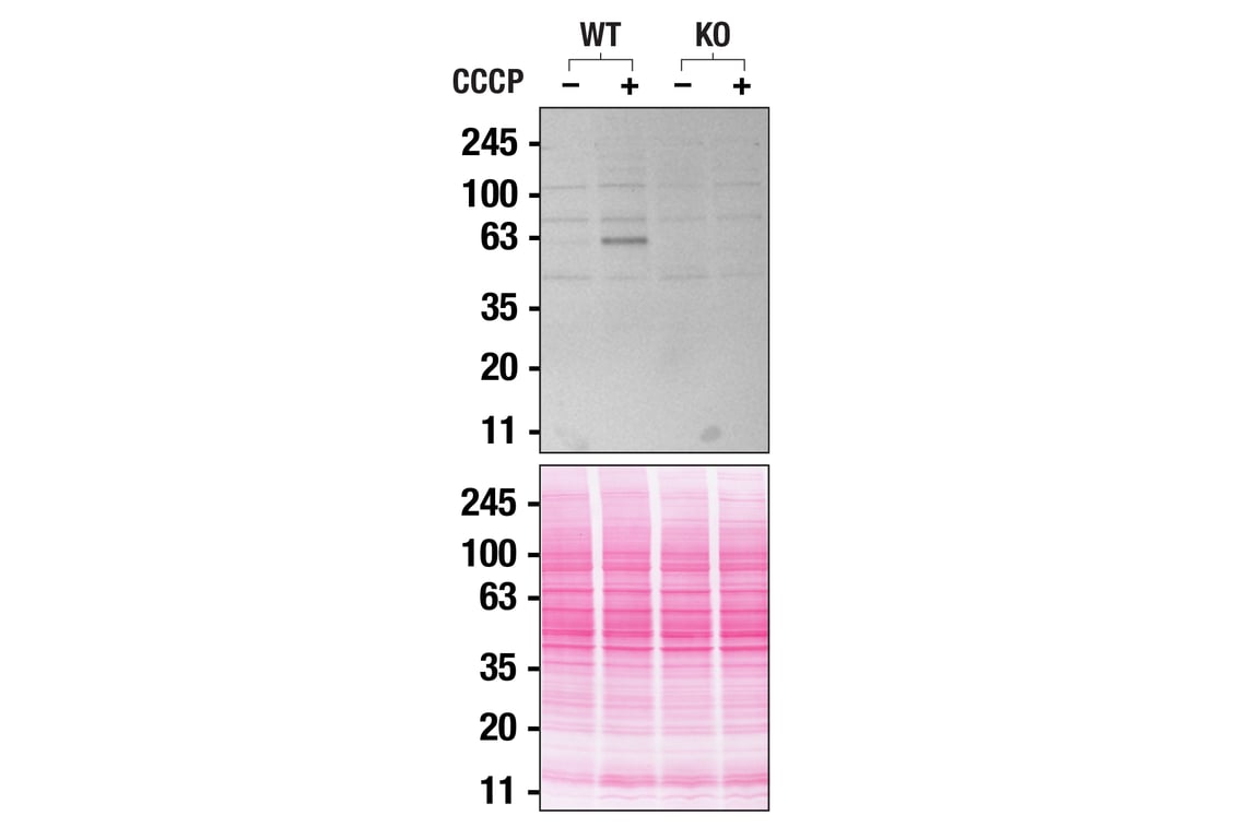
Non-hypoxic induction of mitophagy can be regulated by the PINK1/Parkin pathway, which plays causative roles in neurodegenerative disease, most notably Parkinson’s disease (10, 11). PINK1 is a mitochondrial serine/threonine kinase that is stabilized on the outer mitochondrial membrane of damaged mitochondria. Substrates of PINK1 include the E3 ubiquitin ligase Parkin and ubiquitin itself (12-14). Phosphorylation of Parkin as well as binding to phosphorylated ubiquitin leads to accumulation of ubiquitinated chains on multiple mitochondrial proteins. Ubiquitinated proteins are recognized by selective cargo receptors including SQSTM1/p62, Optineurin, and NDP52 (15-16). Autophagy cargo receptors contain an LC3-interacting region (LIR) required for binding to Atg8/LC3 family members and targeting to the autophagosome (3).
Background References
- Reggiori, F. and Klionsky, D.J. (2002) Eukaryot Cell 1, 11-21.
- Codogno, P. and Meijer, A.J. (2005) Cell Death Differ 12 Suppl 2, 1509-18.
- Birgisdottir, Å.B. et al. (2013) J Cell Sci 126, 3237-47.
- Xu, Z. et al. (2015) Acta Biochim Biophys Sin (Shanghai) 47, 571-80.
- Mancias, J.D. and Kimmelman, A.C. (2016) J Mol Biol 428, 1659-80.
- Liu, L. et al. (2012) Nat Cell Biol 14, 177-85.
- Wu, W. et al. (2014) EMBO Rep 15, 566-75.
- Sowter, H.M. et al. (2001) Cancer Res 61, 6669-73.
- Sandoval, H. et al. (2008) Nature 454, 232-5.
- Kitada, T. et al. (1998) Nature 392, 605-8.
- Valente, E.M. et al. (2004) Science 304, 1158-60.
- Kim, Y. et al. (2008) Biochem Biophys Res Commun 377, 975-80.
- Kane, L.A. et al. (2014) J Cell Biol 205, 143-53.
- Koyano, F. et al. (2014) Nature 510, 162-6.
- Heo, J.M. et al. (2015) Mol Cell 60, 7-20.
- Lazarou, M. et al. (2015) Nature 524, 309-314.
Trademarks and Patents
Cell Signaling Technology is a trademark of Cell Signaling Technology, Inc.
All other trademarks are the property of their respective owners. Visit cellsignal.com/trademarks for more information.
Limited Uses
Except as otherwise expressly agreed in a writing signed by a legally authorized representative of CST, the following terms apply to Products provided by CST, its affiliates or its distributors. Any Customer's terms and conditions that are in addition to, or different from, those contained herein, unless separately accepted in writing by a legally authorized representative of CST, are rejected and are of no force or effect.
Products are labeled with For Research Use Only or a similar labeling statement and have not been approved, cleared, or licensed by the FDA or other regulatory foreign or domestic entity, for any purpose. Customer shall not use any Product for any diagnostic or therapeutic purpose, or otherwise in any manner that conflicts with its labeling statement. Products sold or licensed by CST are provided for Customer as the end-user and solely for research and development uses. Any use of Product for diagnostic, prophylactic or therapeutic purposes, or any purchase of Product for resale (alone or as a component) or other commercial purpose, requires a separate license from CST. Customer shall (a) not sell, license, loan, donate or otherwise transfer or make available any Product to any third party, whether alone or in combination with other materials, or use the Products to manufacture any commercial products, (b) not copy, modify, reverse engineer, decompile, disassemble or otherwise attempt to discover the underlying structure or technology of the Products, or use the Products for the purpose of developing any products or services that would compete with CST products or services, (c) not alter or remove from the Products any trademarks, trade names, logos, patent or copyright notices or markings, (d) use the Products solely in accordance with CST Product Terms of Sale and any applicable documentation, and (e) comply with any license, terms of service or similar agreement with respect to any third party products or services used by Customer in connection with the Products.
Revision 2
Revision 2
Revision 2

Revision 2

Revision 2
Revision 2

Revision 2
Revision 2

Revision 2

Revision 2

Revision 2
Revision 2
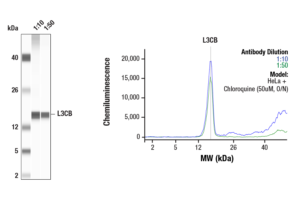
Simple Western™ analysis of lysates (1mg/ml) from HeLa cells treated with Chloroquine (50uM, O/N) using LC3B (D11) XP® Rabbit mAb #3868. The virtual lane view (left) shows the target band (as indicated) at 1:10 and 1:50 dilutions of primary antibody. The corresponding electropherogram view (right) plots chemiluminescence by molecular weight along the capillary at 1:10 (blue line) and 1:50 (green line) dilutions of primary antibody. This experiment was performed under reducing conditions on the Jess™ Simple Western instrument from ProteinSimple, a BioTechne brand, using the 2-40kDa.