Revision 2
#48734
Store at -20C
Mouse Reactive Cell Death and Autophagy Antibody Sampler Kit
1 Kit
(9 x 20 microliters)
877-616-CELL (2355)
877-678-TECH (8324)
3 Trask Lane | Danvers | Massachusetts | 01923 | USA
For Research Use Only. Not for Use in Diagnostic Procedures.
| Product Includes | Product # | Quantity | Mol. Wt | Isotype/Source |
|---|---|---|---|---|
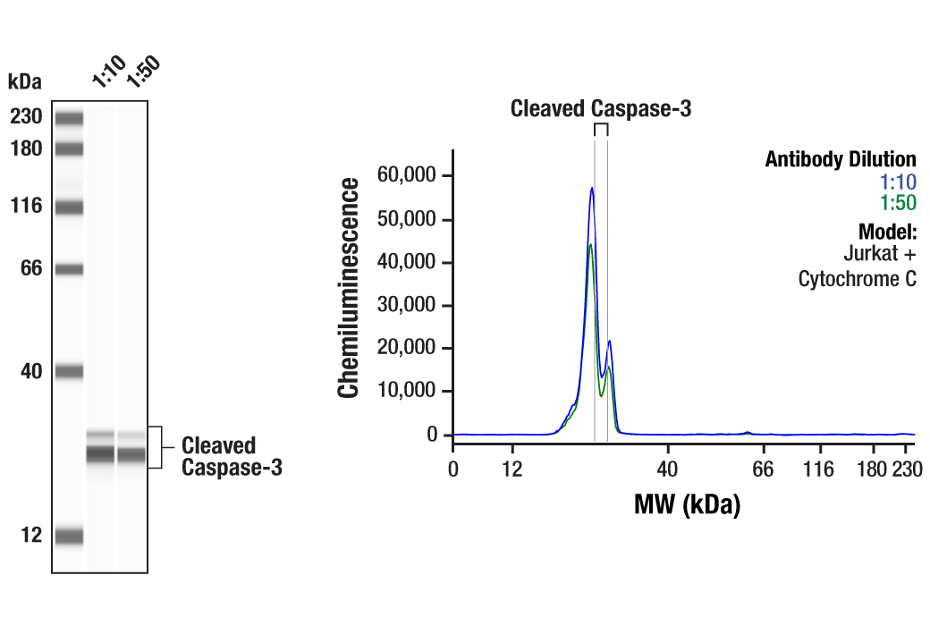
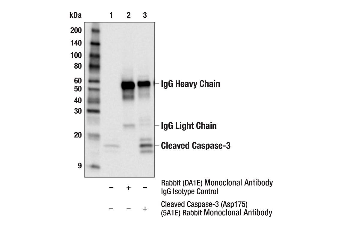
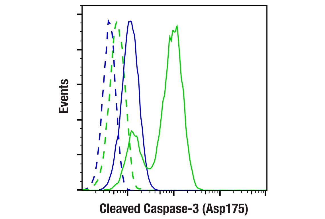
| Cleaved Caspase-3 (Asp175) (5A1E) Rabbit Monoclonal Antibody | 9664 | 20 µl | 17, 19 kDa | Rabbit IgG |
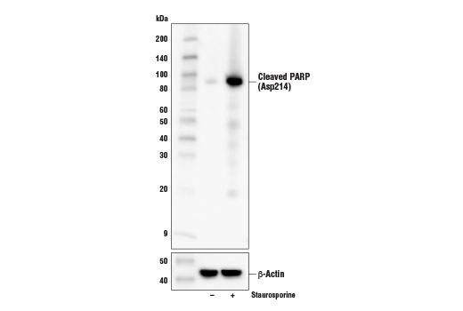
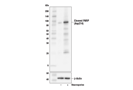
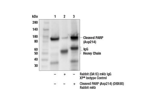
| Cleaved PARP (Asp214) (D6X6X) Rabbit Monoclonal Antibody | 94885 | 20 µl | 89 kDa | Rabbit IgG |
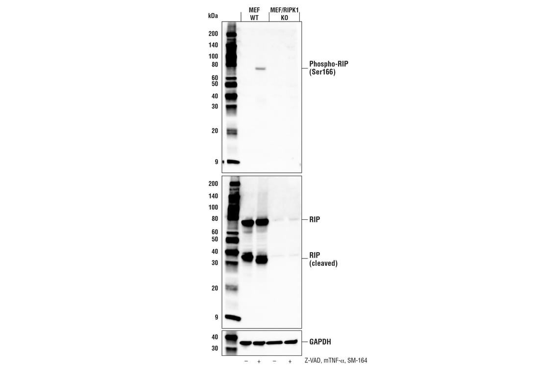
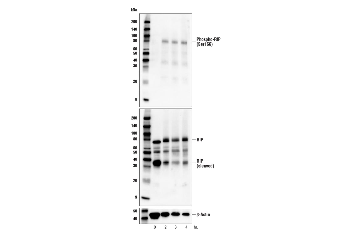
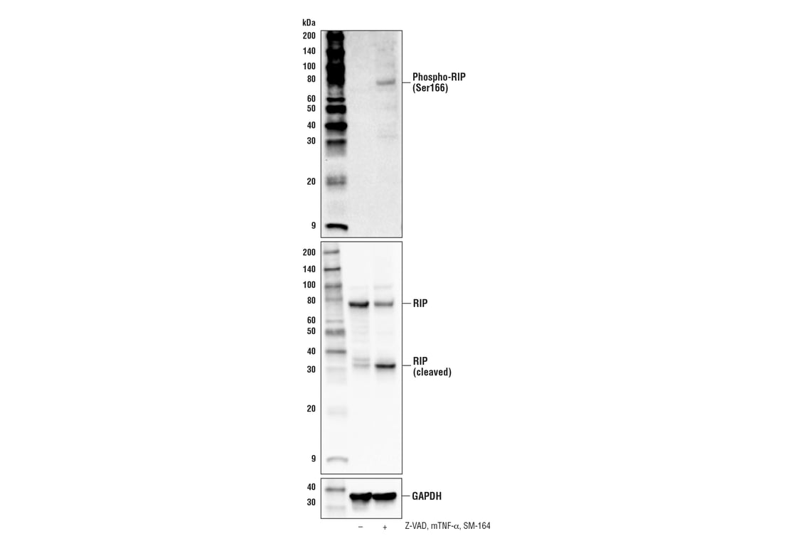
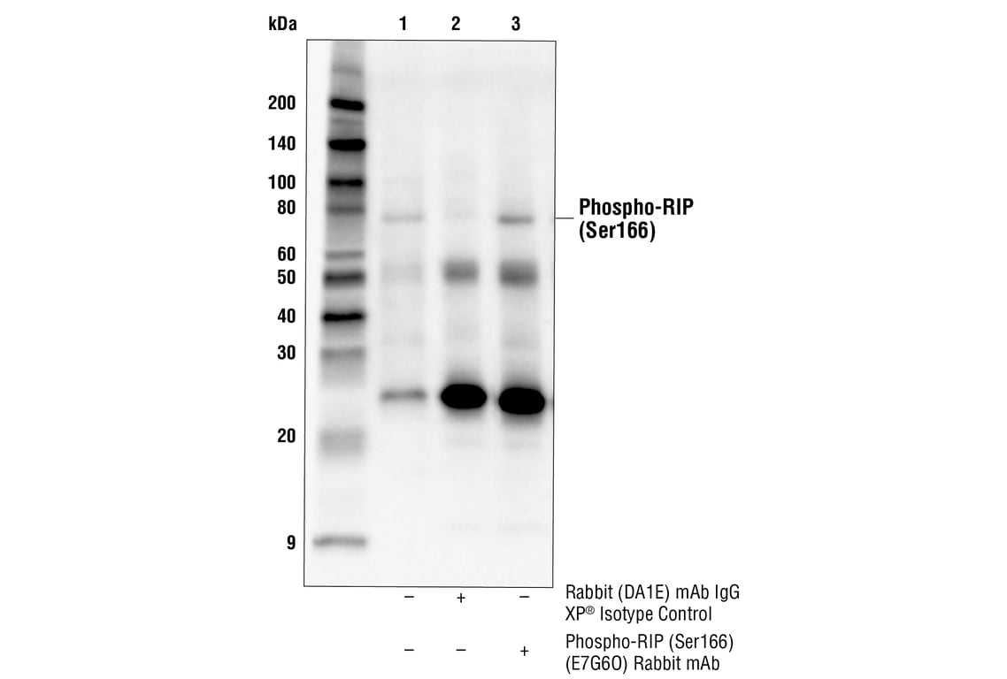
| Phospho-RIP (Ser166) (E7G6O) Rabbit Monoclonal Antibody | 53286 | 20 µl | 78 kDa | Rabbit IgG |
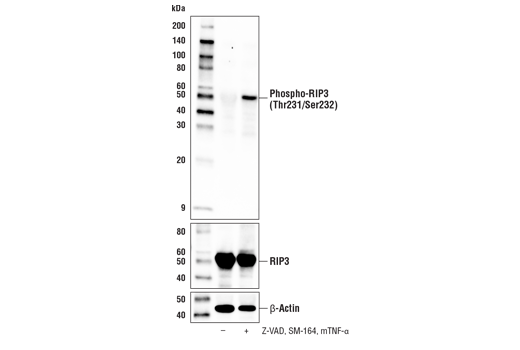
| Phospho-RIP3 (Thr231/Ser232) (E7S1R) Rabbit Monoclonal Antibody | 91702 | 20 µl | 46-62 kDa | Rabbit IgG |
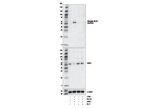
| Phospho-MLKL (Ser345) (D6E3G) Rabbit Monoclonal Antibody | 37333 | 20 µl | 54 kDa | Rabbit IgG |
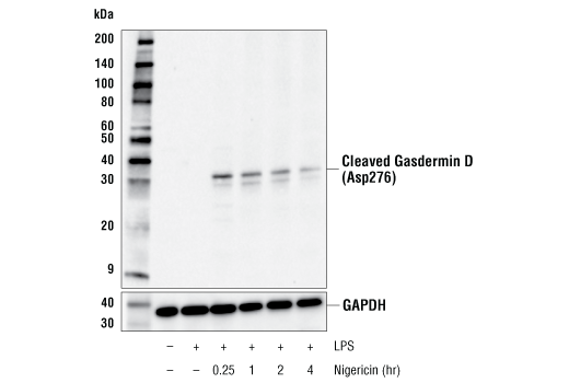
| Cleaved Gasdermin D (Asp276) (E3E3P) Rabbit Monoclonal Antibody | 10137 | 20 µl | 31 kDa | Rabbit IgG |
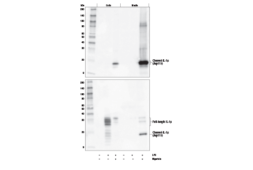
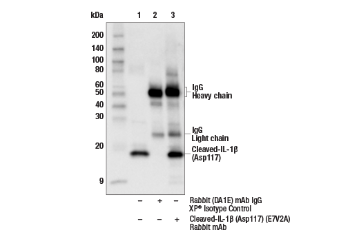
| Cleaved-IL-1 beta (Asp117) (E7V2A) Rabbit Monoclonal Antibody | 63124 | 20 µl | 17 kDa | Rabbit IgG |
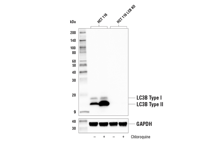
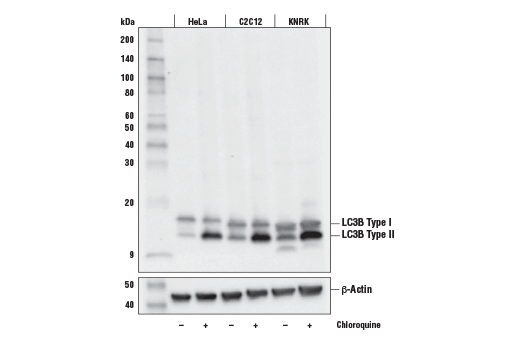
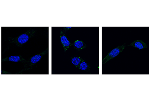
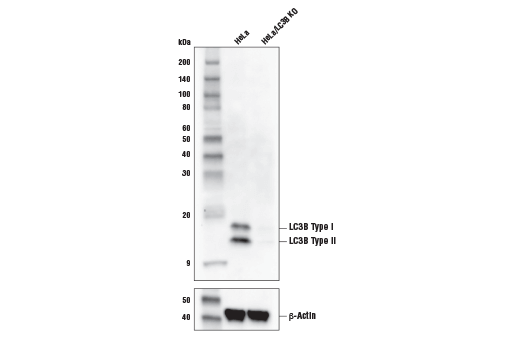
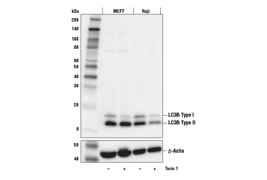
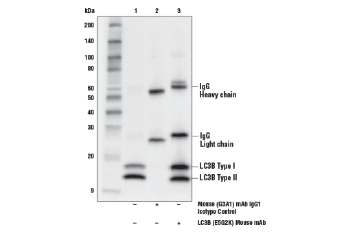
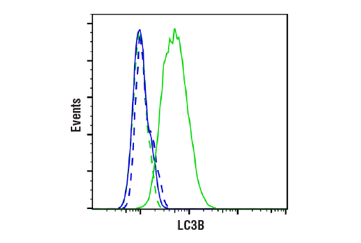
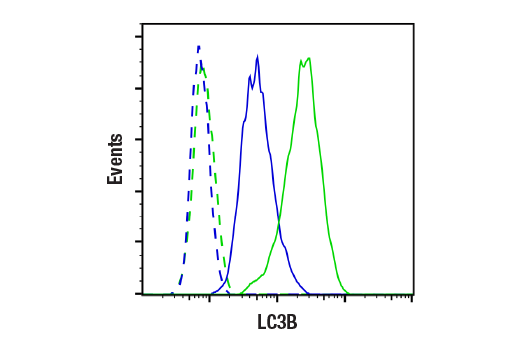
| LC3B (E5Q2K) Mouse Monoclonal Antibody | 83506 | 20 µl | 14, 16 kDa | Mouse IgG2b |
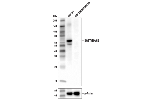
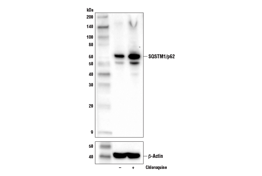
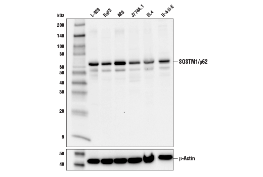
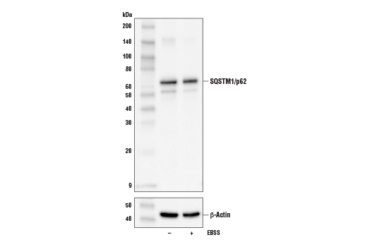
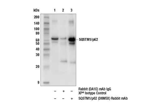
| SQSTM1/p62 (D6M5X) Rabbit Monoclonal Antibody | 23214 | 20 µl | 62 kDa | Rabbit IgG |
| Anti-rabbit IgG, HRP-linked Antibody | 7074 | 100 µl | Goat |
Please visit cellsignal.com for individual component applications, species cross-reactivity, dilutions, protocols, and additional product information.
Description
Storage
Background
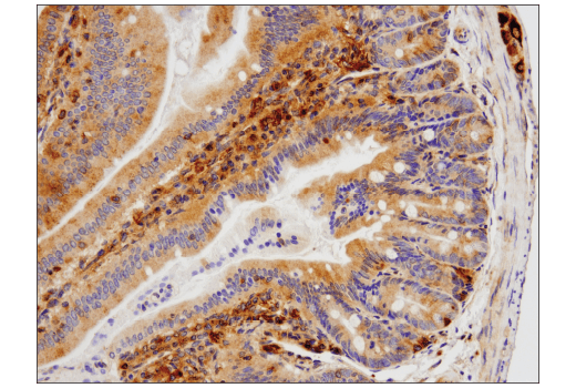
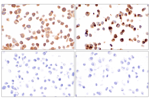
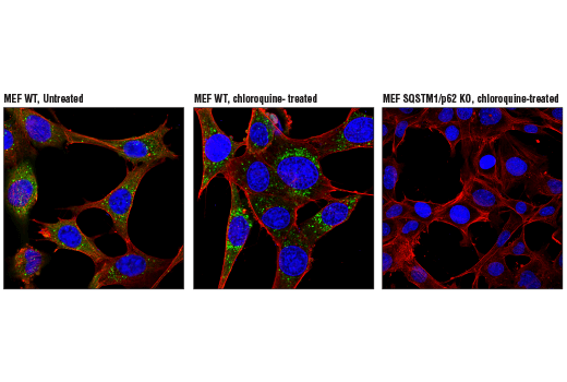
Type II cell death, or autophagy, manifests with extensive cytoplasmic vacuolization, and like apoptosis, can include phagocytic update. Autophagy is a catabolic process for the degradation of cellular components including protein aggregates, damaged organelles, and pathogens (3). The process involves the engulfment of these components into a double membrane structure, the autophagosome, which fuses to the lysosome for degradation. Autophagy requires, and can be monitored by, the conversion of LC3 family members, such as LC3B, from a type I form to a lipidated type II form that is incorporated into the autophagosome membrane and binds to a variety of cargo receptors. Cargo receptors such as SQSTM1/p62 bind LC3 along with ubiquitinated proteins that are targeted for degradation. SQSTM1/p62 is also degraded during this process, and thus its expression is frequently used to monitor this process.
Type III cell death, or necrosis, manifests with plasma membrane permeability with cellular swelling and fragmentation, and lacks a clear phagocytic response which then leads to an inflammatory signaling with the release of damage-associated molecular patterns (DAMPs). Necrosis can be triggered by multiple regulated pathways including necroptosis and pyroptosis. Necroptosis is regulated by the kinase activities of RIP and RIP3 and the pore forming ability of MLKL (4). Necroptosis requires the activation of RIP3 which then phosphorylates MLKL at Ser358 (Ser345 in mouse). Phosphorylation of MLKL leads to generation of a pore complex involved in cell swelling and the secretion of DAMPs. RIP3 activation is triggered through several RIP homotypic interaction motif (RHIM) domain interactions including RIP, TRIF, and ZBP1 and results in the phosphorylation of RIP3 at Ser227 (Thr231/Ser232 in mouse). Canonical necroptosis signaling is mediated by RIP, and this can be inhibited by necrostatins, small molecules that directly inhibit RIP kinase activity. Activation of RIP can be monitored through autophosphorylation sites including Ser166. Pyroptosis is generally induced in cells of the innate immune system, and is characterized by cleavage of Gasdermin D (5). The amino-terminal fragment of Gasdermin D produced following cleavage by inflammatory caspases (Caspase-1, -4, -5), oligomerizes to form a pore. Canonical cleavage of Gasdermin D occurs through a two-step process. The first step involves transcriptional regulation of targets such as NLRP3 and the pro-forms of IL-1β and IL-18. In the second execution step, Caspase-1 is activated through formation of inflammasome complexes. Activated Caspase-1 cleaves Gasdermin D as well as IL-1β and IL-18 to their mature forms, and these active cytokines are secreted through pores formed by Gasdermin D.
Trademarks and Patents
Cell Signaling Technology is a trademark of Cell Signaling Technology, Inc.
All other trademarks are the property of their respective owners. Visit cellsignal.com/trademarks for more information.
Limited Uses
Except as otherwise expressly agreed in a writing signed by a legally authorized representative of CST, the following terms apply to Products provided by CST, its affiliates or its distributors. Any Customer's terms and conditions that are in addition to, or different from, those contained herein, unless separately accepted in writing by a legally authorized representative of CST, are rejected and are of no force or effect.
Products are labeled with For Research Use Only or a similar labeling statement and have not been approved, cleared, or licensed by the FDA or other regulatory foreign or domestic entity, for any purpose. Customer shall not use any Product for any diagnostic or therapeutic purpose, or otherwise in any manner that conflicts with its labeling statement. Products sold or licensed by CST are provided for Customer as the end-user and solely for research and development uses. Any use of Product for diagnostic, prophylactic or therapeutic purposes, or any purchase of Product for resale (alone or as a component) or other commercial purpose, requires a separate license from CST. Customer shall (a) not sell, license, loan, donate or otherwise transfer or make available any Product to any third party, whether alone or in combination with other materials, or use the Products to manufacture any commercial products, (b) not copy, modify, reverse engineer, decompile, disassemble or otherwise attempt to discover the underlying structure or technology of the Products, or use the Products for the purpose of developing any products or services that would compete with CST products or services, (c) not alter or remove from the Products any trademarks, trade names, logos, patent or copyright notices or markings, (d) use the Products solely in accordance with CST Product Terms of Sale and any applicable documentation, and (e) comply with any license, terms of service or similar agreement with respect to any third party products or services used by Customer in connection with the Products.
Revision 2
Revision 2
Revision 2

Revision 2
Revision 2
Revision 2
Revision 2
Revision 2
Revision 2
Revision 2

Revision 2
Revision 2
Revision 2
Revision 2
Revision 2
Revision 2
Revision 2
Revision 2
Revision 2
Revision 2
Revision 2
Revision 2
Revision 2