Revision 1
#9862
Store at -20C
mTOR Substrates Antibody Sampler Kit
1 Kit
(5 x 20 microliters)
877-616-CELL (2355)
877-678-TECH (8324)
3 Trask Lane | Danvers | Massachusetts | 01923 | USA
For Research Use Only. Not for Use in Diagnostic Procedures.
| Product Includes | Product # | Quantity | Mol. Wt | Isotype/Source |
|---|---|---|---|---|
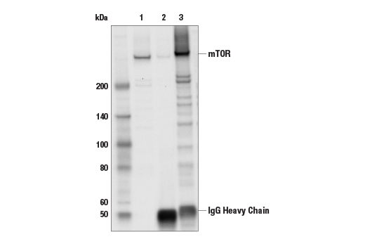
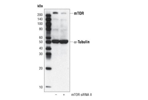
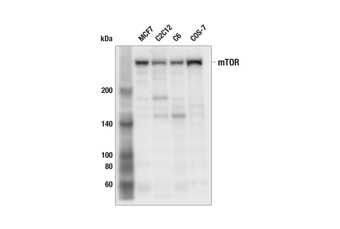
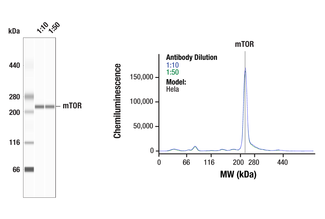
| mTOR (7C10) Rabbit mAb | 2983 | 20 µl | 289 kDa | Rabbit IgG |
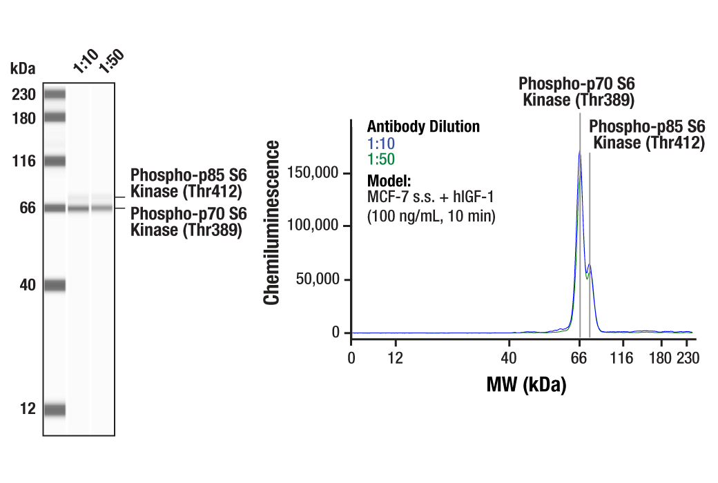
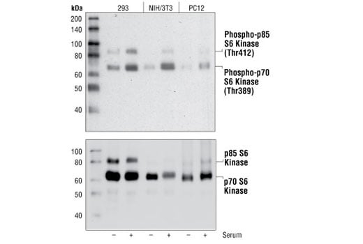
| Phospho-p70 S6 Kinase (Thr389) (108D2) Rabbit mAb | 9234 | 20 µl | 70, 85 kDa | Rabbit IgG |
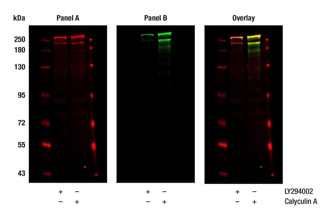
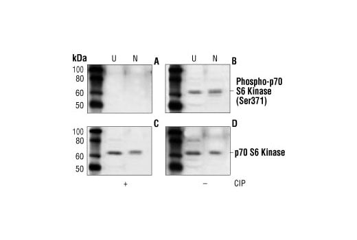
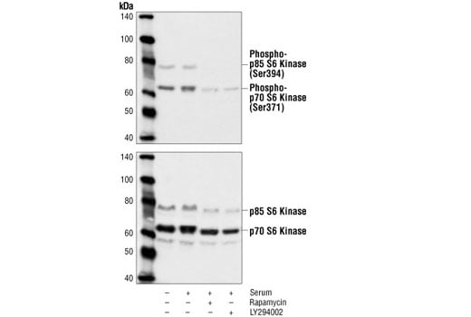
| Phospho-p70 S6 Kinase (Ser371) Antibody | 9208 | 20 µl | 70, 85 kDa | Rabbit |
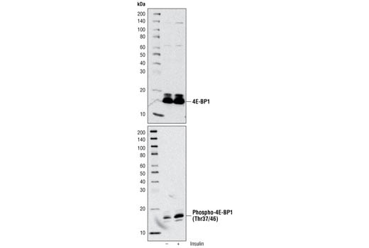
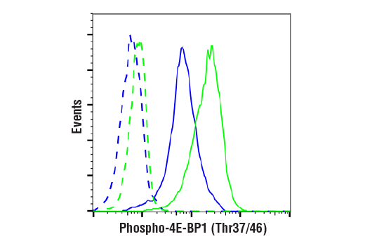
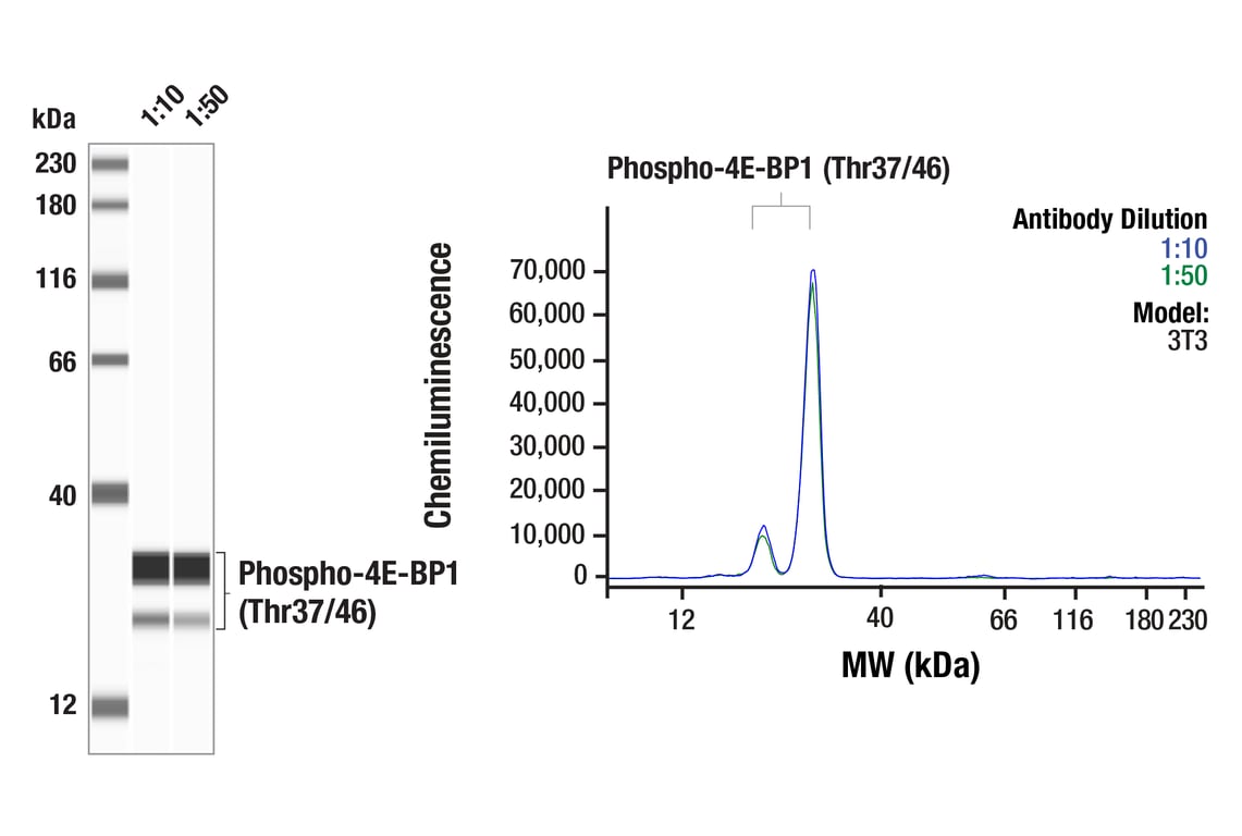
| Phospho-4E-BP1 (Thr37/46) (236B4) Rabbit mAb | 2855 | 20 µl | 15 to 20 kDa | Rabbit IgG |
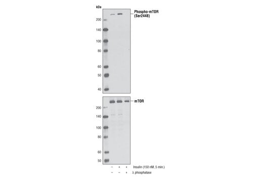
| Phospho-mTOR (Ser2448) (D9C2) XP® Rabbit mAb | 5536 | 20 µl | 289 kDa | Rabbit IgG |
| Anti-rabbit IgG, HRP-linked Antibody | 7074 | 100 µl | Goat |
Please visit cellsignal.com for individual component applications, species cross-reactivity, dilutions, protocols, and additional product information.
Description
Storage
Background
The regulatory associated protein of mTOR (Raptor) interacts with mTOR to mediate mTOR signaling to downstream targets (10,11). Raptor binds to mTOR substrates, such as 4E-BP1 and p70 S6 kinase, through their TOR signaling (TOS) motifs and is required for mTOR-mediated substrate phosphorylation (12,13). Binding of the FKBP12-rapamycin complex to mTOR inhibits mTOR-raptor interaction, which suggests a mechanism for the inhibition of mTOR signaling by rapamycin (14). This mTOR-raptor interaction and its regulation by nutrients and/or rapamycin are dependent on a protein called GβL (15). GβL is part of the rapamycin-insensitive complex between mTOR and rictor (rapamycin-insensitive companion of mTOR) and may mediate rictor-mTOR signaling to PKCα and other downstream targets (16). The rictor-mTOR complex has been identified as the previously elusive PDK2 responsible for the phosphorylation of Akt/PKB at Ser473, which is required for PDK1 phosphorylation of Akt/PKB at Thr308 and full activation of Akt/PKB (17).
Background References
- Sabers, C.J. et al. (1995) J Biol Chem 270, 815-22.
- Brown, E.J. et al. (1994) Nature 369, 756-8.
- Sabatini, D.M. et al. (1994) Cell 78, 35-43.
- Gingras, A.C. et al. (2001) Genes Dev 15, 807-26.
- Dennis, P.B. et al. (2001) Science 294, 1102-5.
- Fang, Y. et al. (2001) Science 294, 1942-5.
- Navé, B.T. et al. (1999) Biochem J 344 Pt 2, 427-31.
- Peterson, R.T. et al. (2000) J Biol Chem 275, 7416-23.
- Huang, S. and Houghton, P.J. (2003) Curr Opin Pharmacol 3, 371-7.
- Hara, K. et al. (2002) Cell 110, 177-89.
- Kim, D.H. et al. (2002) Cell 110, 163-75.
- Beugnet, A. et al. (2003) J Biol Chem 278, 40717-22.
- Nojima, H. et al. (2003) J Biol Chem 278, 15461-4.
- Oshiro, N. et al. (2004) Genes Cells 9, 359-66.
- Kim, D.H. et al. (2003) Mol Cell 11, 895-904.
- Sarbassov, D.D. et al. (2004) Curr Biol 14, 1296-302.
- Sarbassov, D.D. et al. (2005) Science 307, 1098-101.
Trademarks and Patents
Cell Signaling Technology is a trademark of Cell Signaling Technology, Inc.
All other trademarks are the property of their respective owners. Visit cellsignal.com/trademarks for more information.
Limited Uses
Except as otherwise expressly agreed in a writing signed by a legally authorized representative of CST, the following terms apply to Products provided by CST, its affiliates or its distributors. Any Customer's terms and conditions that are in addition to, or different from, those contained herein, unless separately accepted in writing by a legally authorized representative of CST, are rejected and are of no force or effect.
Products are labeled with For Research Use Only or a similar labeling statement and have not been approved, cleared, or licensed by the FDA or other regulatory foreign or domestic entity, for any purpose. Customer shall not use any Product for any diagnostic or therapeutic purpose, or otherwise in any manner that conflicts with its labeling statement. Products sold or licensed by CST are provided for Customer as the end-user and solely for research and development uses. Any use of Product for diagnostic, prophylactic or therapeutic purposes, or any purchase of Product for resale (alone or as a component) or other commercial purpose, requires a separate license from CST. Customer shall (a) not sell, license, loan, donate or otherwise transfer or make available any Product to any third party, whether alone or in combination with other materials, or use the Products to manufacture any commercial products, (b) not copy, modify, reverse engineer, decompile, disassemble or otherwise attempt to discover the underlying structure or technology of the Products, or use the Products for the purpose of developing any products or services that would compete with CST products or services, (c) not alter or remove from the Products any trademarks, trade names, logos, patent or copyright notices or markings, (d) use the Products solely in accordance with CST Product Terms of Sale and any applicable documentation, and (e) comply with any license, terms of service or similar agreement with respect to any third party products or services used by Customer in connection with the Products.
Revision 1
Revision 1

Revision 1
Revision 1
Revision 1
Revision 1
Revision 1
Revision 1
Revision 1