Revision 4
#84406
Store at -20C
877-616-CELL (2355)
877-678-TECH (8324)
3 Trask Lane | Danvers | Massachusetts | 01923 | USA
For Research Use Only. Not for Use in Diagnostic Procedures.
Applications:
W, IP, IF-IC, FC-FP, ChIP, ChIP-seq
Reactivity:
H M
Sensitivity:
Endogenous
MW (kDa):
62
Source/Isotype:
Rabbit IgG
UniProt ID:
#P04198
Entrez-Gene Id:
4613
Product Usage Information
| Application | Dilution |
|---|---|
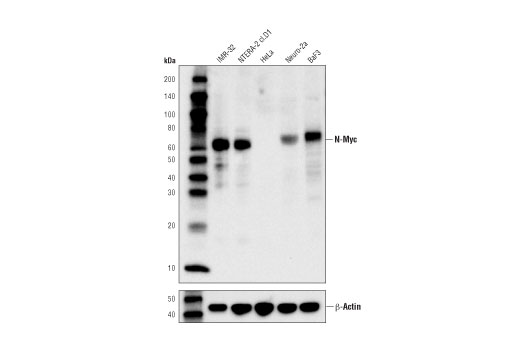
| Western Blotting | 1:1000 |
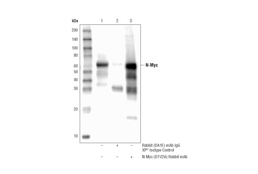
| Immunoprecipitation | 1:200 |
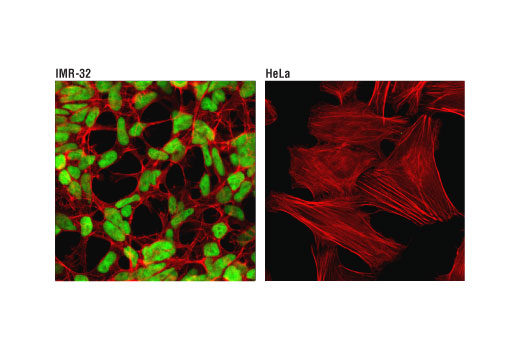
| Immunofluorescence (Immunocytochemistry) | 1:100 - 1:400 |
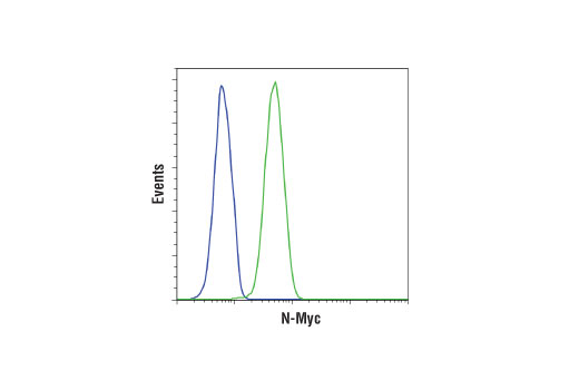
| Flow Cytometry (Fixed/Permeabilized) | 1:200 - 1:800 |
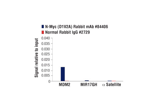
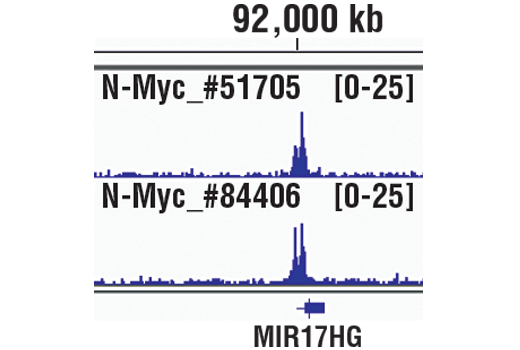
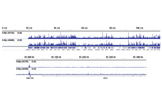
| Chromatin IP | 1:50 |
| Chromatin IP-seq | 1:50 |
Storage
For a carrier free (BSA and azide free) version of this product see product #51063.
Specificity/Sensitivity
Source / Purification
Background
In humans the Myc family consists of 5 genes: c-Myc, N-Myc, L-Myc, R-Myc, and B-Myc. While c-Myc is expressed in many proliferating cells, N-Myc expression is very restricted, with highest levels in during embryonic development and then in the adult during B-cell development. These expression patterns and results from targeted deletion of N-Myc suggest that N-Myc plays an important role in tissue development and differentiation (5). In addition, amplification or overexpression of N-Myc has been found in human neuroblastomas and is associated with rapid progression and poor prognosis (6,7).
Background References
- Baudino, T.A. and Cleveland, J.L. (2001) Mol Cell Biol 21, 691-702.
- Blackwood, E.M. and Eisenman, R.N. (1991) Science 251, 1211-7.
- Henriksson, M. and Lüscher, B. (1996) Adv Cancer Res 68, 109-82.
- Grandori, C. et al. (2000) Annu Rev Cell Dev Biol 16, 653-99.
- Brodeur, G.M. et al. (1984) Science 224, 1121-1124.
Species Reactivity
Species reactivity is determined by testing in at least one approved application (e.g., western blot).
Western Blot Buffer
IMPORTANT: For western blots, incubate membrane with diluted primary antibody in 5% w/v BSA, 1X TBS, 0.1% Tween® 20 at 4°C with gentle shaking, overnight.
Applications Key
W: Western Blotting IP: Immunoprecipitation IF-IC: Immunofluorescence (Immunocytochemistry) FC-FP: Flow Cytometry (Fixed/Permeabilized) ChIP: Chromatin IP ChIP-seq: Chromatin IP-seq
Cross-Reactivity Key
H: Human M: Mouse
Trademarks and Patents
Cell Signaling Technology is a trademark of Cell Signaling Technology, Inc.
Alexa Fluor is a registered trademark of Life Technologies Corporation.
All other trademarks are the property of their respective owners. Visit cellsignal.com/trademarks for more information.
Limited Uses
Except as otherwise expressly agreed in a writing signed by a legally authorized representative of CST, the following terms apply to Products provided by CST, its affiliates or its distributors. Any Customer's terms and conditions that are in addition to, or different from, those contained herein, unless separately accepted in writing by a legally authorized representative of CST, are rejected and are of no force or effect.
Products are labeled with For Research Use Only or a similar labeling statement and have not been approved, cleared, or licensed by the FDA or other regulatory foreign or domestic entity, for any purpose. Customer shall not use any Product for any diagnostic or therapeutic purpose, or otherwise in any manner that conflicts with its labeling statement. Products sold or licensed by CST are provided for Customer as the end-user and solely for research and development uses. Any use of Product for diagnostic, prophylactic or therapeutic purposes, or any purchase of Product for resale (alone or as a component) or other commercial purpose, requires a separate license from CST. Customer shall (a) not sell, license, loan, donate or otherwise transfer or make available any Product to any third party, whether alone or in combination with other materials, or use the Products to manufacture any commercial products, (b) not copy, modify, reverse engineer, decompile, disassemble or otherwise attempt to discover the underlying structure or technology of the Products, or use the Products for the purpose of developing any products or services that would compete with CST products or services, (c) not alter or remove from the Products any trademarks, trade names, logos, patent or copyright notices or markings, (d) use the Products solely in accordance with CST Product Terms of Sale and any applicable documentation, and (e) comply with any license, terms of service or similar agreement with respect to any third party products or services used by Customer in connection with the Products.
Revision 4
Revision 4
Revision 4