| Cat. # | Size | Qty. | Price |
|---|---|---|---|
| 80523T | 1 Kit (5 x 20 microliters) |
|
| Product Includes | Quantity | Applications | Reactivity | MW(kDa) | Isotype |
|---|---|---|---|---|---|
| YTHDF1 (E9P6V) Rabbit mAb 57530 | 20 µl |
|
H M R Mk | 70 | Rabbit IgG |
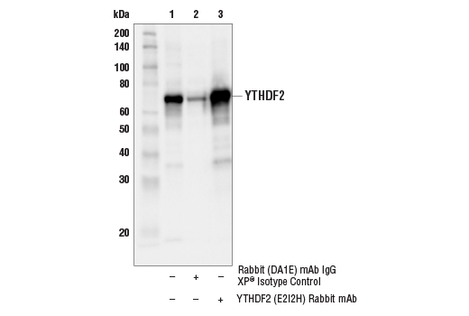
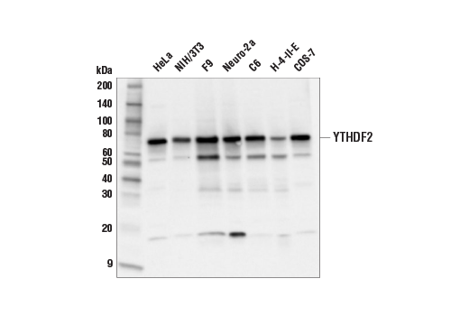
| YTHDF2 (E2I2H) Rabbit mAb 71283 | 20 µl |
|
H M R Mk | 65 | Rabbit IgG |
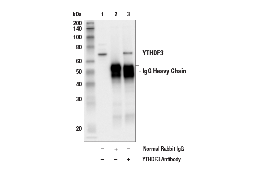
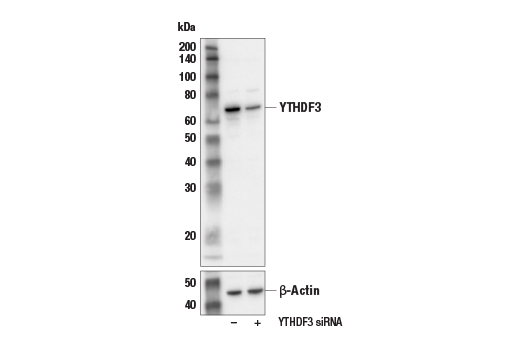
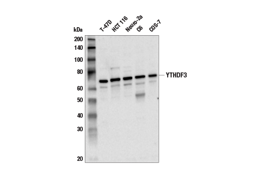
| YTHDF3 Antibody 24206 | 20 µl |
|
H M R Mk | 70 | Rabbit |
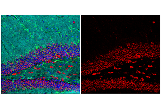
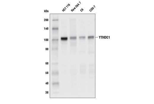
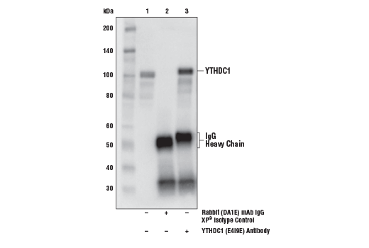
| YTHDC1 (E4I9E) Rabbit mAb 77422 | 20 µl |
|
H M R Mk | 110 | Rabbit IgG |
| YTHDC2 (E6H7U) Rabbit mAb 46324 | 20 µl |
|
H M R Mk | 160 | Rabbit IgG |
| Anti-rabbit IgG, HRP-linked Antibody 7074 | 100 µl |
|
Rab | Goat |
Product Information
Monoclonal antibodies are produced by immunizing animals with a synthetic peptide corresponding to residues surrounding Gly90 of human YTHDF1 protein, Gly167 of human YTHDF2 protein, Phe557 of human YTHDC1 protein, and Gly1243 of human YTHDC2 protein. Polyclonal antibodies are produced by immunizing animals with a synthetic peptide corresponding to residues surrounding Gly169 of human YTHDF3 protein, and are purified by peptide affinity chromatography.
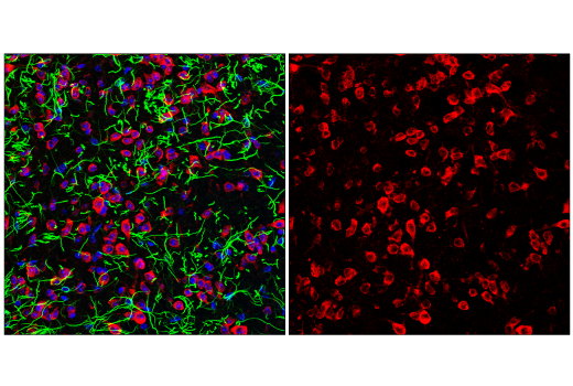
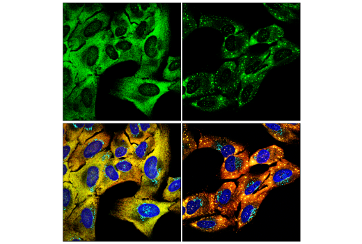
N6-methyladenosine (m6A) is an abundant RNA modification that plays an important role in mRNA splicing, processing, and stability. The m6A modification is specifically recognized by YT521B homology (YTH) domain-containing proteins, consisting of five members in mammals: YTH domain-containing proteins 1 and 2 (YTHDC1 and YTHDC2), and YTH domain-containing family proteins 1, 2, and 3 (YTHDF1, YTHDF2, and YTHDF3) (1). YTHDC1, also known as splicing factor YT521, regulates alternative splicing by functioning as a key regulator of exon-inclusion or exon-skipping. YTHDC1 promotes exon-inclusion by recruiting pre-mRNA splicing factor SRSF3 to regions containing m6A, while repressing exon-skipping by blocking SRSF10 binding to these same regions (2). Increased expression of YTHDC1 promotes malignant endometrial carcinoma (EC) through alternative splicing of vascular endothelial growth factor-A (VEGF-A), resulting in an increase in VEGF-165 isoform and increased EC cell invasion (3). YTHDC2 functions to enhance the translation efficiency of target mRNAs and may play a role in spermatogenesis (4). All three members of the YTHDF family are paralogs that share similar sequence and domain structure, including the conserved C-terminal YTH domain that specifically interacts with m6A (5). Despite these similarities, recent studies suggest that YTHDF proteins are involved in distinct regulatory functions with minimal overlap. Specifically, YTHDF1 binding has been reported to promote enhanced mRNA translation, but has no measurable effect on mRNA stability (6). Conversely, YTHDF2 binding appears to promote mRNA degradation, but has minimal effect on translation efficiency (7). The function of YTHDF3 is less clear, but it has been proposed to function as an auxiliary protein for both YTHDF1 and YTHDF2, helping to promote either increased mRNA translation or decay, respectively (8). Additional studies offer a different viewpoint, suggesting that all three YTHDF proteins initiate mRNA degradation, or mediate increased mRNA stability and protein expression, promoting the idea that these proteins may carry out similar rather than distinct functions (9,10).
Except as otherwise expressly agreed in a writing signed by a legally authorized representative of CST, the following terms apply to Products provided by CST, its affiliates or its distributors. Any Customer's terms and conditions that are in addition to, or different from, those contained herein, unless separately accepted in writing by a legally authorized representative of CST, are rejected and are of no force or effect.
Products are labeled with For Research Use Only or a similar labeling statement and have not been approved, cleared, or licensed by the FDA or other regulatory foreign or domestic entity, for any purpose. Customer shall not use any Product for any diagnostic or therapeutic purpose, or otherwise in any manner that conflicts with its labeling statement. Products sold or licensed by CST are provided for Customer as the end-user and solely for research and development uses. Any use of Product for diagnostic, prophylactic or therapeutic purposes, or any purchase of Product for resale (alone or as a component) or other commercial purpose, requires a separate license from CST. Customer shall (a) not sell, license, loan, donate or otherwise transfer or make available any Product to any third party, whether alone or in combination with other materials, or use the Products to manufacture any commercial products, (b) not copy, modify, reverse engineer, decompile, disassemble or otherwise attempt to discover the underlying structure or technology of the Products, or use the Products for the purpose of developing any products or services that would compete with CST products or services, (c) not alter or remove from the Products any trademarks, trade names, logos, patent or copyright notices or markings, (d) use the Products solely in accordance with CST Product Terms of Sale and any applicable documentation, and (e) comply with any license, terms of service or similar agreement with respect to any third party products or services used by Customer in connection with the Products.