Revision 6
#15035
Store at -20C
877-616-CELL (2355)
877-678-TECH (8324)
3 Trask Lane | Danvers | Massachusetts | 01923 | USA
For Research Use Only. Not for Use in Diagnostic Procedures.
Applications:
W, IF-F
Reactivity:
H M R
Sensitivity:
Endogenous
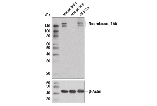
MW (kDa):
140-155
Source/Isotype:
Rabbit IgG
UniProt ID:
#O94856
Entrez-Gene Id:
23114
Product Usage Information
| Application | Dilution |
|---|---|
| Western Blotting | 1:1000 |
| Immunofluorescence (Frozen) | 1:50 |
Storage
For a carrier free (BSA and azide free) version of this product see product #48813.
Specificity/Sensitivity
Source / Purification
Background
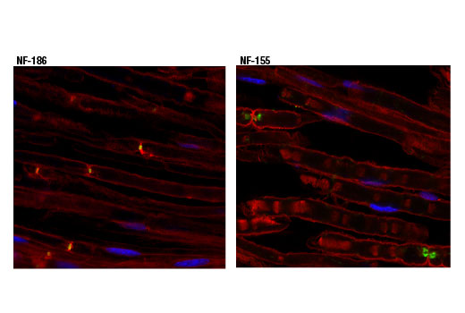
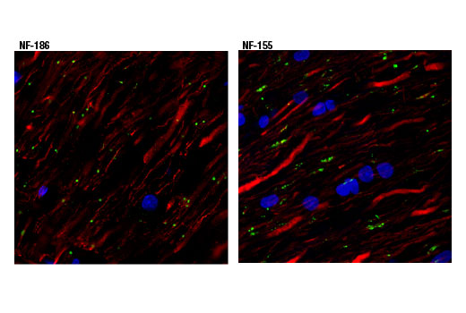
Alternative splicing produces several neurofascin isoforms that differ in temporal and spatial expression. Neurofascin 186 is expressed in axons where it is concentrated at the nodes. Research studies indicate that neurofascin 186 is responsible for nodal assembly and clustering of sodium channels (9). Neurofascin 155 is expressed in glial cells and is localized to myelin paranodes. Interactions between neurofascin 155 and the contactin-associated protein (Caspr) tether the myelin sheath to the axon (10). N-linked glycosylation results in two forms of neurofascin 155 (high and low) that are differentially expressed during development (11).
Background References
- Black, J.A. et al. (1990) Trends Neurosci 13, 48-54.
- Salzer, J.L. (1997) Neuron 18, 843-6.
- Waxman, S.G. et al. (1989) Proc Natl Acad Sci U S A 86, 1406-10.
- Ritchie, J.M. (1992) Trends Neurosci 15, 345-51.
- Berghs, S. et al. (2000) J Cell Biol 151, 985-1002.
- Zhou, D. et al. (1998) J Cell Biol 143, 1295-304.
- Davis, J.Q. et al. (1996) J Cell Biol 135, 1355-67.
- Ratcliffe, C.F. et al. (2001) J Cell Biol 154, 427-34.
- Thaxton, C. et al. (2011) Neuron 69, 244-57.
- Charles, P. et al. (2002) Curr Biol 12, 217-20.
- Pomicter, A.D. et al. (2010) Brain 133, 389-405.
Species Reactivity
Species reactivity is determined by testing in at least one approved application (e.g., western blot).
Western Blot Buffer
IMPORTANT: For western blots, incubate membrane with diluted primary antibody in 5% w/v nonfat dry milk, 1X TBS, 0.1% Tween® 20 at 4°C with gentle shaking, overnight.
Applications Key
W: Western Blotting IF-F: Immunofluorescence (Frozen)
Cross-Reactivity Key
H: Human M: Mouse R: Rat
Trademarks and Patents
Cell Signaling Technology is a trademark of Cell Signaling Technology, Inc.
All other trademarks are the property of their respective owners. Visit cellsignal.com/trademarks for more information.
Limited Uses
Except as otherwise expressly agreed in a writing signed by a legally authorized representative of CST, the following terms apply to Products provided by CST, its affiliates or its distributors. Any Customer's terms and conditions that are in addition to, or different from, those contained herein, unless separately accepted in writing by a legally authorized representative of CST, are rejected and are of no force or effect.
Products are labeled with For Research Use Only or a similar labeling statement and have not been approved, cleared, or licensed by the FDA or other regulatory foreign or domestic entity, for any purpose. Customer shall not use any Product for any diagnostic or therapeutic purpose, or otherwise in any manner that conflicts with its labeling statement. Products sold or licensed by CST are provided for Customer as the end-user and solely for research and development uses. Any use of Product for diagnostic, prophylactic or therapeutic purposes, or any purchase of Product for resale (alone or as a component) or other commercial purpose, requires a separate license from CST. Customer shall (a) not sell, license, loan, donate or otherwise transfer or make available any Product to any third party, whether alone or in combination with other materials, or use the Products to manufacture any commercial products, (b) not copy, modify, reverse engineer, decompile, disassemble or otherwise attempt to discover the underlying structure or technology of the Products, or use the Products for the purpose of developing any products or services that would compete with CST products or services, (c) not alter or remove from the Products any trademarks, trade names, logos, patent or copyright notices or markings, (d) use the Products solely in accordance with CST Product Terms of Sale and any applicable documentation, and (e) comply with any license, terms of service or similar agreement with respect to any third party products or services used by Customer in connection with the Products.
Revision 6