Revision 1

#4767

Store at -20C

NF-kappaB p65 Antibody Sampler Kit

1 Kit

(5 x 20 microliters)

CST Logo
Orders:

877-616-CELL (2355)

[email protected]

Support:

877-678-TECH (8324)

3 Trask Lane | Danvers | Massachusetts | 01923 | USA

For Research Use Only. Not for Use in Diagnostic Procedures.

Product Includes Product # Quantity Mol. Wt Isotype/Source
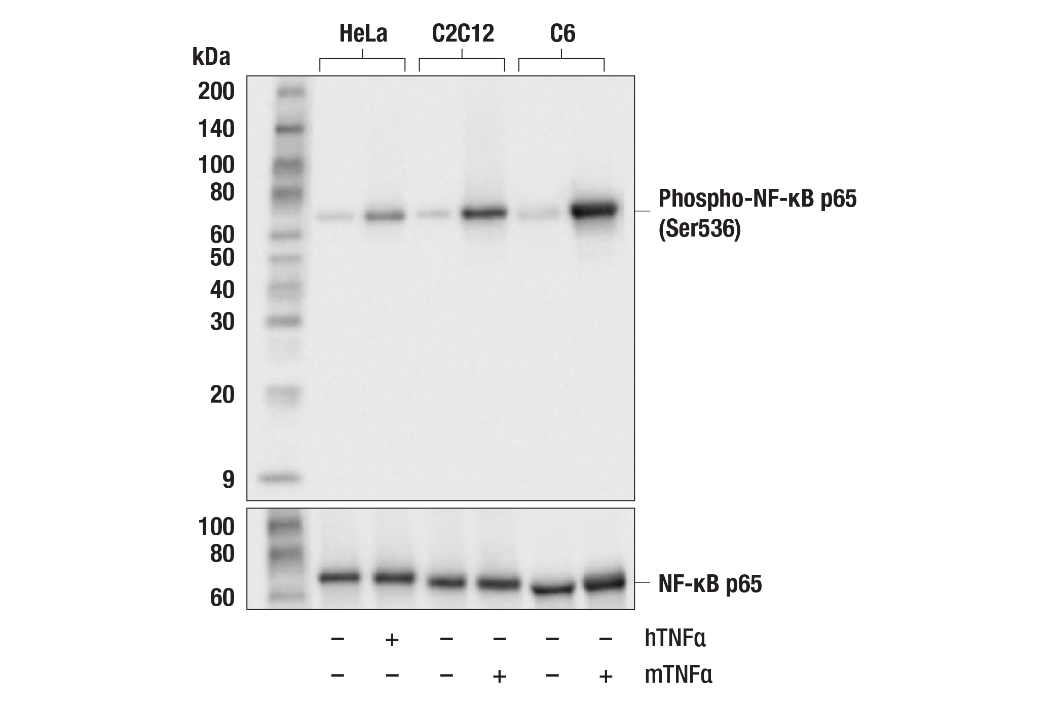
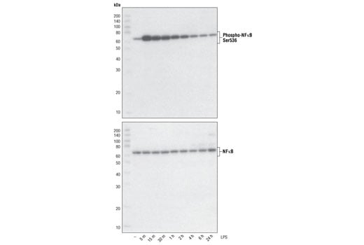
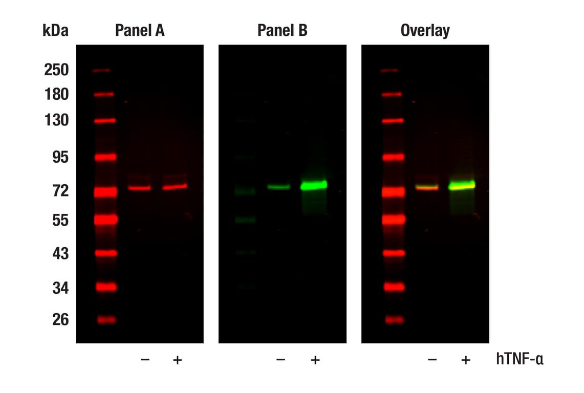
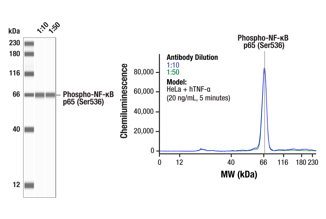
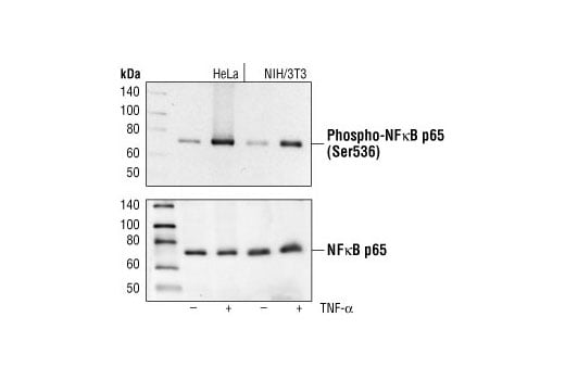
Phospho-NF-kappaB p65 (Ser536) (93H1) Rabbit Monoclonal Antibody303320 µl65 kDaRabbit IgG
Acetyl-NF-kappaB p65 (Lys310) (D2S3J) Rabbit Monoclonal Antibody1262920 µl65 kDaRabbit IgG
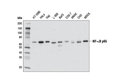
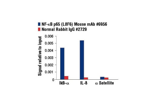
NF-kappaB p65 (L8F6) Mouse Monoclonal Antibody695620 µl65 kDaMouse IgG2b
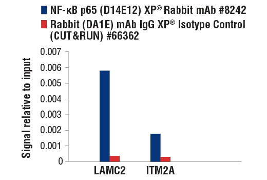
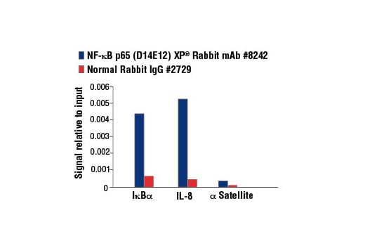
NF-kappaB p65 (D14E12) Rabbit Monoclonal Antibody824220 µl65 kDaRabbit IgG
Phospho-NF-kappaB p65 (Ser468) Antibody303920 µl65 kDaRabbit
Anti-rabbit IgG, HRP-linked Antibody7074100 µlGoat
Anti-mouse IgG, HRP-linked Antibody7076100 µlHorse

Please visit cellsignal.com for individual component applications, species cross-reactivity, dilutions, protocols, and additional product information.

Description

The NF-κB p65 Antibody Sampler Kit contains reagents to examine NF-κB p65/RelA phosphorylation at Ser468 and Ser536; acetylation at Lys310; and total p65 levels.

Storage

Supplied in 10 mM sodium HEPES (pH 7.5), 150 mM NaCl, 100 µg/ml BSA, 50% glycerol and less than 0.02% sodium azide. Store at –20°C. Do not aliquot the antibody.

Background

Transcription factors of the nuclear factor κB (NF-κB)/Rel family play a pivotal role in inflammatory and immune responses (1,2). There are five family members in mammals: RelA, c-Rel, RelB, NF-κB1 (p105/p50), and NF-κB2 (p100/p52). Both p105 and p100 are proteolytically processed by the proteasome to produce p50 and p52, respectively. Rel proteins bind p50 and p52 to form dimeric complexes that bind DNA and regulate transcription. In unstimulated cells, NF-κB is sequestered in the cytoplasm by IκB inhibitory proteins (3-5). NF-κB-activating agents can induce the phosphorylation of IκB proteins, targeting them for rapid degradation through the ubiquitin-proteasome pathway and releasing NF-κB to enter the nucleus where it regulates gene expression (6-8). NIK and IKKα (IKK1) regulate the phosphorylation and processing of NF-κB2 (p100) to produce p52, which translocates to the nucleus (9-11).
RelA/p65 is a subunit of the NF-κB transcription complex, which plays a crucial role in inflammatory and immune responses. The complex is composed of various homodimeric and heterodimeric Rel family member combinations, the activity of which is modulated by post-translational modifications including phosphorylation and acetylation. p65 phosphorylation by PKA and/or MSK1 at Ser276 allows for increased interaction with the transcriptional coactivator p300/CBP to enhance transcriptional activity. NF-κB dimer assembly with IκB, as well as its DNA binding and transcriptional activities, are regulated by p300/CBP acetyltransferases that principally target Lys218, Lys221 and Lys310 (12-14). This process is reciprocally regulated by histone deacetylases (HDACs); several HDAC inhibitors have been shown to activate NF-κB (12-14). T-cell co-stimulation and Calyculin A have both been shown to increase Ser468 phosphorylation (15,16). IKKβ (but not IKKα) and GSK-3β both target this site (16,17), which appears to have a negative regulatory role not involving inhibition of nuclear translocation after TNF-α or IL-1β stimulation (17). p65 phosphorylation at Ser536 regulates activation, nuclear localization, protein-protein interactions, transcriptional activity, and apoptosis (18-22).

Trademarks and Patents

Cell Signaling Technology is a trademark of Cell Signaling Technology, Inc.

All other trademarks are the property of their respective owners. Visit cellsignal.com/trademarks for more information.

Limited Uses

Except as otherwise expressly agreed in a writing signed by a legally authorized representative of CST, the following terms apply to Products provided by CST, its affiliates or its distributors. Any Customer's terms and conditions that are in addition to, or different from, those contained herein, unless separately accepted in writing by a legally authorized representative of CST, are rejected and are of no force or effect.

Products are labeled with For Research Use Only or a similar labeling statement and have not been approved, cleared, or licensed by the FDA or other regulatory foreign or domestic entity, for any purpose. Customer shall not use any Product for any diagnostic or therapeutic purpose, or otherwise in any manner that conflicts with its labeling statement. Products sold or licensed by CST are provided for Customer as the end-user and solely for research and development uses. Any use of Product for diagnostic, prophylactic or therapeutic purposes, or any purchase of Product for resale (alone or as a component) or other commercial purpose, requires a separate license from CST. Customer shall (a) not sell, license, loan, donate or otherwise transfer or make available any Product to any third party, whether alone or in combination with other materials, or use the Products to manufacture any commercial products, (b) not copy, modify, reverse engineer, decompile, disassemble or otherwise attempt to discover the underlying structure or technology of the Products, or use the Products for the purpose of developing any products or services that would compete with CST products or services, (c) not alter or remove from the Products any trademarks, trade names, logos, patent or copyright notices or markings, (d) use the Products solely in accordance with CST Product Terms of Sale and any applicable documentation, and (e) comply with any license, terms of service or similar agreement with respect to any third party products or services used by Customer in connection with the Products.

Orders: 877-616-CELL (2355) [email protected] Support: 877-678-TECH (8324) [email protected] Web: cellsignal.com
For Research Use Only. Not for Use in Diagnostic Procedures.

Revision 1

Cell Signaling Technology Logo
Western blot analysis of extracts from 293T cells, mock transfected (-) or transfected with constructs expressing Myc/DDK-tagged human NF-κB p65 (hNF-κB p65; +) and HA-tagged human p300 (hp300-HA; +), using Acetyl-NF-κB p65 (Lys310) (D2S3J) Rabbit mAb (upper) or NF-κB p65 (D14E12) XP® Rabbit mAb #8242 (lower).
undefined Image 1: NF-kappaB p65 Antibody Sampler Kit
Western blot analysis of extracts from HeLa, C2C12, and C6 cells, untreated (-) or treated (+) as indicated with human TNF-α (hTNF⍺; 20ng/ml, 5min) or mouse TNF-α (mTNF⍺; 20ng/mL, 5min) using Phospho-NF-κB p65 (Ser536) (93H1) Rabbit mAb (upper) or NF-κB p65 (D14E12) XP® Rabbit mAb #8242 (lower). Phospho-NF-κB p65 is induced by human TNF-⍺ or mouse TNF-⍺ treatment as expected.
undefined Image 2: NF-kappaB p65 Antibody Sampler Kit
Western blot analysis of extracts from HeLa cells treated for 5 minutes with TNF-alpha #2169 (20 ng/ml), Calyculin A #9902 (50 nM), or both compounds, using 
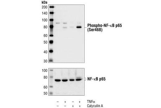
Phospho-NF-kappaB p65 (Ser468) Antibody (top) or NF-kappaB p65 Antibody #3034 (bottom).
undefined Image 3: NF-kappaB p65 Antibody Sampler Kit
Orders: 877-616-CELL (2355) [email protected] Support: 877-678-TECH (8324) [email protected] Web: cellsignal.com
For Research Use Only. Not for Use in Diagnostic Procedures.

Revision 1

Cell Signaling Technology Logo
Western blot analysis of extracts from control HeLa cells (lane 1) or NF-κB p65 knockout HeLa cells (lane 2) using NF-κB p65 (L8F6) Mouse mAb #6956 (upper) or β-actin (13E5) Rabbit mAb #4970 (lower). The absence of signal in the NF-κB p65 knockout HeLa cells confirms the specificity of the antibody for NF-κB p65.
undefined Image 4: NF-kappaB p65 Antibody Sampler Kit
After the primary antibody is bound to the target protein, a complex with HRP-linked secondary antibody is formed. The LumiGLO® is added and emits light during enzyme catalyzed decomposition.
undefined Image 5: NF-kappaB p65 Antibody Sampler Kit
After the primary antibody is bound to the target protein, a complex with HRP-linked secondary antibody is formed. The LumiGLO* is added and emits light during enzyme catalyzed decomposition.
undefined Image 6: NF-kappaB p65 Antibody Sampler Kit
Orders: 877-616-CELL (2355) [email protected] Support: 877-678-TECH (8324) [email protected] Web: cellsignal.com
For Research Use Only. Not for Use in Diagnostic Procedures.

Revision 1

Cell Signaling Technology Logo
Western blot analysis of extracts from various cell lines using NF-κB p65 (D14E12) XP® Rabbit mAb.
undefined Image 7: NF-kappaB p65 Antibody Sampler Kit
Immunohistochemical analysis of paraffin-embedded human endometrioid carcinoma using NF-κB p65 (D14E12) XP® Rabbit mAb.
undefined Image 8: NF-kappaB p65 Antibody Sampler Kit
Immunoprecipitation of acetyl-NF-κB p65 (Lys310) from 293T cells, cotransfected with Myc/DDK-tagged human NF-κB p65 and HA-tagged human p300, using Rabbit (DA1E) mAb IgG XP® Isotype Control #3900 (lane 2) or Acetyl-NF-κB p65 (Lys310) (D2S3J) Rabbit mAb (lane 3). Lane 1 is 10% input. Western blot analysis was performed using Acetyl-NF-κB p65 (Lys310) (D2S3J) Rabbit mAb.
undefined Image 9: NF-kappaB p65 Antibody Sampler Kit
Orders: 877-616-CELL (2355) [email protected] Support: 877-678-TECH (8324) [email protected] Web: cellsignal.com
For Research Use Only. Not for Use in Diagnostic Procedures.

Revision 1

Cell Signaling Technology Logo
Western blot analysis of extracts from HeLa and NIH/3T3 cells, untreated or TNF-α treated (#2169, 20 ng/ml for 5 minutes), using Phospho-NF-κB p65 (Ser536) (93H1) Rabbit mAb (upper) or NF-κB p65 Antibody #3034 (lower).
undefined Image 10: NF-kappaB p65 Antibody Sampler Kit
Western blot analysis of extracts from HeLa cells, transfected with 100 nM SignalSilence® Control siRNA (Unconjugated) #6568 (-) or SignalSilence® NF-κB p65 siRNA I #6261 (+), using NF-κB p65 (L8F6) Mouse mAb (upper) or α-Tubulin (11Η10) Rabbit mAb #2125 (lower). The NF-κB p65 (L8F6) Mouse mAb confirms silencing of NF-κB p65 expression, while the α-Tubulin (11Η10) Rabbit mAb is used as a loading control.
undefined Image 11: NF-kappaB p65 Antibody Sampler Kit
Immunohistochemical analysis of paraffin-embedded human gastric carcinoma using NF-κB p65 (D14E12) XP® Rabbit mAb.
undefined Image 12: NF-kappaB p65 Antibody Sampler Kit
Orders: 877-616-CELL (2355) [email protected] Support: 877-678-TECH (8324) [email protected] Web: cellsignal.com
For Research Use Only. Not for Use in Diagnostic Procedures.

Revision 1

Cell Signaling Technology Logo
Western blot analysis of extracts from THP-1 cells, differentiated with TPA (#9905, 80 nM for 24h) and treated with 1 μg/ml LPS for the indicated times, using Phospho-NF-κB p65 (Ser536) (93H1) Rabbit mAb (upper) and NF-κB p65 (C22B4) Rabbit mAb #4764 (lower).
undefined Image 13: NF-kappaB p65 Antibody Sampler Kit
Western blot analysis of extracts from various cell lines using NF-κB p65 (L8F6) Mouse mAb.
undefined Image 14: NF-kappaB p65 Antibody Sampler Kit
Immunohistochemical analysis of paraffin-embedded human thymoma using NF-κB p65 (D14E12) XP® Rabbit mAb.
undefined Image 15: NF-kappaB p65 Antibody Sampler Kit
Orders: 877-616-CELL (2355) [email protected] Support: 877-678-TECH (8324) [email protected] Web: cellsignal.com
For Research Use Only. Not for Use in Diagnostic Procedures.

Revision 1

Cell Signaling Technology Logo
Immunohistochemical analysis of paraffin-embedded human chronic cholecystitis using NF-κB p65 (D14E12) XP® Rabbit mAb.
undefined Image 16: NF-kappaB p65 Antibody Sampler Kit
Immunohistochemical analysis of paraffin-embedded human lung adenocarcinoma using NF-κB p65 (D14E12) XP® Rabbit mAb.
undefined Image 17: NF-kappaB p65 Antibody Sampler Kit
Confocal immunofluorescent analysis of HeLa cells, serum starved (left) or TNF-α treated (#8902 at 20 ng/ml for 20 min, right), using Phospho-NF-κB p65 (Ser536) (93H1) Rabbit mAb (green). Actin filaments have been labeled with Alexa Fluor® phalloidin 555 (red).
undefined Image 18: NF-kappaB p65 Antibody Sampler Kit
Orders: 877-616-CELL (2355) [email protected] Support: 877-678-TECH (8324) [email protected] Web: cellsignal.com
For Research Use Only. Not for Use in Diagnostic Procedures.

Revision 1

Cell Signaling Technology Logo
Confocal immunofluorescent analysis of HT-1080 cells, untreated (left) or treated with hTNF-α #8902 (20 ng/ml, 20 min) (right), using NF-κB p65 (D14E12) XP® Rabbit mAb (green). Actin filaments were labeled with DY-554 phalloidin (red). Blue pseudocolor = DRAQ5® #4084 (fluorescent DNA dye).
undefined Image 19: NF-kappaB p65 Antibody Sampler Kit
Immunohistochemical analysis of paraffin-embedded normal human small intestine using NF-κB p65 (D14E12) XP® Rabbit mAb.
undefined Image 20: NF-kappaB p65 Antibody Sampler Kit
Flow cytometric analysis of HeLa cells, untreated (blue) or treated with Human Tumor Necrosis Factor-α (hTNF-α) #8902 and Calyculin A #9902 (20 ng/ml and 100 nM, 15 min; green), using Phospho-NF-κB p65 (Ser536) (93H1) Rabbit mAb (solid lines) or concentration-matched Rabbit (DA1E) mAb IgG XP® Isotype Control #3900 (dashed lines). Anti-rabbit IgG (H+L), F(ab')2 Fragment (Alexa Fluor® 488 Conjugate) #4412 was used as a secondary antibody.
undefined Image 21: NF-kappaB p65 Antibody Sampler Kit
Orders: 877-616-CELL (2355) [email protected] Support: 877-678-TECH (8324) [email protected] Web: cellsignal.com
For Research Use Only. Not for Use in Diagnostic Procedures.

Revision 1

Cell Signaling Technology Logo
Flow cytometric analysis of HeLa cells using NF-κB p65 (D14E12) XP® Rabbit mAb (blue) compared to concentration matched Rabbit (DA1E) mAb IgG XP® Isotype Control #3900 (red).
undefined Image 22: NF-kappaB p65 Antibody Sampler Kit
Immunohistochemical analysis of paraffin-embedded human adenoid cystic carcinoma of the salivary gland (left), squamous cell lung carcinoma (middle) or normal colon (right) using NF-κB p65 (D14E12) XP® Rabbit mAb (top) or an
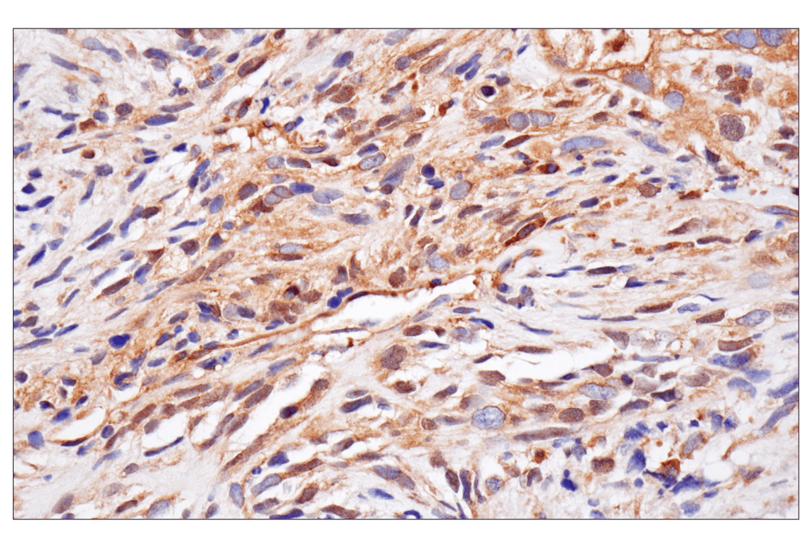
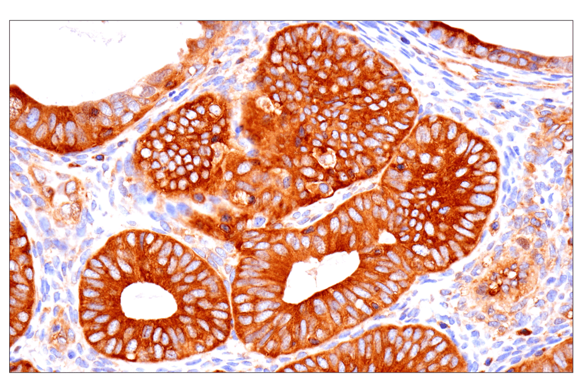
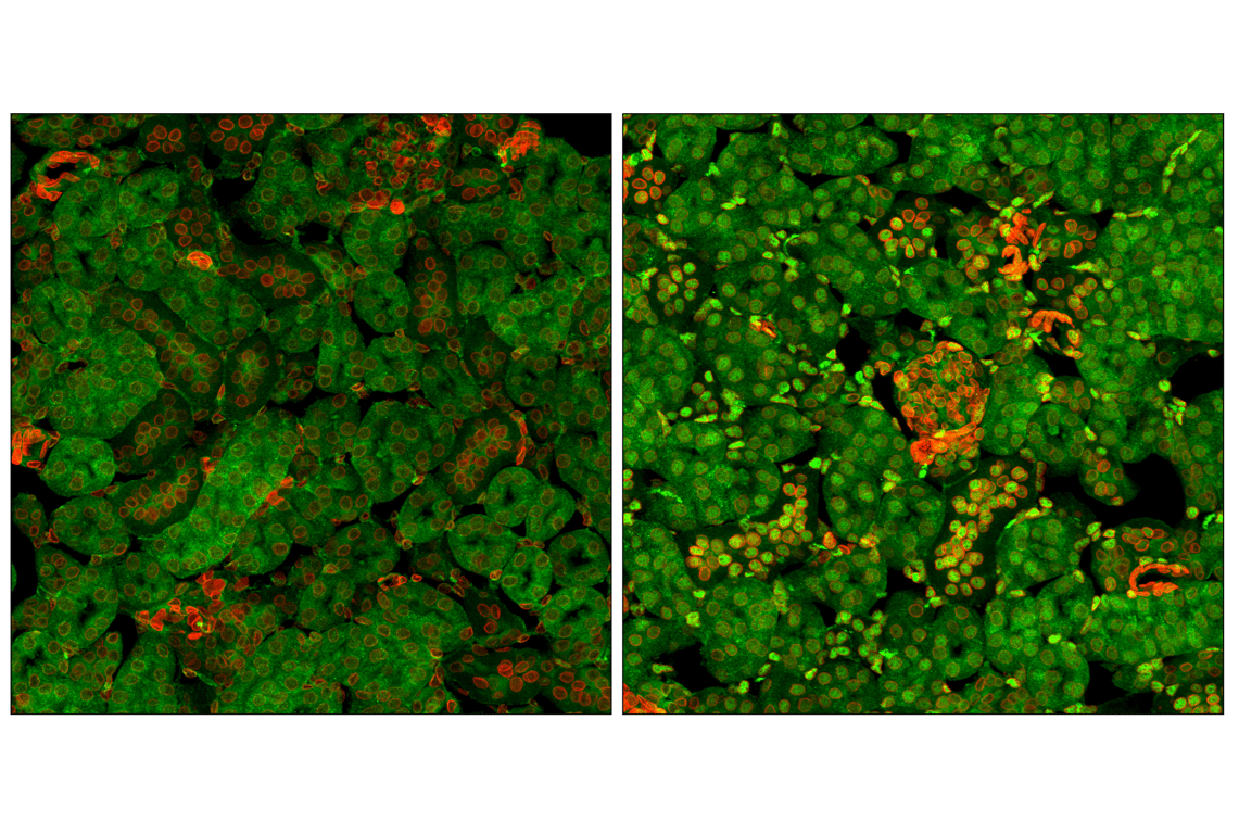
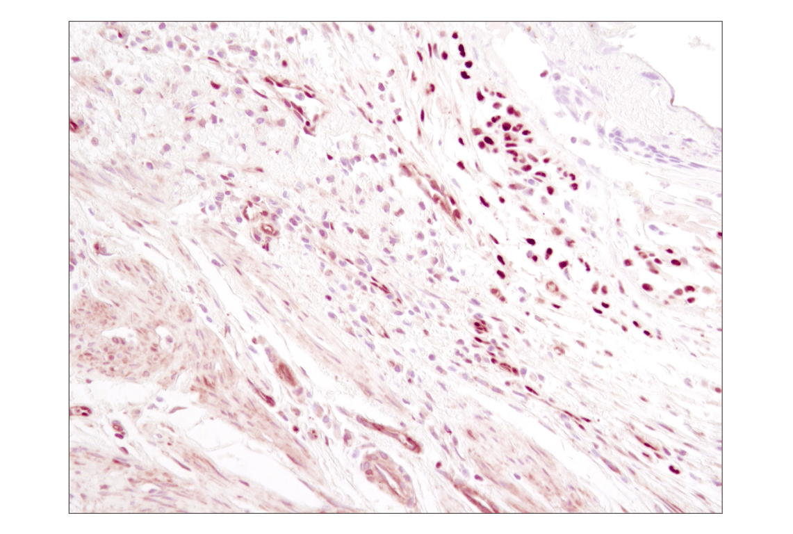
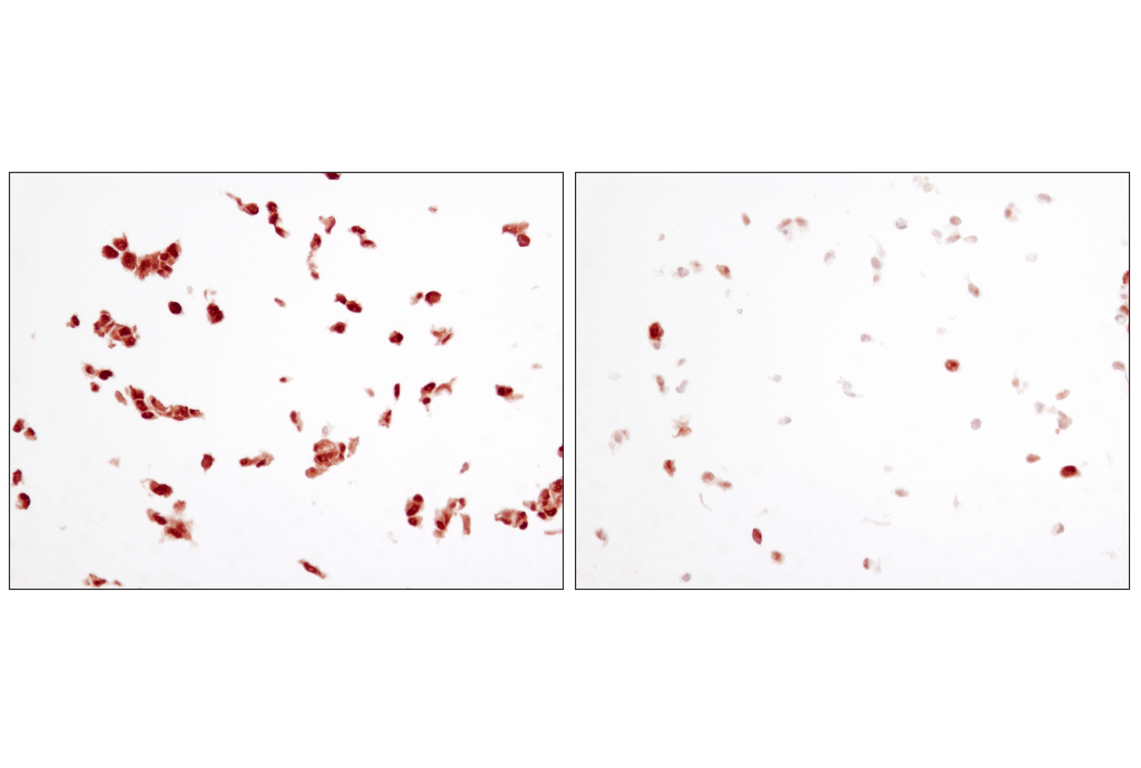
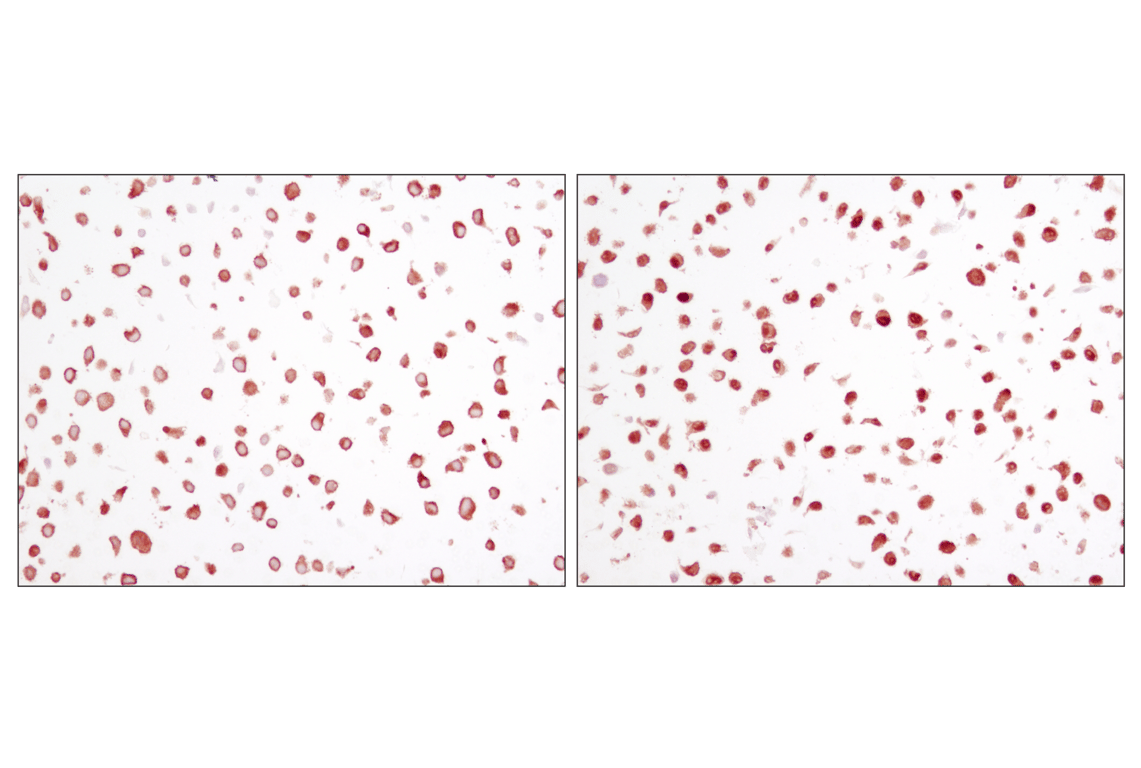
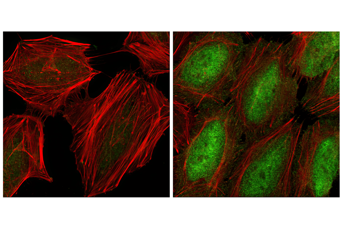
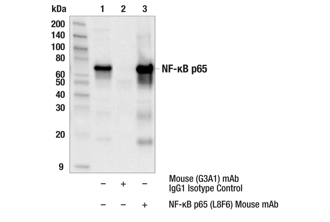
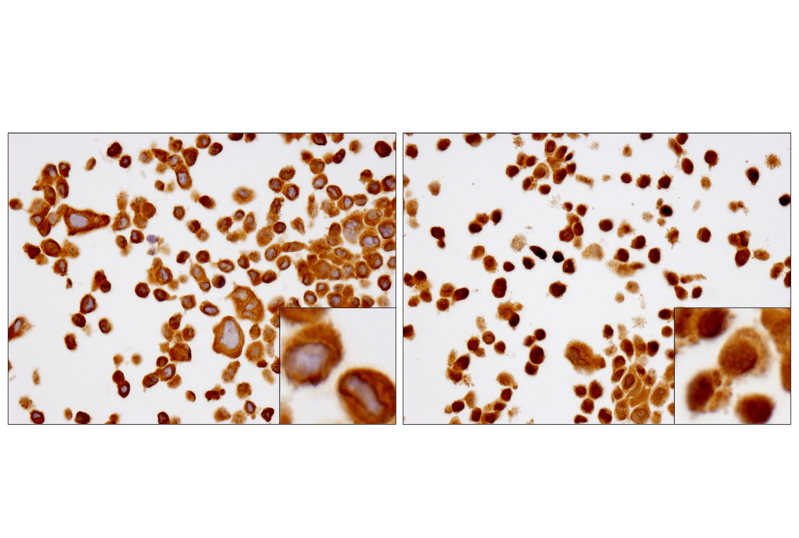
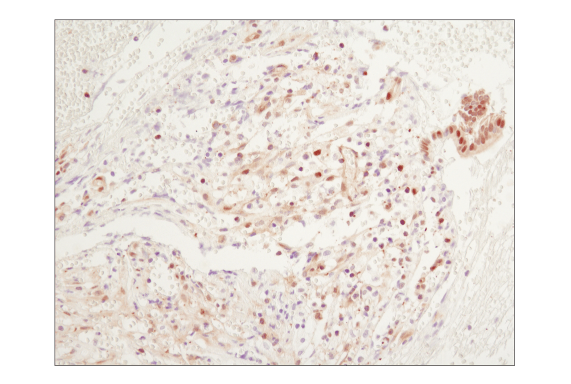
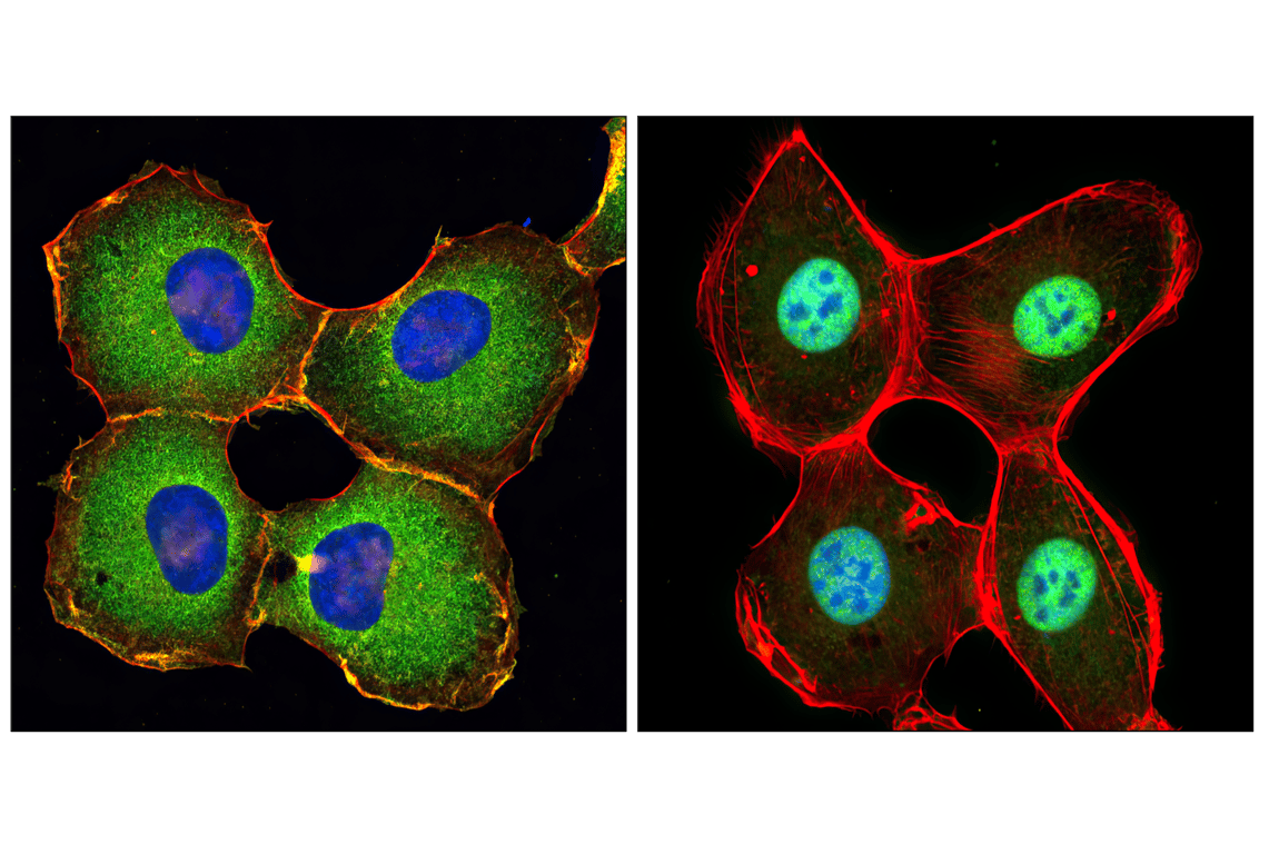
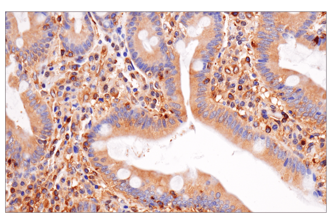
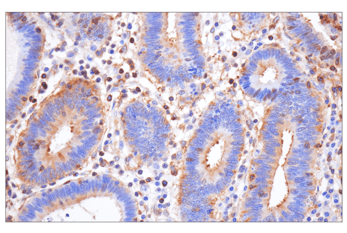
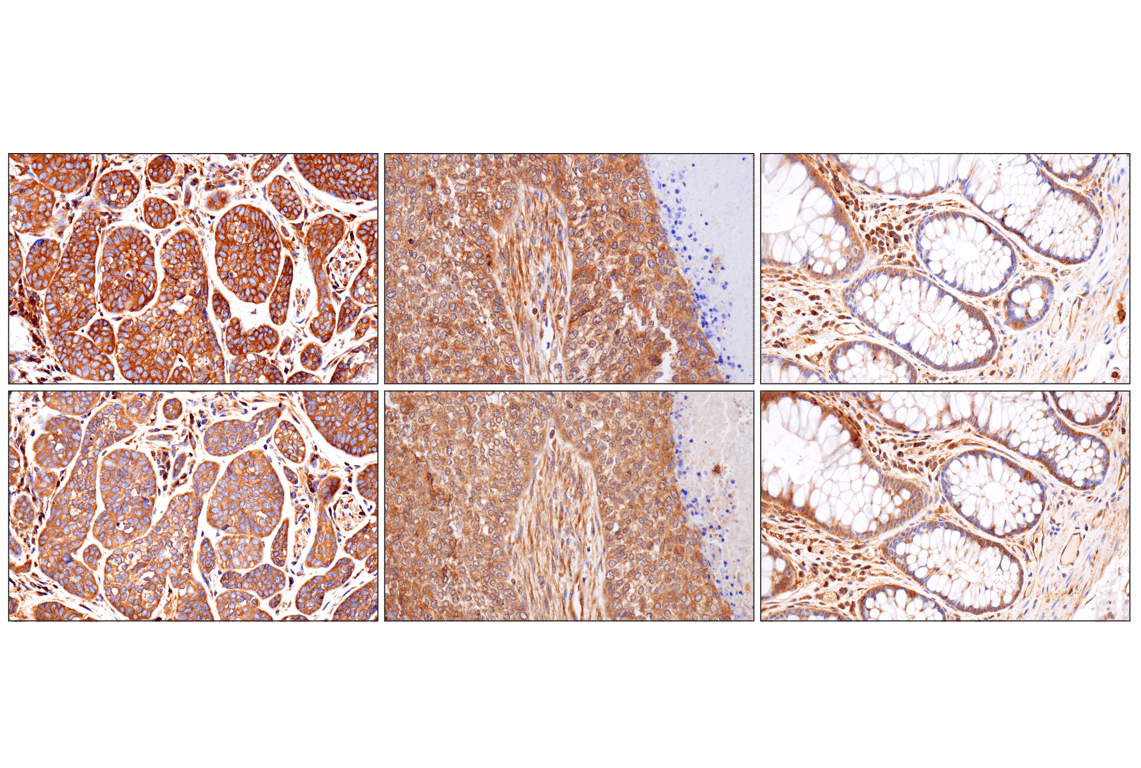
NF-κB p65 Mouse mAb (bottom). These two antibodies detect unique, non-overlapping epitopes on human NF-κB p65. The similar staining patterns obtained with both antibodies help to confirm the specificity of the staining.
undefined Image 23: NF-kappaB p65 Antibody Sampler Kit
Confocal immunofluorescent analysis of HeLa cells, untreated (left) or treated with Human Tumor Necrosis Factor-α (hTNF-α) #8902 (20 ng/mL, 20 min; right), using NF-κB p65 (L8F6) Mouse mAb (green). Actin filaments were labeled with DY-554 phalloidin (red). Blue pseudocolor = DRAQ5® #4084 (fluorescent DNA dye).
undefined Image 24: NF-kappaB p65 Antibody Sampler Kit
Orders: 877-616-CELL (2355) [email protected] Support: 877-678-TECH (8324) [email protected] Web: cellsignal.com
For Research Use Only. Not for Use in Diagnostic Procedures.

Revision 1

Cell Signaling Technology Logo
Chromatin immunoprecipitations were performed with cross-linked chromatin from HeLa cells treated with hTNF-α #8902 (30 ng/ml, 1 hr) and NF-κB p65 (D14E12) XP® Rabbit mAb, using SimpleChIP® Enzymatic Chromatin IP Kit (Magnetic Beads) #9005. DNA Libraries were prepared using DNA Library Prep Kit for Illumina® (ChIP-seq, CUT&RUN) #56795. The figure shows binding across IL-8, a known target gene of NFκB (see additional figure containing ChIP-qPCR data). For additional ChIP-seq tracks, please download the product datasheet.
undefined Image 25: NF-kappaB p65 Antibody Sampler Kit
Immunohistochemical analysis using NF-κB p65 (D14E12) XP® Rabbit mAb on SignalSlide® NF-κB p65 IHC Controls #12873 (paraffin-embedded HCT116 cells, untreated (left) or treated with hTNF-α #8902 (right)).
undefined Image 26: NF-kappaB p65 Antibody Sampler Kit
Flow cytometric analysis of MCF7 cells using NF-kB p65 (L8F6) Mouse mAb (solid line) compared to a concentration-matched Mouse (G3A1) mAb IgG1 Isotype Control #5415 (dashed lines). Anti-mouse IgG (H+L), F(ab')2 Fragment (Alexa Fluor® 488 Conjugate) #4408 was used as a secondary antibody.
undefined Image 27: NF-kappaB p65 Antibody Sampler Kit
Orders: 877-616-CELL (2355) [email protected] Support: 877-678-TECH (8324) [email protected] Web: cellsignal.com
For Research Use Only. Not for Use in Diagnostic Procedures.

Revision 1

Cell Signaling Technology Logo
Chromatin immunoprecipitations were performed with cross-linked chromatin from HeLa cells treated with hTNF-α #8902 (30 ng/ml, 1 hr) and NF-κB p65 (D14E12) XP® Rabbit mAb, using SimpleChIP® Enzymatic Chromatin IP Kit (Magnetic Beads) #9005. DNA Libraries were prepared using DNA Library Prep Kit for Illumina® (ChIP-seq, CUT&RUN) #56795. The figure shows binding across chromosome 4 (upper), including IL-8 (lower), a known target gene of NFκB (see additional figure containing ChIP-qPCR data).
undefined Image 28: NF-kappaB p65 Antibody Sampler Kit
Chromatin immunoprecipitations were performed with cross-linked chromatin from HeLa cells treated with Human Tumor Necrosis Factor-α (hTNF-α) #8902 (30 ng/ml, 1 hr) and either NF-κB p65 (L8F6) Mouse mAb or Normal Rabbit IgG #2729 using SimpleChIP® Enzymatic Chromatin IP Kit (Magnetic Beads) #9003. The enriched DNA was quantified by real-time PCR using SimpleChIP® Human IκBα Promoter Primers #5552, human IL-8 promoter primers, and SimpleChIP® Human α Satellite Repeat Primers #4486. The amount of immunoprecipitated DNA in each sample is represented as signal relative to the total amount of input chromatin, which is equivalent to one.
undefined Image 29: NF-kappaB p65 Antibody Sampler Kit
Chromatin immunoprecipitations were performed with cross-linked chromatin from HeLa cells treated with hTNF-α #8902 (30 ng/ml, 1 hr) and either NF-κB p65 (D14E12) XP® Rabbit mAb or Normal Rabbit IgG #2729 using SimpleChIP® Enzymatic Chromatin IP Kit (Magnetic Beads) #9003. The enriched DNA was quantified by Real-Time PCR using SimpleChIP® Human IκBα Promoter Primers #5552, human IL-8 promoter primers, and SimpleChIP® Human α Satellite Repeat Primers #4486. The amount of immunoprecipitated DNA in each sample is represented as signal relative to the total amount of input chromatin, which is equivalent to one.
undefined Image 30: NF-kappaB p65 Antibody Sampler Kit
Orders: 877-616-CELL (2355) [email protected] Support: 877-678-TECH (8324) [email protected] Web: cellsignal.com
For Research Use Only. Not for Use in Diagnostic Procedures.

Revision 1

Cell Signaling Technology Logo
CUT&RUN was performed with HeLa cells treated with hTNF-α #8902 (30 ng/ml, 1 hr) and NF-κB p65 (D14E12) XP® Rabbit mAb, using CUT&RUN Assay Kit #86652. DNA Libraries were prepared using DNA Library Prep Kit for Illumina® (ChIP-seq, CUT&RUN) #56795. The figure shows binding across LAMC2, a known target gene of NF-κB p65 (see additional figure containing CUT&RUN-qPCR data).
undefined Image 31: NF-kappaB p65 Antibody Sampler Kit
CUT&RUN was performed with HeLa cells treated with hTNF-α #8902 (30 ng/ml, 1 hr) and NF-κB p65 (D14E12) XP® Rabbit mAb, using CUT&RUN Assay Kit #86652. DNA Libraries were prepared using DNA Library Prep Kit for Illumina® (ChIP-seq, CUT&RUN) #56795. The figures show binding across chromosome 1 (upper), including LAMC2 (lower), a known target gene of NF-κB p65 (see additional figure containing CUT&RUN-qPCR data).
undefined Image 32: NF-kappaB p65 Antibody Sampler Kit
CUT&RUN was performed with HeLa cells treated with hTNF-α #8902 (30 ng/ml, 1 hr) and either NF-κB p65 (D14E12) XP® Rabbit mAb or Rabbit (DA1E) mAb IgG XP® Isotype Control (CUT&RUN) #66362, using CUT&RUN Assay Kit #86652. The enriched DNA was quantified by real-time PCR using human LAMC2 upstream primers, and human ITM2A upstream primers. The amount of immunoprecipitated DNA in each sample is represented as signal relative to the total amount of input chromatin, which is equivalent to one.
undefined Image 33: NF-kappaB p65 Antibody Sampler Kit
Orders: 877-616-CELL (2355) [email protected] Support: 877-678-TECH (8324) [email protected] Web: cellsignal.com
For Research Use Only. Not for Use in Diagnostic Procedures.

Revision 1

Cell Signaling Technology Logo
Immunoprecipitation of NF-kB p65 from CHO cell extracts. Lane 1 is 10% input, lane 2 is precipitated with Rabbit (DA1E) mAb IgG XP® Isotype Control #3900, and lane 3 is NF-κB p65 (D14E12) XP® Rabbit mAb, #8242. Western blot was performed using NF-κB p65 (L8F6) Mouse mAb, #6956.
undefined Image 34: NF-kappaB p65 Antibody Sampler Kit
Simple Western™ analysis of lysates (1.0 mg/mL) from HeLa cells treated with hTNF-α (20 ng/mL, 5 minutes) using Phospho-NF-κB p65 (Ser536) (93H1) Rabbit mAb #3033. The virtual lane view (left) shows a single target band (as indicated) at 1:10 and 1:50 dilutions of primary antibody. The corresponding electropherogram view (right) plots chemiluminescence by molecular weight along the capillary at 1:10 (blue line) and 1:50 (green line) dilutions of primary antibody. This experiment was performed under reducing conditions on the Jess™ Simple Western instrument from ProteinSimple, a BioTechne brand, using the 12-230 kDa separation module.
undefined Image 35: NF-kappaB p65 Antibody Sampler Kit
Immunoprecipitation of Phospho-NF-κB p65 (Ser536) from HeLa extracts treated with hTNF-α #8902 (20 ng/ml, 5 min). Lane 1 is 10% input, lane 2 is Rabbit (DA1E) mAb IgG XP® Isotype Control #3900, and lane 3 is Phospho-NF-κB p65 (Ser536) (93H1) Rabbit mAb. Western blot analysis was performed using Phospho-NF-κB p65 (Ser536) (93H1) Rabbit mAb. Anti-rabbit IgG, HRP-linked Antibody #7074 was used as a secondary antibody.
undefined Image 36: NF-kappaB p65 Antibody Sampler Kit
Orders: 877-616-CELL (2355) [email protected] Support: 877-678-TECH (8324) [email protected] Web: cellsignal.com
For Research Use Only. Not for Use in Diagnostic Procedures.

Revision 1

Cell Signaling Technology Logo
Western blot analysis of extracts from HeLa cells, untreated (-) or treated with hTNF-⍺ (20 ng/ml, 5 min; +), using NF-kB p65 (L8F6) Mouse mAb #6956 (Panel A) and Phospho-NF-kB p65 (Ser536) (93H1) Rabbit mAb #3033 (Panel B). Anti-mouse IgG (H+L) (DyLight 680 Conjugate) #5470 (red) and Anti-rabbit IgG (H+L) (DyLight 800 4X PEG Conjugate) #5151 (green) were used as secondary antibodies.
undefined Image 37: NF-kappaB p65 Antibody Sampler Kit
Immunoprecipitation of NF-kB p65 from HeLa cell extracts. Lane 1 is 10% input, lane 2 is precipitated with Mouse (G3A1) mAb IgG1 Isotype Control #5415, and lane 3 is NF-κB p65 (L8F6) Mouse mAb, #6956. Western blot was performed using NF-κB p65 (D14E12) XP® Rabbit mAb, #8242.
undefined Image 38: NF-kappaB p65 Antibody Sampler Kit
Western blot analysis of extracts from HeLa cells, untreated (-) or treated with hTNF-⍺ (20 ng/ml, 5 min; +), using NF-kB p65 (L8F6) Mouse mAb #6956 (Panel A) and Phospho-NF-kB p65 (Ser536) (93H1) Rabbit mAb #3033 (Panel B). Anti-mouse IgG (H+L) (DyLight 680 Conjugate) #5470 (red) and Anti-rabbit IgG (H+L) (DyLight 800 4X PEG Conjugate) #5151 (green) were used as secondary antibodies.
undefined Image 39: NF-kappaB p65 Antibody Sampler Kit
Orders: 877-616-CELL (2355) [email protected] Support: 877-678-TECH (8324) [email protected] Web: cellsignal.com
For Research Use Only. Not for Use in Diagnostic Procedures.

Revision 1

Cell Signaling Technology Logo
Simple Western™ analysis of lysates (1 mg/mL) from HeLa cells using NF-κB p65 (D14E12) XP® Rabbit mAb #8242. The virtual lane view (left) shows a single target band (as indicated) at 1:10 and 1:50 dilutions of primary antibody. The corresponding electropherogram view (right) plots chemiluminescence by molecular weight along the capillary at 1:10 (blue line) and 1:50 (green line) dilutions of primary antibody. This experiment was performed under reducing conditions on the Jess™ Simple Western instrument from ProteinSimple, a BioTechne brand, using the 12-230 kDa separation module.
undefined Image 40: NF-kappaB p65 Antibody Sampler Kit
Confocal immunofluorescent analysis of fixed frozen kidney from untreated (left) or LPS-treated mice (2 hours, 5 mg/kg) (right) using NF-κB p65 (D14E12) XP® Rabbit mAb (green) and Lamin A/C (4C11) Mouse mAb (Alexa Fluor® 647 Conjugate) #41357 (red).
undefined Image 41: NF-kappaB p65 Antibody Sampler Kit
Orders: 877-616-CELL (2355) [email protected] Support: 877-678-TECH (8324) [email protected] Web: cellsignal.com
For Research Use Only. Not for Use in Diagnostic Procedures.