Revision 1
#55764
Store at -20C
NF-kappaB Family Antibody Sampler Kit II
1 Kit
(6 x 20 microliters)
877-616-CELL (2355)
877-678-TECH (8324)
3 Trask Lane | Danvers | Massachusetts | 01923 | USA
For Research Use Only. Not for Use in Diagnostic Procedures.
| Product Includes | Product # | Quantity | Mol. Wt | Isotype/Source |
|---|---|---|---|---|
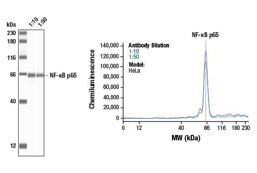
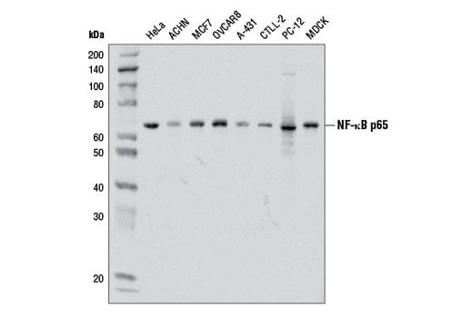
| NF-kappaB p65 (D14E12) Rabbit Monoclonal Antibody | 8242 | 20 µl | 65 kDa | Rabbit IgG |
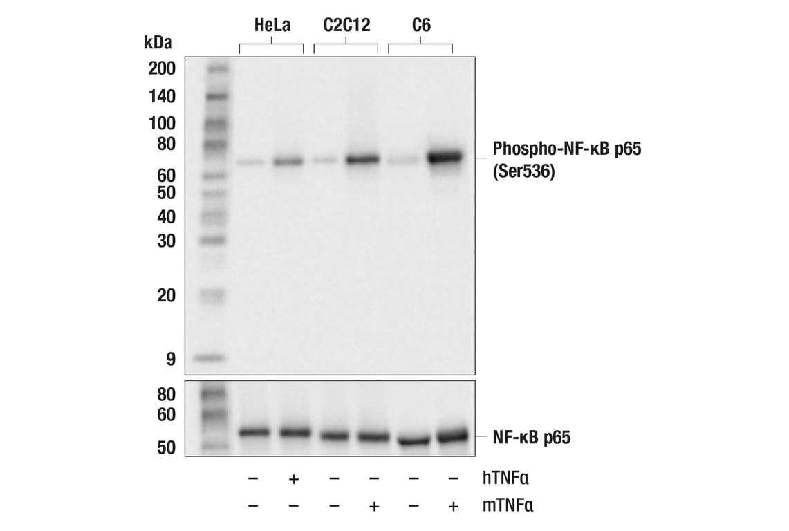
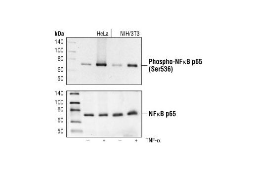
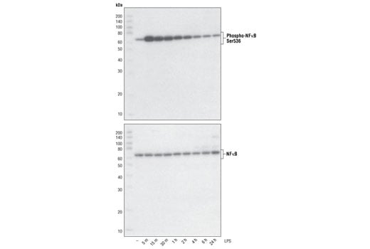
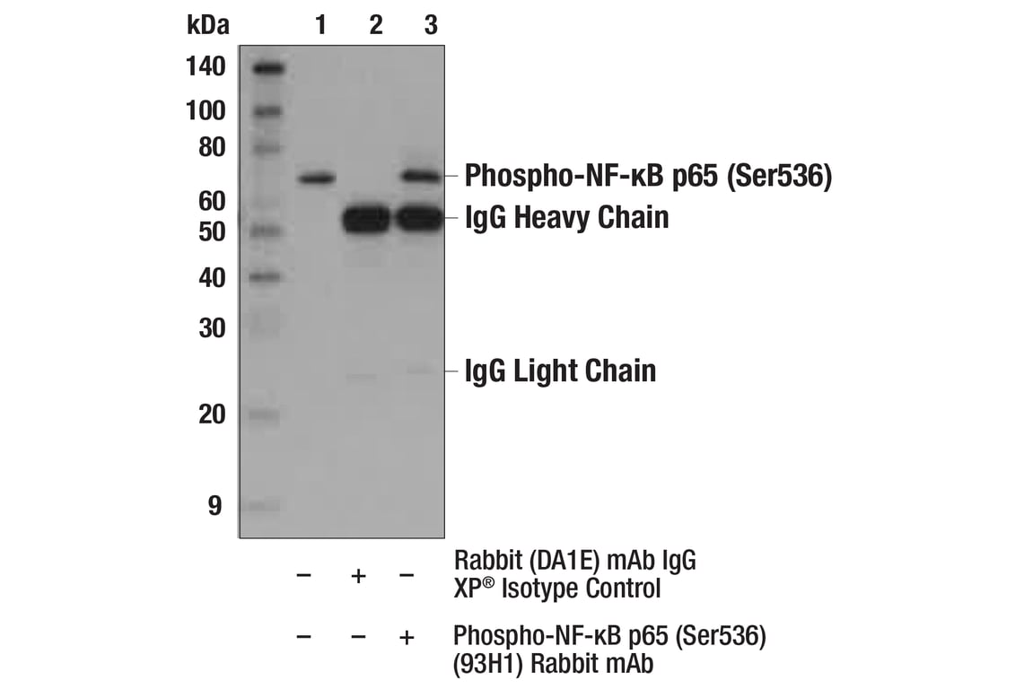
| Phospho-NF-kappaB p65 (Ser536) (93H1) Rabbit Monoclonal Antibody | 3033 | 20 µl | 65 kDa | Rabbit IgG |
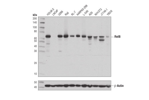
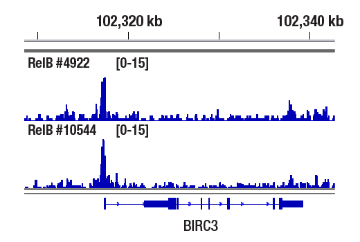
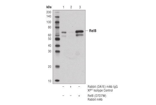
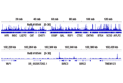
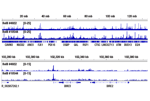
| RelB (D7D7W) Rabbit Monoclonal Antibody | 10544 | 20 µl | 70 kDa | Rabbit IgG |
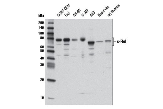
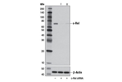
| c-Rel (D4Y6M) Rabbit Monoclonal Antibody | 12707 | 20 µl | 68-78 kDa | Rabbit IgG |
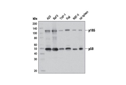
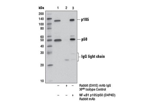
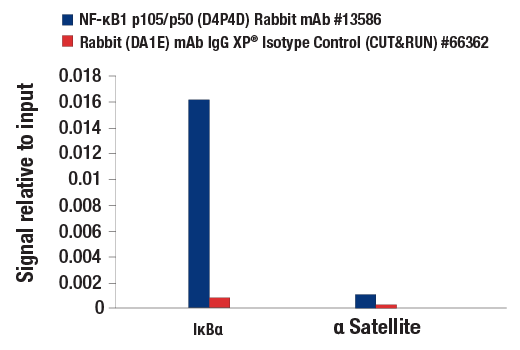
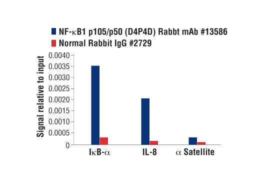
| NF-kappaB1 p105/p50 (D4P4D) Rabbit Monoclonal Antibody | 13586 | 20 µl | 50 Active form. 120 Precursor kDa | Rabbit IgG |
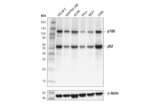
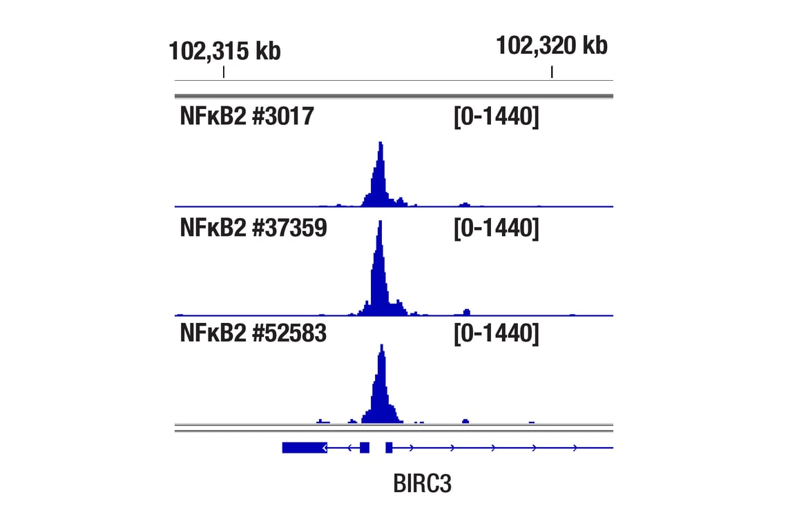
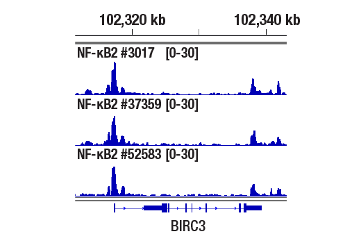
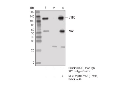
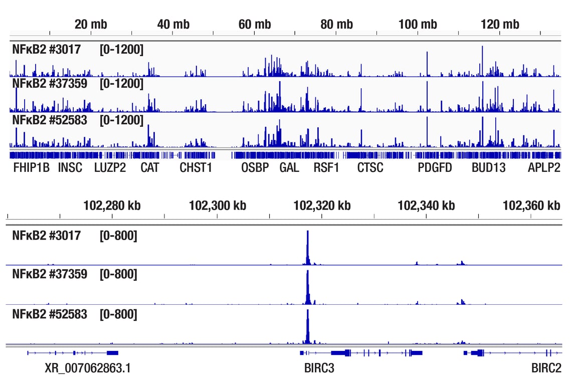
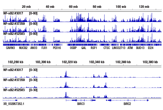
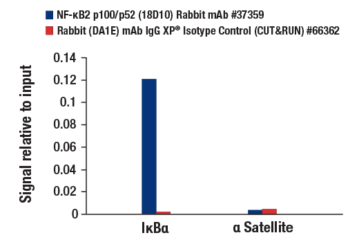
| NF-kappaB2 p100/p52 (D7A9K) Rabbit Monoclonal Antibody | 37359 | 20 µl | 120, 52 kDa | Rabbit IgG |
| Anti-rabbit IgG, HRP-linked Antibody | 7074 | 100 µl | Goat |
Please visit cellsignal.com for individual component applications, species cross-reactivity, dilutions, protocols, and additional product information.
Description
Storage
Background
Background References
- Baeuerle, P.A. and Henkel, T. (1994) Annu Rev Immunol 12, 141-79.
- Baeuerle, P.A. and Baltimore, D. (1996) Cell 87, 13-20.
- Haskill, S. et al. (1991) Cell 65, 1281-9.
- Thompson, J.E. et al. (1995) Cell 80, 573-82.
- Whiteside, S.T. et al. (1997) EMBO J 16, 1413-26.
- Traenckner, E.B. et al. (1995) EMBO J 14, 2876-83.
- Scherer, D.C. et al. (1995) Proc Natl Acad Sci USA 92, 11259-63.
- Chen, Z.J. et al. (1996) Cell 84, 853-62.
- Senftleben, U. et al. (2001) Science 293, 1495-9.
- Coope, H.J. et al. (2002) EMBO J 21, 5375-85.
- Xiao, G. et al. (2001) Mol Cell 7, 401-9.
Trademarks and Patents
Cell Signaling Technology is a trademark of Cell Signaling Technology, Inc.
All other trademarks are the property of their respective owners. Visit cellsignal.com/trademarks for more information.
Limited Uses
Except as otherwise expressly agreed in a writing signed by a legally authorized representative of CST, the following terms apply to Products provided by CST, its affiliates or its distributors. Any Customer's terms and conditions that are in addition to, or different from, those contained herein, unless separately accepted in writing by a legally authorized representative of CST, are rejected and are of no force or effect.
Products are labeled with For Research Use Only or a similar labeling statement and have not been approved, cleared, or licensed by the FDA or other regulatory foreign or domestic entity, for any purpose. Customer shall not use any Product for any diagnostic or therapeutic purpose, or otherwise in any manner that conflicts with its labeling statement. Products sold or licensed by CST are provided for Customer as the end-user and solely for research and development uses. Any use of Product for diagnostic, prophylactic or therapeutic purposes, or any purchase of Product for resale (alone or as a component) or other commercial purpose, requires a separate license from CST. Customer shall (a) not sell, license, loan, donate or otherwise transfer or make available any Product to any third party, whether alone or in combination with other materials, or use the Products to manufacture any commercial products, (b) not copy, modify, reverse engineer, decompile, disassemble or otherwise attempt to discover the underlying structure or technology of the Products, or use the Products for the purpose of developing any products or services that would compete with CST products or services, (c) not alter or remove from the Products any trademarks, trade names, logos, patent or copyright notices or markings, (d) use the Products solely in accordance with CST Product Terms of Sale and any applicable documentation, and (e) comply with any license, terms of service or similar agreement with respect to any third party products or services used by Customer in connection with the Products.
Revision 1
Revision 1
Revision 1
Revision 1

Revision 1
Revision 1
Revision 1

Revision 1
Revision 1
Revision 1
Revision 1

Revision 1

Revision 1
Revision 1
Revision 1
Revision 1
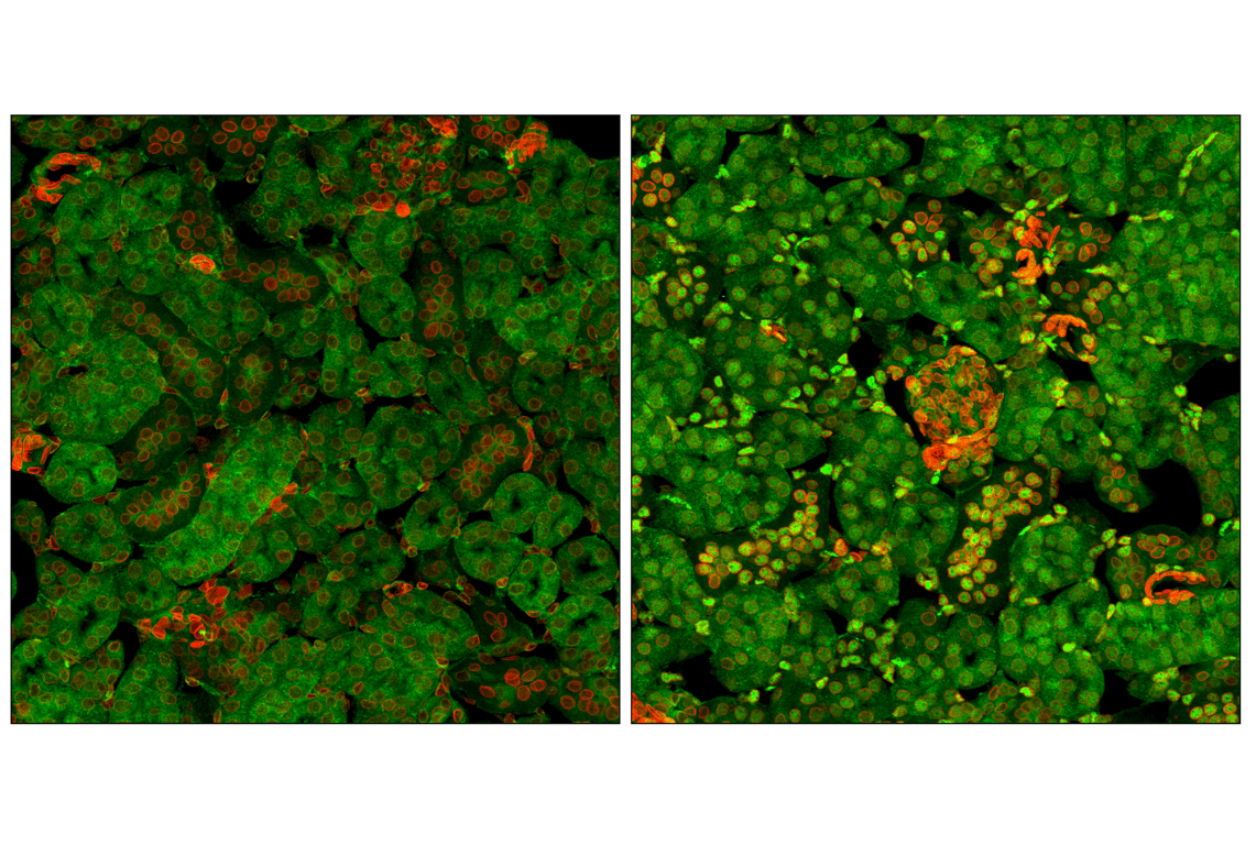
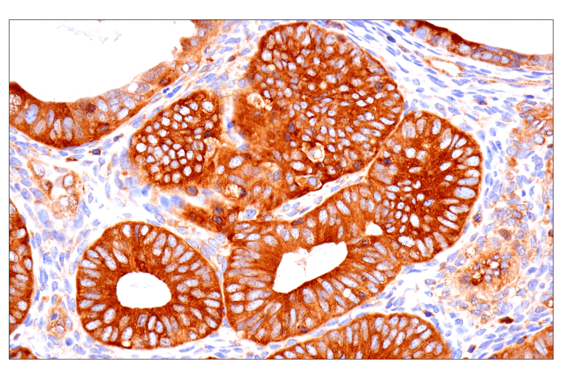
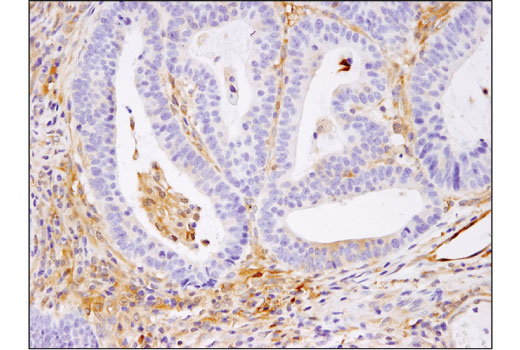
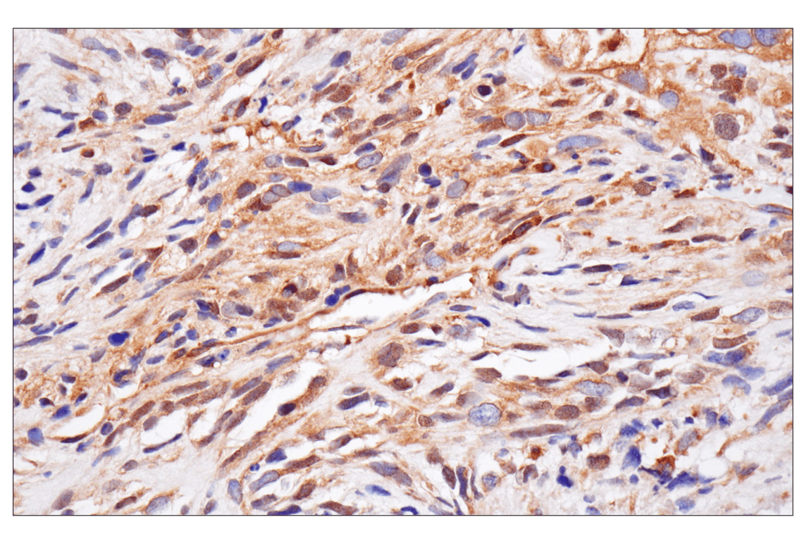
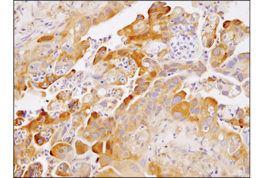
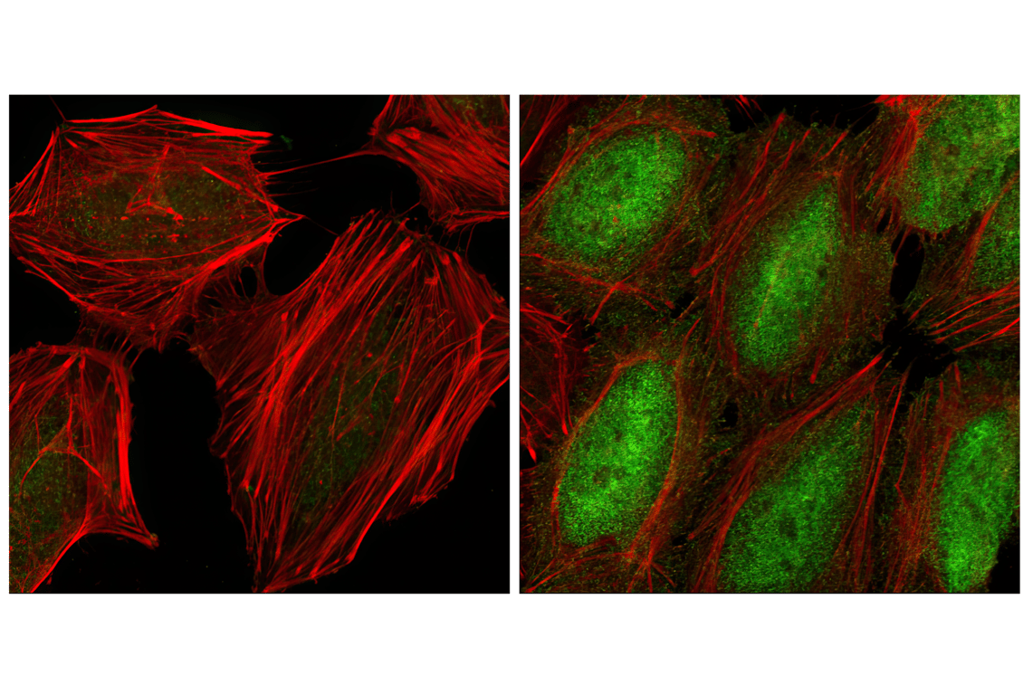
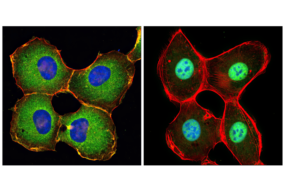
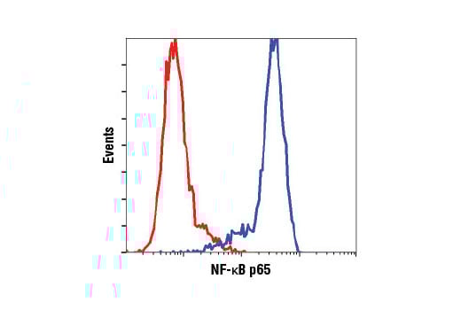
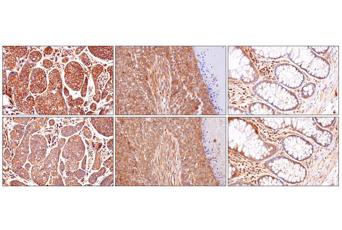
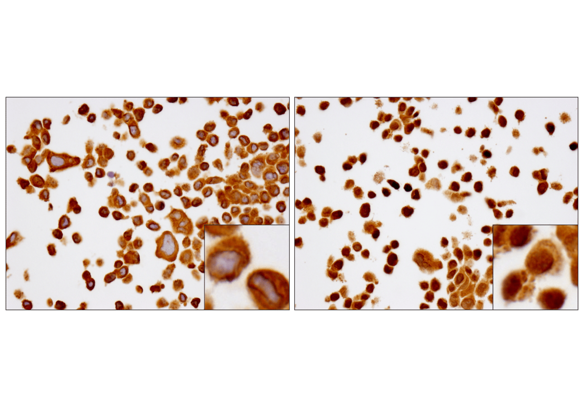
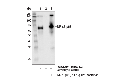
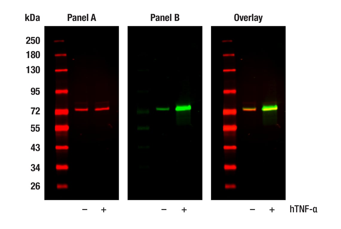
NF-κB p65 Mouse mAb (bottom). These two antibodies detect unique, non-overlapping epitopes on human NF-κB p65. The similar staining patterns obtained with both antibodies help to confirm the specificity of the staining.
Revision 1
Revision 1
Revision 1
Revision 1
Revision 1