Revision 1
#4888
Store at -20C
NF-kappaB Non-Canonical Pathway Antibody Sampler Kit
1 Kit
(8 x 20 microliters)
877-616-CELL (2355)
877-678-TECH (8324)
3 Trask Lane | Danvers | Massachusetts | 01923 | USA
For Research Use Only. Not for Use in Diagnostic Procedures.
| Product Includes | Product # | Quantity | Mol. Wt | Isotype/Source |
|---|---|---|---|---|
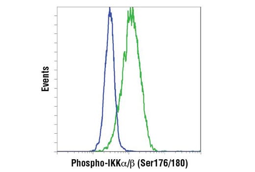
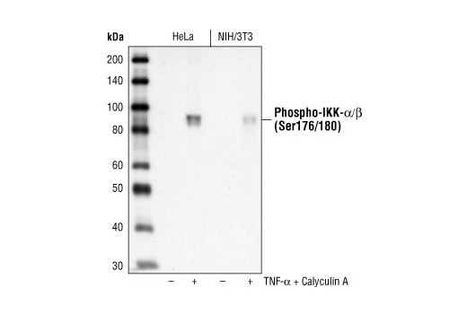
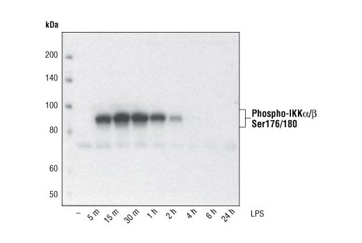
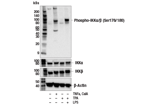
| Phospho-IKK alpha/beta (Ser176/180) (16A6) Rabbit Monoclonal Antibody | 2697 | 20 µl | 85 IKK-alpha 87 IKK-beta kDa | Rabbit IgG |
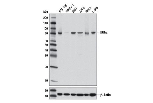
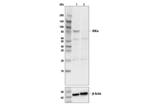
| IKK alpha (3G12) Mouse Monoclonal Antibody | 11930 | 20 µl | 85 kDa | Mouse IgG1 |
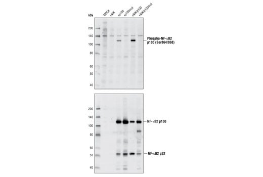
| Phospho-NF-kappaB2 p100 (Ser866/870) Antibody | 4810 | 20 µl | 110 kDa | Rabbit |
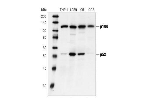
| NF-kappaB2 p100/p52 Antibody | 4882 | 20 µl | 52 (mature). 120 (precursor). kDa | Rabbit |
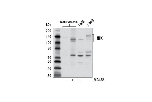
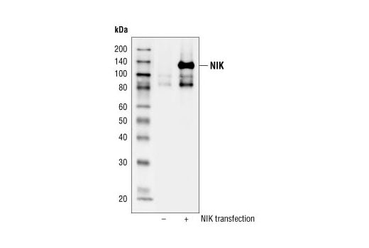
| NIK Antibody | 4994 | 20 µl | 125 kDa | Rabbit |
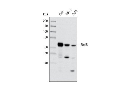
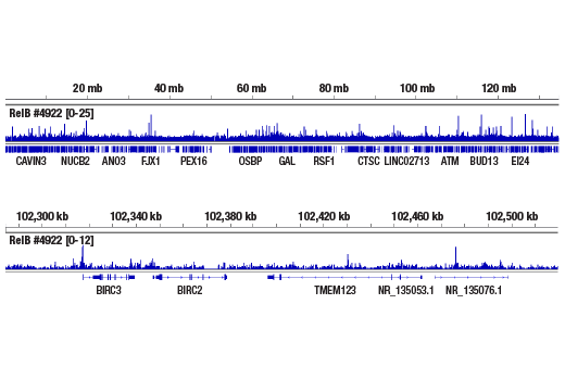
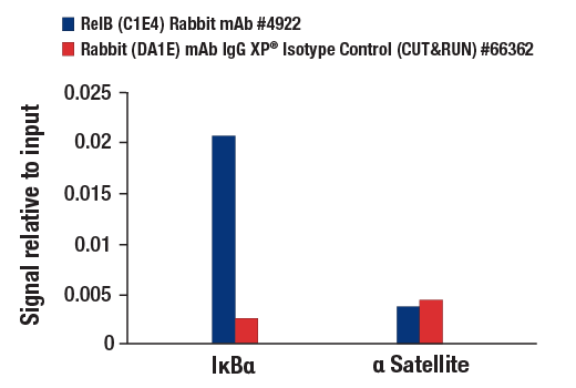
| RelB (C1E4) Rabbit Monoclonal Antibody | 4922 | 20 µl | 70 kDa | Rabbit IgG |
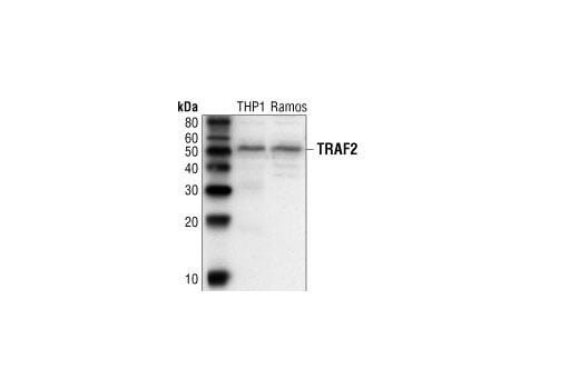
| TRAF2 Antibody | 4712 | 20 µl | 53 kDa | Rabbit |
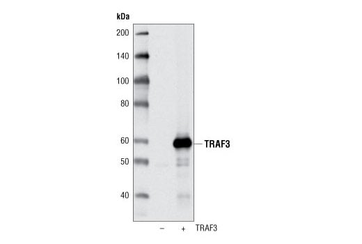
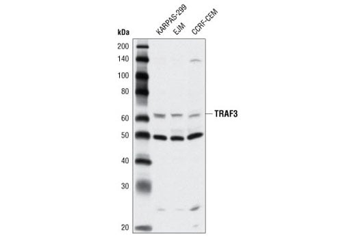
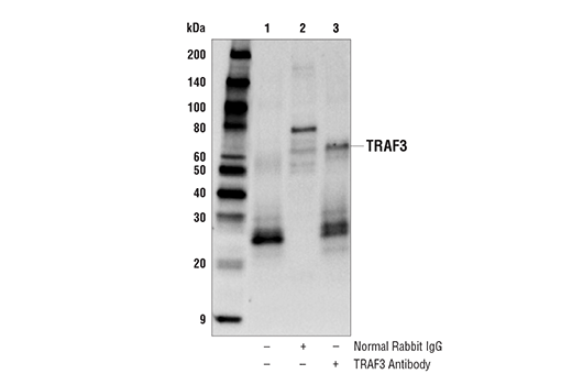
| TRAF3 Antibody | 4729 | 20 µl | 62 kDa | Rabbit |
| Anti-rabbit IgG, HRP-linked Antibody | 7074 | 100 µl | Goat | |
| Anti-mouse IgG, HRP-linked Antibody | 7076 | 100 µl | Horse |
Please visit cellsignal.com for individual component applications, species cross-reactivity, dilutions, protocols, and additional product information.
Description
Storage
Background
The noncanonical pathway, triggered by BAFF, CD40L, and certain other stimuli, is based on the inducible phosphorylation and proteasome-mediated partial degradation of NF-κB2 p100 to p52, a process regulated by the NF-κB Inducing Kinase (NIK) and IKKα, but not IKKβ or IKKγ (4-6). NIK phosphorylates IKKα at Ser176/180 (6) and p100 at Ser866/870, then recruits IKKα to p100 where IKKα phosphorylates additional residues in the N- and C-terminus (8), leading to the ubiquitination and processing of p100 (9). The TNF Receptor Associated Factor molecules TRAF2 and TRAF3 have been shown to be negative regulators of the noncanonical pathway (10, 11), and their differential binding to receptors may also play a role in determining whether transduced signals activate the canonical pathway, noncanonical pathway, or both (12). TRAF3 promotes the rapid turnover of NIK in resting cells, and its activation-induced degradation is a key regulatory point in the pathway (13). This pathway is required for B cell maturation and activation, proper architecture of peripheral lymphoid tissue, and safeguards against autoimmunity (14).
Background References
- Baeuerle, P.A. and Henkel, T. (1994) Annu Rev Immunol 12, 141-79.
- Baeuerle, P.A. and Baltimore, D. (1996) Cell 87, 13-20.
- Ghosh, S. and Karin, M. (2002) Cell 109 Suppl, S81-96.
- Xiao, G. et al. (2001) Mol Cell 7, 401-9.
- Senftleben, U. et al. (2001) Science 293, 1495-9.
- Xiao, G. et al. (2001) EMBO J 20, 6805-15.
- Ling, L. et al. (1998) Proc Natl Acad Sci USA 95, 3792-7.
- Xiao, G. et al. (2004) J Biol Chem 279, 30099-105.
- Liang, C. et al. (2006) Cell Signal 18, 1309-17.
- Xia, Z.P. and Chen, Z.J. (2005) Sci STKE 2005, pe7.
- Liao, G. et al. (2004) J Biol Chem 279, 26243-50.
- Morrison, M.D. et al. (2005) J Biol Chem 280, 10018-24.
- Qing, G. et al. (2005) J Biol Chem 280, 40578-82.
- Xiao, G. et al. (2006) Cytokine Growth Factor Rev 17, 281-93.
Trademarks and Patents
Cell Signaling Technology is a trademark of Cell Signaling Technology, Inc.
All other trademarks are the property of their respective owners. Visit cellsignal.com/trademarks for more information.
Limited Uses
Except as otherwise expressly agreed in a writing signed by a legally authorized representative of CST, the following terms apply to Products provided by CST, its affiliates or its distributors. Any Customer's terms and conditions that are in addition to, or different from, those contained herein, unless separately accepted in writing by a legally authorized representative of CST, are rejected and are of no force or effect.
Products are labeled with For Research Use Only or a similar labeling statement and have not been approved, cleared, or licensed by the FDA or other regulatory foreign or domestic entity, for any purpose. Customer shall not use any Product for any diagnostic or therapeutic purpose, or otherwise in any manner that conflicts with its labeling statement. Products sold or licensed by CST are provided for Customer as the end-user and solely for research and development uses. Any use of Product for diagnostic, prophylactic or therapeutic purposes, or any purchase of Product for resale (alone or as a component) or other commercial purpose, requires a separate license from CST. Customer shall (a) not sell, license, loan, donate or otherwise transfer or make available any Product to any third party, whether alone or in combination with other materials, or use the Products to manufacture any commercial products, (b) not copy, modify, reverse engineer, decompile, disassemble or otherwise attempt to discover the underlying structure or technology of the Products, or use the Products for the purpose of developing any products or services that would compete with CST products or services, (c) not alter or remove from the Products any trademarks, trade names, logos, patent or copyright notices or markings, (d) use the Products solely in accordance with CST Product Terms of Sale and any applicable documentation, and (e) comply with any license, terms of service or similar agreement with respect to any third party products or services used by Customer in connection with the Products.
Revision 1
Revision 1
Revision 1

Revision 1


Revision 1
Revision 1
Revision 1
Revision 1
Revision 1