Revision 2
#3640
Store at -20C
Notch Isoform Antibody Sampler Kit
1 Kit
(4 x 20 microliters)
877-616-CELL (2355)
877-678-TECH (8324)
3 Trask Lane | Danvers | Massachusetts | 01923 | USA
For Research Use Only. Not for Use in Diagnostic Procedures.
| Product Includes | Product # | Quantity | Mol. Wt | Isotype/Source |
|---|---|---|---|---|
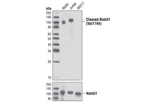
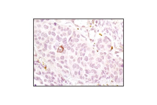
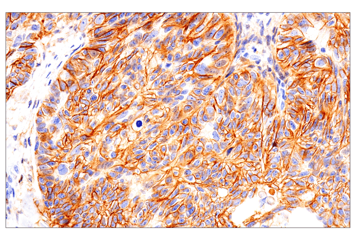
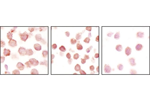
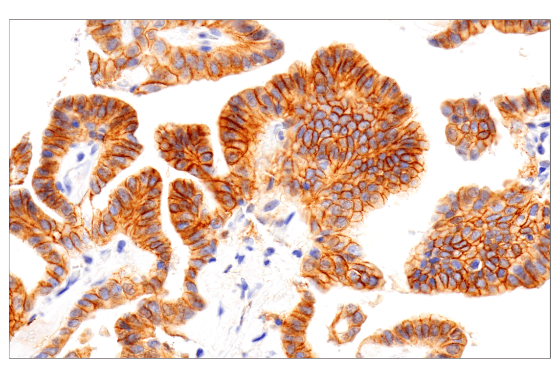
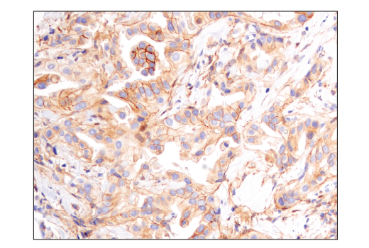
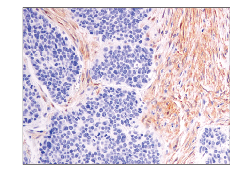
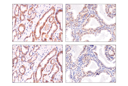
| Cleaved Notch1 (Val1744) (D3B8) Rabbit Monoclonal Antibody | 4147 | 20 µl | 110 kDa | Rabbit IgG |
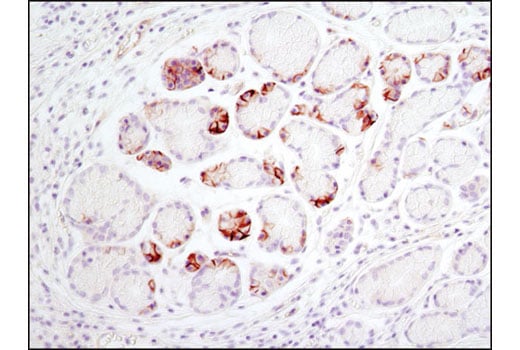
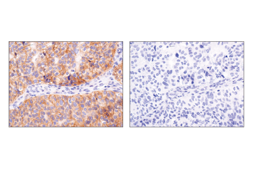
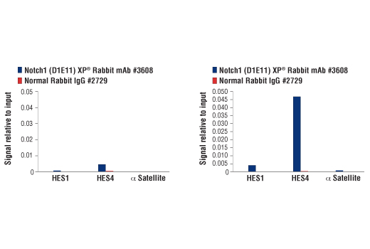
| Notch1 (D1E11) Rabbit Monoclonal Antibody | 3608 | 20 µl | 120, 300 kDa | Rabbit IgG |
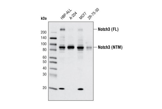
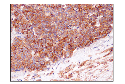
| Notch3 (D11B8) Rabbit Monoclonal Antibody | 5276 | 20 µl | 90, 270 kDa | Rabbit IgG |
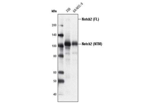
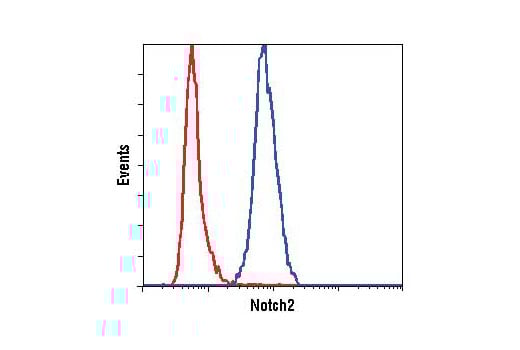
| Notch2 (D76A6) Rabbit Monoclonal Antibody | 5732 | 20 µl | 110, 300 kDa | Rabbit |
| Anti-rabbit IgG, HRP-linked Antibody | 7074 | 100 µl | Goat |
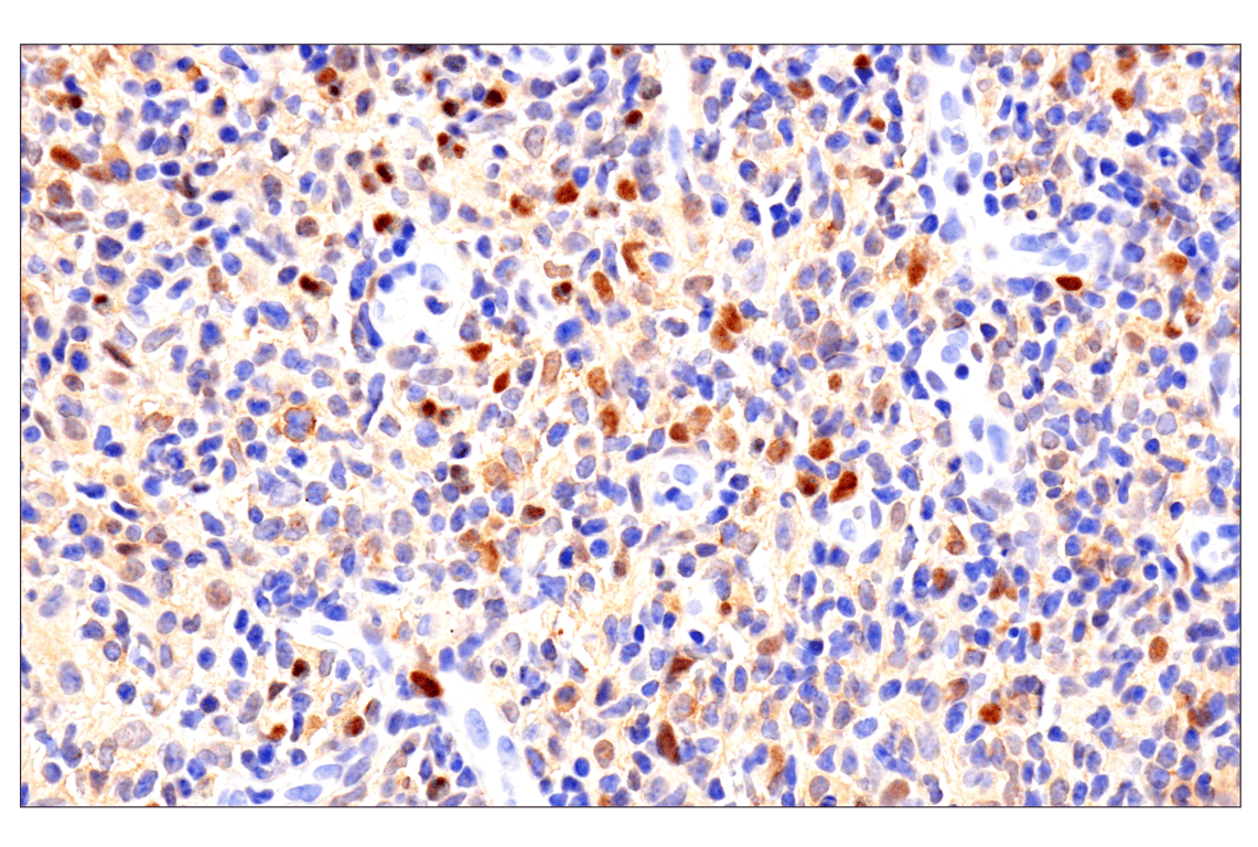
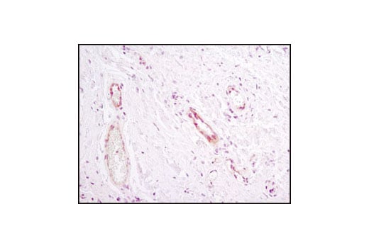
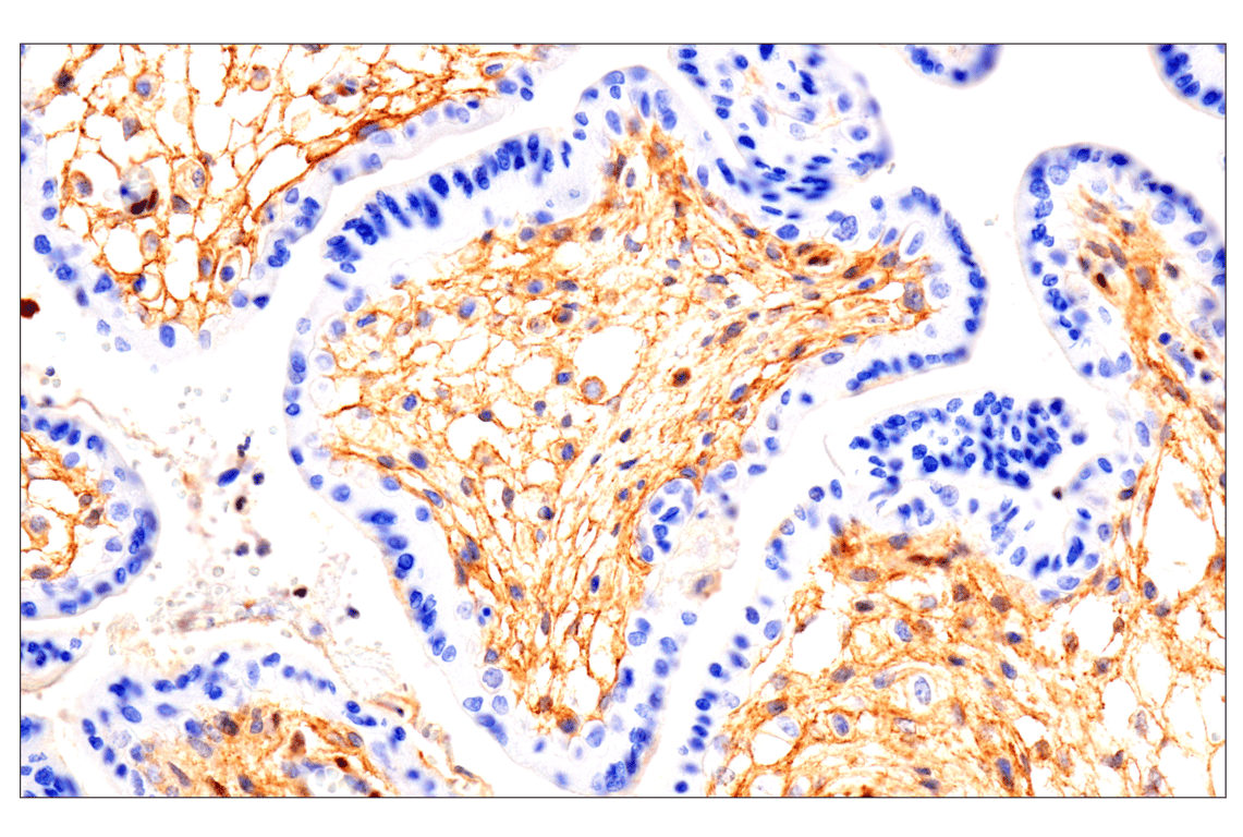
Please visit cellsignal.com for individual component applications, species cross-reactivity, dilutions, protocols, and additional product information.
Description
Storage
Background
Constitutively activated Notch1 signaling is associated with the majority of cases of T cell acute lymphoblastic leukemia (T-ALL). The activation is either due to mutations in Notch1 itself or in the components of ubiquitin ligase complex, namely FBW7 (5-6). Notch2 is a member of the Notch family and mutation in Notch2 is associated with Alagille syndrome (7). Notch3 is a member of the Notch family and is processed similar to Notch1 (8). It is expressed primarily in arterial smooth muscle cells (SMC). Mutations altering the number of cysteine residues in the notch3 extracellular region are associated with cerebral autosomal dominant arteriopathy with subcortical infarcts and leukoencephalopathy (CADASIL), a hereditary angiopathy leading to strokes and dementia in adults (9-11). Recent studies indicate that Notch3 is overexpressed in many types of cancer (12-14).
Background References
- Artavanis-Tsakonas, S. et al. (1999) Science 284, 770-6.
- Chan, Y.M. and Jan, Y.N. (1998) Cell 94, 423-6.
- Schroeter, E.H. et al. (1998) Nature 393, 382-6.
- Rand, M.D. et al. (2000) Mol Cell Biol 20, 1825-35.
- Weng, A.P. et al. (2004) Science 306, 269-71.
- Thompson, B.J. et al. (2007) J Exp Med 204, 1825-35.
- McDaniell, R. et al. (2006) Am J Hum Genet 79, 169-73.
- Baron, M. (2003) Semin Cell Dev Biol 14, 113-9.
- Kalimo, H. et al. (2002) Brain Pathol 12, 371-84.
- Karlström, H. et al. (2002) Proc Natl Acad Sci U S A 99, 17119-24.
- Monet, M. et al. (2007) Hum Mol Genet 16, 982-92.
- Park, J.T. et al. (2006) Cancer Res 66, 6312-8.
- Gramantieri, L. et al. (2007) Liver Int 27, 997-1007.
- Yamaguchi, N. et al. (2008) Cancer Res 68, 1881-8.
Trademarks and Patents
Cell Signaling Technology is a trademark of Cell Signaling Technology, Inc.
All other trademarks are the property of their respective owners. Visit cellsignal.com/trademarks for more information.
Limited Uses
Except as otherwise expressly agreed in a writing signed by a legally authorized representative of CST, the following terms apply to Products provided by CST, its affiliates or its distributors. Any Customer's terms and conditions that are in addition to, or different from, those contained herein, unless separately accepted in writing by a legally authorized representative of CST, are rejected and are of no force or effect.
Products are labeled with For Research Use Only or a similar labeling statement and have not been approved, cleared, or licensed by the FDA or other regulatory foreign or domestic entity, for any purpose. Customer shall not use any Product for any diagnostic or therapeutic purpose, or otherwise in any manner that conflicts with its labeling statement. Products sold or licensed by CST are provided for Customer as the end-user and solely for research and development uses. Any use of Product for diagnostic, prophylactic or therapeutic purposes, or any purchase of Product for resale (alone or as a component) or other commercial purpose, requires a separate license from CST. Customer shall (a) not sell, license, loan, donate or otherwise transfer or make available any Product to any third party, whether alone or in combination with other materials, or use the Products to manufacture any commercial products, (b) not copy, modify, reverse engineer, decompile, disassemble or otherwise attempt to discover the underlying structure or technology of the Products, or use the Products for the purpose of developing any products or services that would compete with CST products or services, (c) not alter or remove from the Products any trademarks, trade names, logos, patent or copyright notices or markings, (d) use the Products solely in accordance with CST Product Terms of Sale and any applicable documentation, and (e) comply with any license, terms of service or similar agreement with respect to any third party products or services used by Customer in connection with the Products.
Revision 2
Revision 2
Revision 2

Revision 2

Revision 2
Revision 2
Revision 2

Revision 2

Revision 2