Revision 4
#80471
Store at -20C
877-616-CELL (2355)
877-678-TECH (8324)
3 Trask Lane | Danvers | Massachusetts | 01923 | USA
For Research Use Only. Not for Use in Diagnostic Procedures.
Applications:
W, W-S, IP
Reactivity:
H M R
Sensitivity:
Endogenous
MW (kDa):
80-100
Source/Isotype:
Rabbit IgG
UniProt ID:
#O60313
Entrez-Gene Id:
4976
Product Usage Information
| Application | Dilution |
|---|---|
| Western Blotting | 1:1000 |
| Simple Western™ | 1:50 - 1:250 |
| Immunoprecipitation | 1:100 |
Storage
Specificity/Sensitivity
Source / Purification
Background
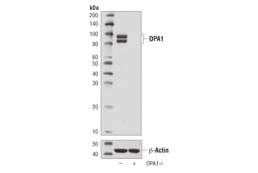
OPA1, or Optic Atrophy 1, was originally identified as a genetic cause for Autosomal Dominant Optic Atrophy, a neuropathy resulting in progressive visual loss (2,3). OPA1 is a widely expressed protein localized to the inner mitochondrial membrane, which regulates mitochondrial fusion and cristae morphology and protects against apoptosis (4-6). OPA1 activity is tightly regulated through alternative splicing and post-translational modifications including complex proteolytic processing by multiple proteases (7-12). In addition, OPA1 expression can be induced under conditions of metabolic demand through a pathway involving Parkin induced NF-κB activation (13).
Background References
- Kasahara, A. and Scorrano, L. (2014) Trends Cell Biol 24, 761-70.
- Delettre, C. et al. (2000) Nat Genet 26, 207-10.
- Alexander, C. et al. (2000) Nat Genet 26, 211-5.
- Frezza, C. et al. (2006) Cell 126, 177-89.
- Olichon, A. et al. (2003) J Biol Chem 278, 7743-6.
- Griparic, L. et al. (2004) J Biol Chem 279, 18792-8.
- Delettre, C. et al. (2001) Hum Genet 109, 584-91.
- Olichon, A. et al. (2007) Cell Death Differ 14, 682-92.
- Ishihara, N. et al. (2006) EMBO J 25, 2966-77.
- Cipolat, S. et al. (2006) Cell 126, 163-75.
- Griparic, L. et al. (2007) J Cell Biol 178, 757-64.
- Merkwirth, C. et al. (2008) Genes Dev 22, 476-88.
- Müller-Rischart, A.K. et al. (2013) Mol Cell 49, 908-21.
Species Reactivity
Species reactivity is determined by testing in at least one approved application (e.g., western blot).
Western Blot Buffer
IMPORTANT: For western blots, incubate membrane with diluted primary antibody in 5% w/v BSA, 1X TBS, 0.1% Tween® 20 at 4°C with gentle shaking, overnight.
Applications Key
W: Western Blotting W-S: Simple Western™ IP: Immunoprecipitation
Cross-Reactivity Key
H: Human M: Mouse R: Rat
Trademarks and Patents
Cell Signaling Technology is a trademark of Cell Signaling Technology, Inc.
SignalSilence is a registered trademark of Cell Signaling Technology, Inc.
All other trademarks are the property of their respective owners. Visit cellsignal.com/trademarks for more information.
Limited Uses
Except as otherwise expressly agreed in a writing signed by a legally authorized representative of CST, the following terms apply to Products provided by CST, its affiliates or its distributors. Any Customer's terms and conditions that are in addition to, or different from, those contained herein, unless separately accepted in writing by a legally authorized representative of CST, are rejected and are of no force or effect.
Products are labeled with For Research Use Only or a similar labeling statement and have not been approved, cleared, or licensed by the FDA or other regulatory foreign or domestic entity, for any purpose. Customer shall not use any Product for any diagnostic or therapeutic purpose, or otherwise in any manner that conflicts with its labeling statement. Products sold or licensed by CST are provided for Customer as the end-user and solely for research and development uses. Any use of Product for diagnostic, prophylactic or therapeutic purposes, or any purchase of Product for resale (alone or as a component) or other commercial purpose, requires a separate license from CST. Customer shall (a) not sell, license, loan, donate or otherwise transfer or make available any Product to any third party, whether alone or in combination with other materials, or use the Products to manufacture any commercial products, (b) not copy, modify, reverse engineer, decompile, disassemble or otherwise attempt to discover the underlying structure or technology of the Products, or use the Products for the purpose of developing any products or services that would compete with CST products or services, (c) not alter or remove from the Products any trademarks, trade names, logos, patent or copyright notices or markings, (d) use the Products solely in accordance with CST Product Terms of Sale and any applicable documentation, and (e) comply with any license, terms of service or similar agreement with respect to any third party products or services used by Customer in connection with the Products.
Revision 4


Revision 4


