Revision 2
#8648
Store at -20C
Parkinson's Research Antibody Sampler Kit
1 Kit
(5 x 20 microliters)
877-616-CELL (2355)
877-678-TECH (8324)
3 Trask Lane | Danvers | Massachusetts | 01923 | USA
For Research Use Only. Not for Use in Diagnostic Procedures.
| Product Includes | Product # | Quantity | Mol. Wt | Isotype/Source |
|---|---|---|---|---|
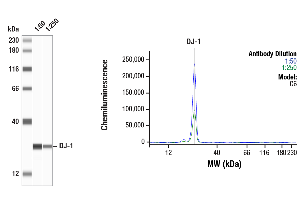
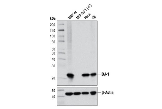
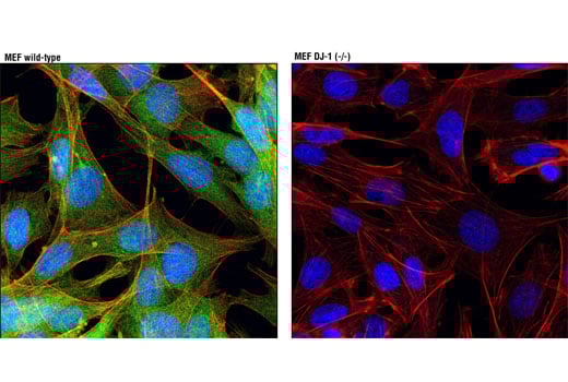
| DJ-1 (D29E5) Rabbit Monoclonal Antibody | 5933 | 20 µl | 22 kDa | Rabbit IgG |
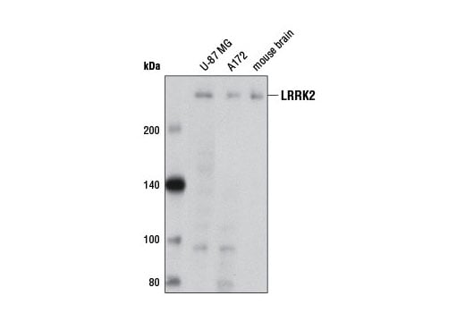
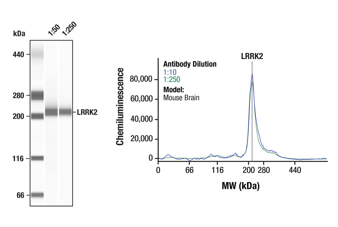
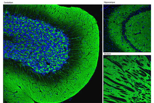
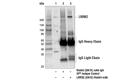
| LRRK2 (D18E12) Rabbit Monoclonal Antibody | 13046 | 20 µl | 290 kDa | Rabbit IgG |
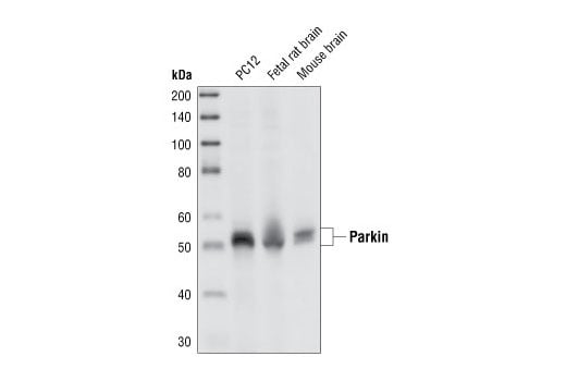
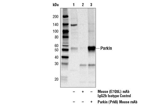
| Parkin (Prk8) Mouse Monoclonal Antibody | 4211 | 20 µl | 50 kDa | Mouse IgG2b |
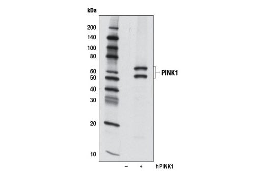
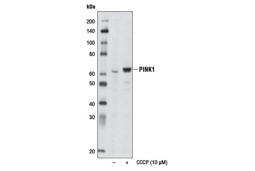
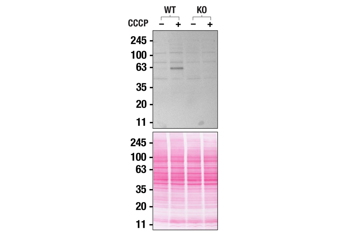
| PINK1 (D8G3) Rabbit Monoclonal Antibody | 6946 | 20 µl | 60, 50 kDa | Rabbit IgG |
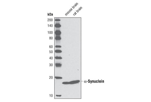
| alpha-Synuclein (D37A6) Rabbit Monoclonal Antibody | 4179 | 20 µl | 18 kDa | Rabbit IgG |
| Anti-rabbit IgG, HRP-linked Antibody | 7074 | 100 µl | Goat | |
| Anti-mouse IgG, HRP-linked Antibody | 7076 | 100 µl | Horse |
Please visit cellsignal.com for individual component applications, species cross-reactivity, dilutions, protocols, and additional product information.
Description
Storage
Background
α-Synuclein, a 140 amino acid protein expressed abundantly in the brain, is a major component of aggregates found in Lewy bodies (3). Parkin is involved in protein degradation through the ubiquitin-proteasome pathway, and investigators have shown that mutations in Parkin cause early onset of PD (4). In the case of autosomal recessive juvenile Parkinsonism (AR-JP), deletions have been found on chromosome 6 in the Parkin gene (5). PTEN induced putative kinase 1 (PINK1) is a mitochondrial serine/threonine kinase involved in the normal function and integrity of mitochondria, as well as a reduction of cytochrome c release from mitochondria (6-8). PINK1 phosphorylates Parkin and promotes its translocation to mitochondria (7). Mutations of PINK1 are associated with loss of protective function, mitrochondrial dysfunction, aggregation of α-synuclein, and proteasome dysfunction (6,8). DJ-1 is involved in multiple cellular functions; it has been shown to cooperate with Ras to increase cell transformation, to regulate transcription of the androgen receptor, and may function as an indicator of oxidative stress, while loss-of-function mutations in DJ-1 cause early onset of PD (9-12). Dopamine D2 receptor-mediated functions are greatly impaired in DJ-1 (-/-) mice, resulting in reduced long-term depression (13). Leucine-rich repeat kinase 2 (LRRK2) contains amino-terminal leucine-rich repeats (LRR), a Ras-like small GTP binding protein-like (ROC) domain, an MLK protein kinase domain, and a carboxy-terminal WD40-repeat. At least 20 LRRK2 mutations have been linked to PD (14). The most prevalent mutation, G2019S, causes increased LRRK2 kinase activity, leading to progressive neurite loss and decreased neuronal survival (15).
Background References
- Fahn, S. (2003) Ann N Y Acad Sci 991, 1-14.
- Moore, D.J. et al. (2005) Annu Rev Neurosci 28, 57-87.
- Goldberg, M.S. and Lansbury, P.T. (2000) Nat Cell Biol 2, E115-9.
- Borrelli, E. (2005) Neuron 45, 479-81.
- Polymeropoulos, M.H. et al. (1997) Science 276, 2045-7.
- Liu, W. et al. (2009) PLoS One 4, e4597.
- Kim, Y. et al. (2008) Biochem Biophys Res Commun 377, 975-80.
- Petit, A. et al. (2005) J Biol Chem 280, 34025-32.
- Bonifati, V. et al. (2003) Science 299, 256-9.
- Nagakubo, D. et al. (1997) Biochem Biophys Res Commun 231, 509-13.
- Takahashi, K. et al. (2001) J Biol Chem 276, 37556-63.
- Mitsumoto, A. and Nakagawa, Y. (2001) Free Radic Res 35, 885-93.
- Goldberg, M.S. et al. (2005) Neuron 45, 489-96.
- Mata, I.F. et al. (2006) Trends Neurosci 29, 286-93.
- MacLeod, D. et al. (2006) Neuron 52, 587-93.
Trademarks and Patents
Cell Signaling Technology is a trademark of Cell Signaling Technology, Inc.
All other trademarks are the property of their respective owners. Visit cellsignal.com/trademarks for more information.
Limited Uses
Except as otherwise expressly agreed in a writing signed by a legally authorized representative of CST, the following terms apply to Products provided by CST, its affiliates or its distributors. Any Customer's terms and conditions that are in addition to, or different from, those contained herein, unless separately accepted in writing by a legally authorized representative of CST, are rejected and are of no force or effect.
Products are labeled with For Research Use Only or a similar labeling statement and have not been approved, cleared, or licensed by the FDA or other regulatory foreign or domestic entity, for any purpose. Customer shall not use any Product for any diagnostic or therapeutic purpose, or otherwise in any manner that conflicts with its labeling statement. Products sold or licensed by CST are provided for Customer as the end-user and solely for research and development uses. Any use of Product for diagnostic, prophylactic or therapeutic purposes, or any purchase of Product for resale (alone or as a component) or other commercial purpose, requires a separate license from CST. Customer shall (a) not sell, license, loan, donate or otherwise transfer or make available any Product to any third party, whether alone or in combination with other materials, or use the Products to manufacture any commercial products, (b) not copy, modify, reverse engineer, decompile, disassemble or otherwise attempt to discover the underlying structure or technology of the Products, or use the Products for the purpose of developing any products or services that would compete with CST products or services, (c) not alter or remove from the Products any trademarks, trade names, logos, patent or copyright notices or markings, (d) use the Products solely in accordance with CST Product Terms of Sale and any applicable documentation, and (e) comply with any license, terms of service or similar agreement with respect to any third party products or services used by Customer in connection with the Products.
Revision 2
Revision 2

Revision 2

Revision 2
Revision 2
Revision 2