Revision 7
#60433
Store at -20C
877-616-CELL (2355)
877-678-TECH (8324)
3 Trask Lane | Danvers | Massachusetts | 01923 | USA
For Research Use Only. Not for Use in Diagnostic Procedures.
Applications:
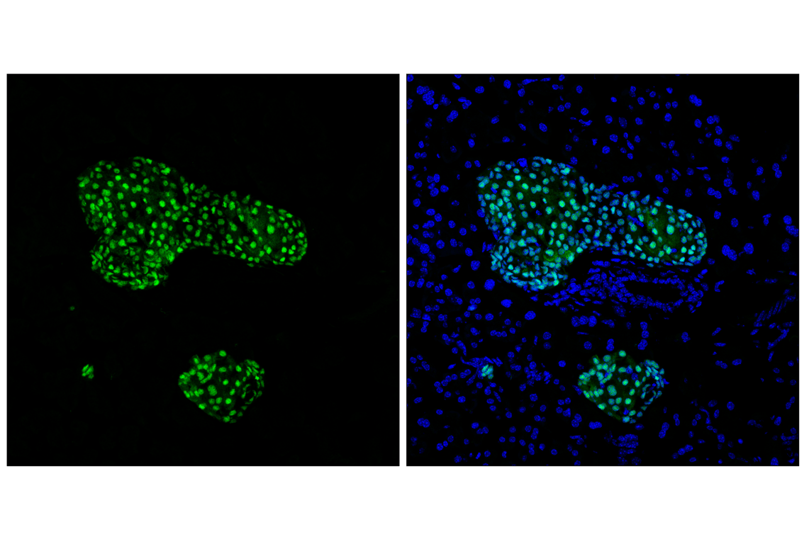
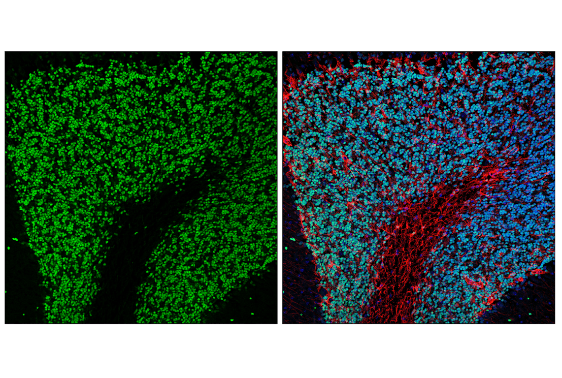
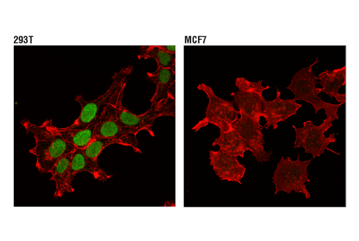
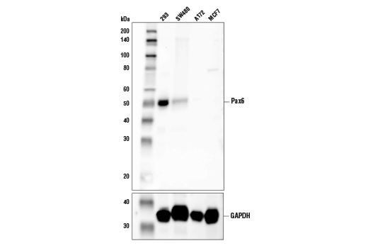
W, IP, IHC-P, IF-F, IF-IC
Reactivity:
H
Sensitivity:
Endogenous
MW (kDa):
50
Source/Isotype:
Rabbit IgG
UniProt ID:
#P26367
Entrez-Gene Id:
5080
Product Usage Information
| Application | Dilution |
|---|---|
| Western Blotting | 1:1000 |
| Immunoprecipitation | 1:200 |
| Immunohistochemistry (Paraffin) | 1:250 - 1:1000 |
| Immunofluorescence (Frozen) | 1:3200 |
| Immunofluorescence (Immunocytochemistry) | 1:100 - 1:400 |
Storage
Specificity/Sensitivity
Species predicted to react based on 100% sequence homology
Source / Purification
Background
Pax6 has important functions in organ development. It is a key regulator of eye development (5), and mutations in Pax6 have been associated with some forms of aniridia, a congenital malformation of the eye (6). Pax6 is also involved in neuronal development, which plays an especially important role in corticogenesis (7). Within its role in the adult brain, it has recently been associated with aging, with gene occupancy studies showing increased association of Pax6 with genes associated with many aging processes in mice (8,9). Pax6 also plays another important function in development and maintenance of pancreatic β-cells (10). Driven by expression of Pdx1, both Pax6 and NGN3 expression is required for β-cell identity (11).
Background References
- Lang, D. et al. (2007) Biochem Pharmacol 73, 1-14.
- Robson, E.J. et al. (2006) Nat Rev Cancer 6, 52-62.
- Wang, Q. et al. (2008) J Cell Mol Med 12, 2281-94.
- Blake, J.A. et al. (2008) Dev Dyn 237, 2791-803.
- Shaham, O. et al. (2012) Prog Retin Eye Res 31, 351-76.
- Hingorani, M. et al. (2012) Eur J Hum Genet 20, 1011-7.
- Manuel, M.N. et al. (2015) Front Cell Neurosci 9, 70.
- Maurya, S.K. and Mishra, R. (2017) Ann Neurosci 24, 20-25.
- Maurya, S.K. and Mishra, R. (2017) J Chem Neuroanat 82, 60-64.
- Swisa, A. et al. (2017) Front Genet 8, 21.
- Mitchell, R.K. et al. (2017) J Biol Chem 292, 8892-8906.
Species Reactivity
Species reactivity is determined by testing in at least one approved application (e.g., western blot).
Western Blot Buffer
IMPORTANT: For western blots, incubate membrane with diluted primary antibody in 5% w/v BSA, 1X TBS, 0.1% Tween® 20 at 4°C with gentle shaking, overnight.
Applications Key
W: Western Blotting IP: Immunoprecipitation IHC-P: Immunohistochemistry (Paraffin) IF-F: Immunofluorescence (Frozen) IF-IC: Immunofluorescence (Immunocytochemistry)
Cross-Reactivity Key
H: Human
Trademarks and Patents
Cell Signaling Technology is a trademark of Cell Signaling Technology, Inc.
SignalStain is a registered trademark of Cell Signaling Technology, Inc.
All other trademarks are the property of their respective owners. Visit cellsignal.com/trademarks for more information.
Limited Uses
Except as otherwise expressly agreed in a writing signed by a legally authorized representative of CST, the following terms apply to Products provided by CST, its affiliates or its distributors. Any Customer's terms and conditions that are in addition to, or different from, those contained herein, unless separately accepted in writing by a legally authorized representative of CST, are rejected and are of no force or effect.
Products are labeled with For Research Use Only or a similar labeling statement and have not been approved, cleared, or licensed by the FDA or other regulatory foreign or domestic entity, for any purpose. Customer shall not use any Product for any diagnostic or therapeutic purpose, or otherwise in any manner that conflicts with its labeling statement. Products sold or licensed by CST are provided for Customer as the end-user and solely for research and development uses. Any use of Product for diagnostic, prophylactic or therapeutic purposes, or any purchase of Product for resale (alone or as a component) or other commercial purpose, requires a separate license from CST. Customer shall (a) not sell, license, loan, donate or otherwise transfer or make available any Product to any third party, whether alone or in combination with other materials, or use the Products to manufacture any commercial products, (b) not copy, modify, reverse engineer, decompile, disassemble or otherwise attempt to discover the underlying structure or technology of the Products, or use the Products for the purpose of developing any products or services that would compete with CST products or services, (c) not alter or remove from the Products any trademarks, trade names, logos, patent or copyright notices or markings, (d) use the Products solely in accordance with CST Product Terms of Sale and any applicable documentation, and (e) comply with any license, terms of service or similar agreement with respect to any third party products or services used by Customer in connection with the Products.
Revision 7

Revision 7
Revision 7