Revision 1
#12651
Store at -20C
PDGF Receptor Activation Antibody Sampler Kit
1 Kit
(8 x 20 microliters)
877-616-CELL (2355)
877-678-TECH (8324)
3 Trask Lane | Danvers | Massachusetts | 01923 | USA
For Research Use Only. Not for Use in Diagnostic Procedures.
| Product Includes | Product # | Quantity | Mol. Wt | Isotype/Source |
|---|---|---|---|---|
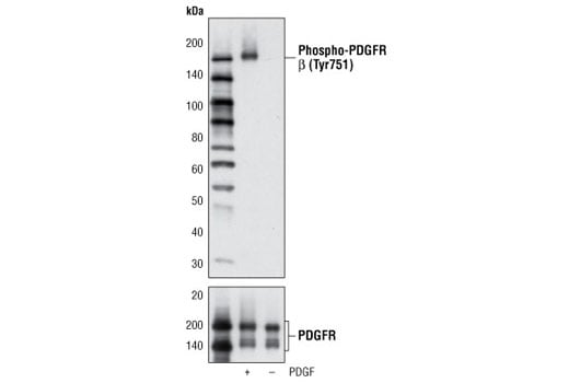
| Phospho-PDGF Receptor beta (Tyr751) (C63G6) Rabbit Monoclonal Antibody | 4549 | 20 µl | 190 kDa | Rabbit IgG |
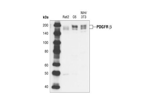
| PDGF Receptor beta (28E1) Rabbit Monoclonal Antibody | 3169 | 20 µl | 190 kDa | Rabbit IgG |
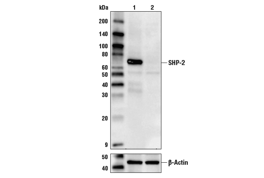
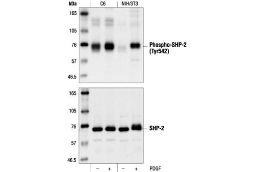
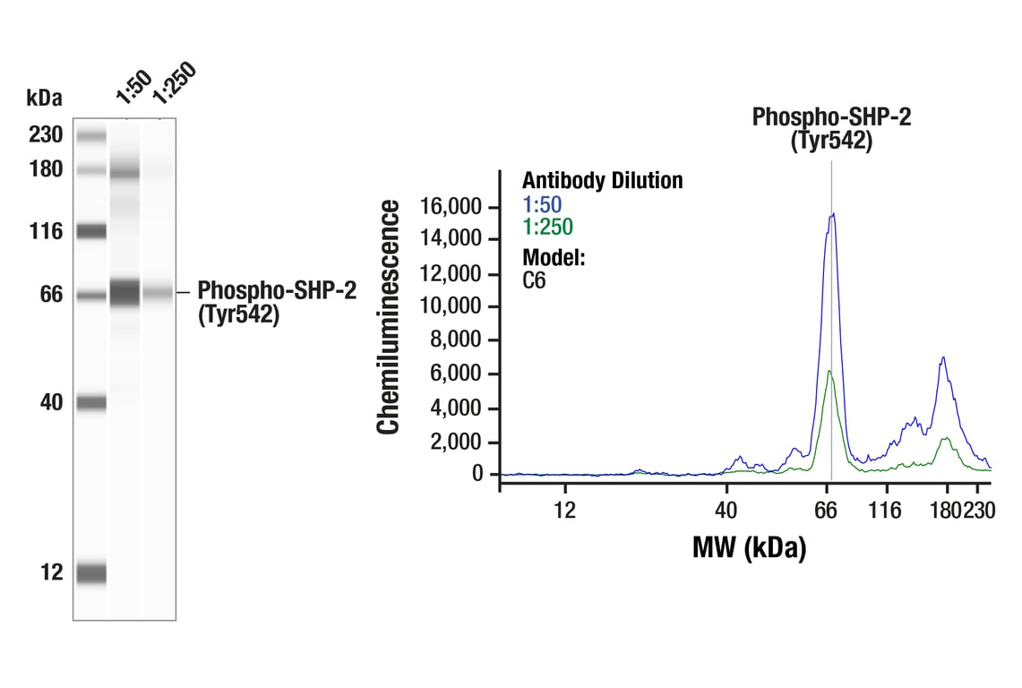
| Phospho-SHP-2 (Tyr542) Antibody | 3751 | 20 µl | 72 kDa | Rabbit |
| SHP-2 (D50F2) Rabbit Monoclonal Antibody | 3397 | 20 µl | 72 kDa | Rabbit IgG |
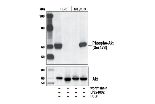
| Phospho-Akt (Ser473) (D9E) Rabbit Monoclonal Antibody | 4060 | 20 µl | 60 kDa | Rabbit IgG |
| Akt (pan) (C67E7) Rabbit Monoclonal Antibody | 4691 | 20 µl | 60 kDa | Rabbit IgG |
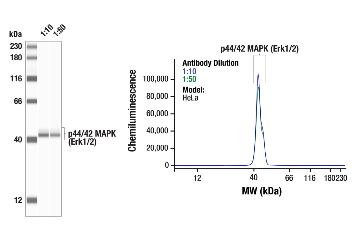
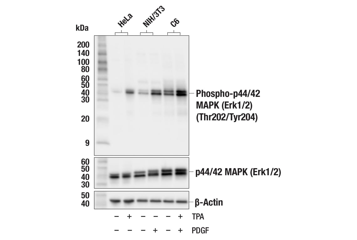
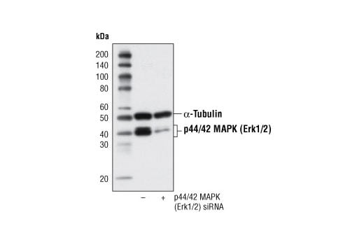
| Phospho-p44/42 MAPK (Erk1/2) (Thr202/Tyr204) (D13.14.4E) Rabbit Monoclonal Antibody | 4370 | 20 µl | 44, 42 kDa | Rabbit IgG |
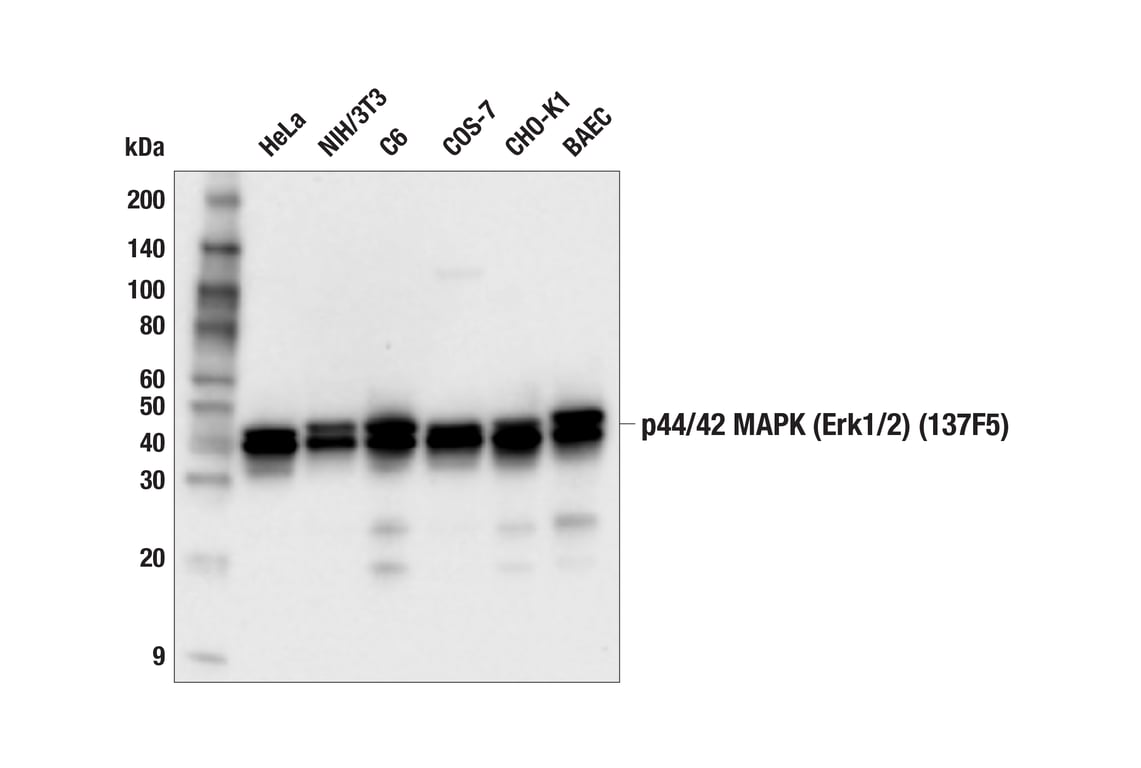
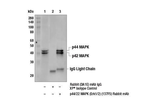
| p44/42 MAPK (Erk1/2) (137F5) Rabbit Monoclonal Antibody | 4695 | 20 µl | 42, 44 kDa | Rabbit IgG |
| Anti-rabbit IgG, HRP-linked Antibody | 7074 | 100 µl | Goat |
Please visit cellsignal.com for individual component applications, species cross-reactivity, dilutions, protocols, and additional product information.
Description
Storage
Background
SHP-2 (PTPN11) is a nonreceptor protein tyrosine phosphatase that participates in signaling pathways that control cell growth, differentiation, migration, and death (5). Activation of SHP-2 and its association with Gab1 is critical for sustained Erk activation downstream of growth factor receptors and cytokines (6). Phosphorylation of SHP-2 at Tyr542 and Tyr580 in response to growth factor receptor activation is thought to relieve basal inhibition and stimulate SHP-2 tyrosine phosphatase activity (7,8).
Insulin and various growth/survival factors activate Akt, a kinase that acts in a wortmannin-sensitive pathway involving PI3 kinase to help control survival and apoptosis (9-11). Akt is activated by phospholipid binding and activation loop phosphorylation at Thr308 by PDK1 (12) and by phosphorylation within the carboxy terminus at Ser473.
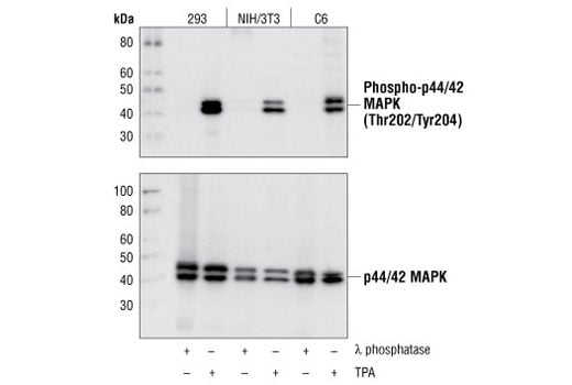
The p44/42 MAPK (Erk1/2) signaling pathway is activated in response to extracellular stimuli including mitogens, growth factors, and cytokines (13-15). Research suggests that this pathway is an important target in cancer diagnosis and treatment (16). External stimuli lead to activation of a kinase cascade that results in the activation of p44 and p42 by a MAP kinase. MEK1 and MEK2 activate p44 and p42 through phosphorylation of activation loop residues Thr202/Tyr204 and Thr185/Tyr187, respectively.
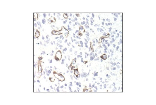
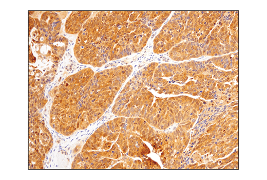
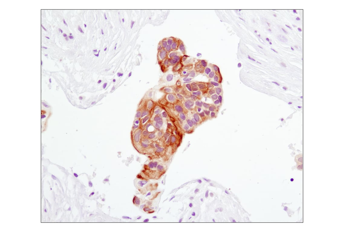
Clinical studies describe PDGF expression in a number of different solid tumors, from glioblastomas to prostate carcinomas. The biological role of PDGF signaling in these tumors varies from autocrine stimulation of cancer cell growth to more subtle paracrine interactions involving adjacent stroma and even angiogenesis. Targeting PDGF signaling may be an effective way for tumor treatment (17).
Background References
- Deuel, T.F. et al. (1988) Biofactors 1, 213-7.
- Ostman, A. and Heldin, C.H. (2001) Adv Cancer Res 80, 1-38.
- Betsholtz, C. et al. (2001) Bioessays 23, 494-507.
- Ramalingam, K. et al. (1995) Bioorg Med Chem 3, 1263-72.
- Qu, C.K. (2000) Cell Res 10, 279-88.
- Maroun, C.R. et al. (2000) Mol Cell Biol 20, 8513-25.
- Bennett, A.M. et al. (1994) Proc Natl Acad Sci U S A 91, 7335-9.
- Lu, W. et al. (2001) Mol Cell 8, 759-69.
- Franke, T.F. et al. (1997) Cell 88, 435-7.
- Burgering, B.M. and Coffer, P.J. (1995) Nature 376, 599-602.
- Franke, T.F. et al. (1995) Cell 81, 727-36.
- Alessi, D.R. et al. (1996) EMBO J 15, 6541-51.
- Roux, P.P. and Blenis, J. (2004) Microbiol Mol Biol Rev 68, 320-44.
- Baccarini, M. (2005) FEBS Lett 579, 3271-7.
- Meloche, S. and Pouysségur, J. (2007) Oncogene 26, 3227-39.
- Roberts, P.J. and Der, C.J. (2007) Oncogene 26, 3291-310.
- George, D. (2001) Semin Oncol 28, 27-33.
Trademarks and Patents
Cell Signaling Technology is a trademark of Cell Signaling Technology, Inc.
U.S. Patent No. 7,429,487, foreign equivalents, and child patents deriving therefrom.
All other trademarks are the property of their respective owners. Visit cellsignal.com/trademarks for more information.
Limited Uses
Except as otherwise expressly agreed in a writing signed by a legally authorized representative of CST, the following terms apply to Products provided by CST, its affiliates or its distributors. Any Customer's terms and conditions that are in addition to, or different from, those contained herein, unless separately accepted in writing by a legally authorized representative of CST, are rejected and are of no force or effect.
Products are labeled with For Research Use Only or a similar labeling statement and have not been approved, cleared, or licensed by the FDA or other regulatory foreign or domestic entity, for any purpose. Customer shall not use any Product for any diagnostic or therapeutic purpose, or otherwise in any manner that conflicts with its labeling statement. Products sold or licensed by CST are provided for Customer as the end-user and solely for research and development uses. Any use of Product for diagnostic, prophylactic or therapeutic purposes, or any purchase of Product for resale (alone or as a component) or other commercial purpose, requires a separate license from CST. Customer shall (a) not sell, license, loan, donate or otherwise transfer or make available any Product to any third party, whether alone or in combination with other materials, or use the Products to manufacture any commercial products, (b) not copy, modify, reverse engineer, decompile, disassemble or otherwise attempt to discover the underlying structure or technology of the Products, or use the Products for the purpose of developing any products or services that would compete with CST products or services, (c) not alter or remove from the Products any trademarks, trade names, logos, patent or copyright notices or markings, (d) use the Products solely in accordance with CST Product Terms of Sale and any applicable documentation, and (e) comply with any license, terms of service or similar agreement with respect to any third party products or services used by Customer in connection with the Products.
Revision 1
Revision 1
Revision 1
Revision 1


Revision 1
Revision 1
Revision 1
Revision 1

Revision 1

Revision 1
Revision 1
Revision 1

Revision 1
Revision 1
Revision 1
Revision 1
Revision 1
Revision 1
Revision 1
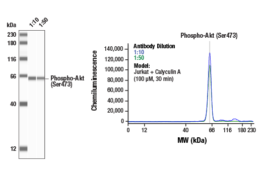
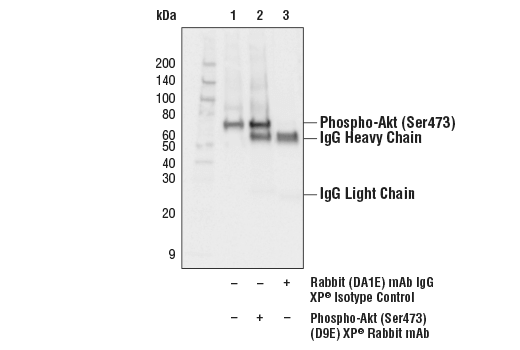
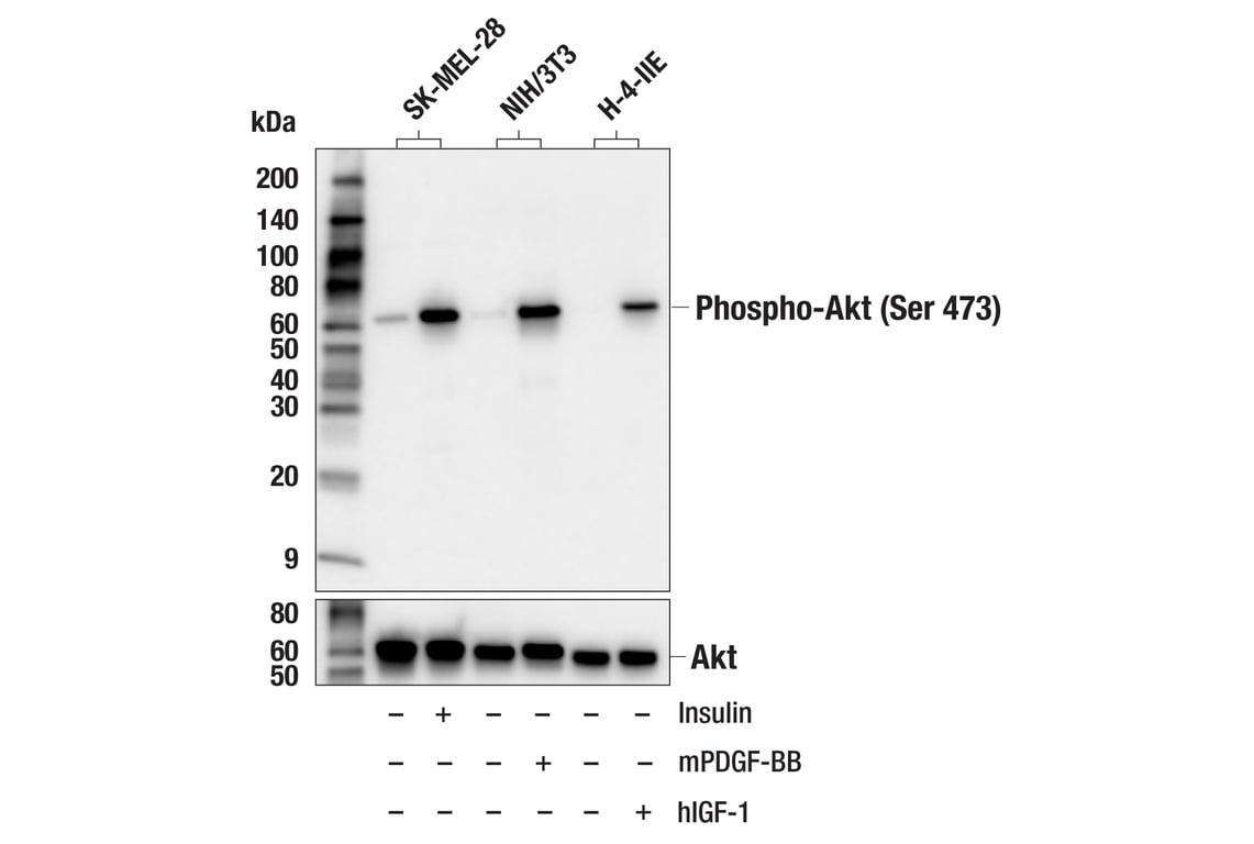
Western blot analysis of extracts from various cell lines, untreated (-) or treated (+) as indicated with human insulin (100 nM, 20 min), mouse PDGF-BB (100 ng/ml, 20 min), or human Insulin-like Growth Factor I (hIGF-I) (100 ng/ml; 5 min), using Phospho-Akt (Ser473) (D9E) XP® Rabbit mAb (upper) or Akt (pan) (C67E7) Rabbit mAb #4691 (lower).
Revision 1
Revision 1