| Cat. # | Size | Qty. | Price |
|---|---|---|---|
| 77111S | 100 µl |
|
| REACTIVITY | H |
| SENSITIVITY | Endogenous |
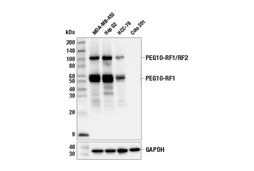
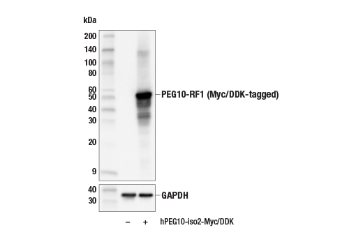
| MW (kDa) | 50-60, 110 |
| SOURCE | Rabbit |
Product Information
| Application | Dilution |
|---|---|
| Western Blotting | 1:1000 |
For western blots, incubate membrane with diluted primary antibody in 5% w/v BSA, 1X TBS, 0.1% Tween® 20 at 4°C with gentle shaking, overnight.
NOTE: Please refer to primary antibody product webpage for recommended antibody dilution.
From sample preparation to detection, the reagents you need for your Western Blot are now in one convenient kit: #12957 Western Blotting Application Solutions Kit
NOTE: Prepare solutions with reverse osmosis deionized (RODI) or equivalent grade water.
Load 20 µl onto SDS-PAGE gel (10 cm x 10 cm).
NOTE: Loading of prestained molecular weight markers (#59329, 10 µl/lane) to verify electrotransfer and biotinylated protein ladder (#7727, 10 µl/lane) to determine molecular weights are recommended.
NOTE: Volumes are for 10 cm x 10 cm (100 cm2) of membrane; for different sized membranes, adjust volumes accordingly.
* Avoid repeated exposure to skin.
posted June 2005
revised June 2020
Protocol Id: 10
Human
Polyclonal antibodies are produced by immunizing animals with a synthetic peptide corresponding to residues surrounding Lys85 of human PEG10 protein. Antibodies are purified by peptide affinity chromatography.
PEG10 (paternally expressed gene 10) is an imprinted gene thought to be derived from a Ty3/Gypsy long terminal repeat (LTR) retrotransposon family encoding Gag- and Pol-like domains (1). Deletion of PEG10 in mice leads to embryonic lethality due to defects in placental formation (2). Similarly, PEG10 deficient trophoblast stem cells exhibited impaired differentiation into placental lineages (3). PEG10 is aberrantly expressed in several cancer types, including hepatocellular carcinoma, and contributes to tumorigenesis by affecting cell proliferation, apoptosis, and metastasis (4). The PEG10 gene has two overlapping open reading frames that are regulated by the programmed process of -1 frameshifting (5). The first encoded protein, ORF1 (or RF1), has a Gag domain with coiled-coil domain and zinc finger domains, while ORF1-2 (or RF1/RF2) is produced by -1 frameshifting and creates a fusion of the ORF1 Gag domain with a carboxyl-terminal Pol-like protease domain. PEG10 has retained the ability to form virus-like particles (VLPs) that are secreted as small extracellular vesicles delivered to distant sites (6). Studies have also shown that PEG10 can bind to mRNA and that PEG10 mRNA can be incorporated into VLPs formed by the PEG10 protein (3,6). Those discoveries have led to a potentially new technique in which unrelated genes can be modified with a region from the PEG10 untranslated region (UTR), allowing genes of interest to be delivered via secreted vesicles (6).
Except as otherwise expressly agreed in a writing signed by a legally authorized representative of CST, the following terms apply to Products provided by CST, its affiliates or its distributors. Any Customer's terms and conditions that are in addition to, or different from, those contained herein, unless separately accepted in writing by a legally authorized representative of CST, are rejected and are of no force or effect.
Products are labeled with For Research Use Only or a similar labeling statement and have not been approved, cleared, or licensed by the FDA or other regulatory foreign or domestic entity, for any purpose. Customer shall not use any Product for any diagnostic or therapeutic purpose, or otherwise in any manner that conflicts with its labeling statement. Products sold or licensed by CST are provided for Customer as the end-user and solely for research and development uses. Any use of Product for diagnostic, prophylactic or therapeutic purposes, or any purchase of Product for resale (alone or as a component) or other commercial purpose, requires a separate license from CST. Customer shall (a) not sell, license, loan, donate or otherwise transfer or make available any Product to any third party, whether alone or in combination with other materials, or use the Products to manufacture any commercial products, (b) not copy, modify, reverse engineer, decompile, disassemble or otherwise attempt to discover the underlying structure or technology of the Products, or use the Products for the purpose of developing any products or services that would compete with CST products or services, (c) not alter or remove from the Products any trademarks, trade names, logos, patent or copyright notices or markings, (d) use the Products solely in accordance with CST Product Terms of Sale and any applicable documentation, and (e) comply with any license, terms of service or similar agreement with respect to any third party products or services used by Customer in connection with the Products.