Revision 1
#67748
Store at -20C
877-616-CELL (2355)
877-678-TECH (8324)
3 Trask Lane | Danvers | Massachusetts | 01923 | USA
For Research Use Only. Not for Use in Diagnostic Procedures.
Applications:
W
Reactivity:
H
Sensitivity:
Endogenous
MW (kDa):
20
Source/Isotype:
Rabbit
UniProt ID:
#P20963
Entrez-Gene Id:
919
Product Usage Information
| Application | Dilution |
|---|---|
| Western Blotting | 1:1000 |
Storage
Specificity/Sensitivity
Species predicted to react based on 100% sequence homology
Source / Purification
Background
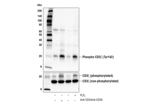
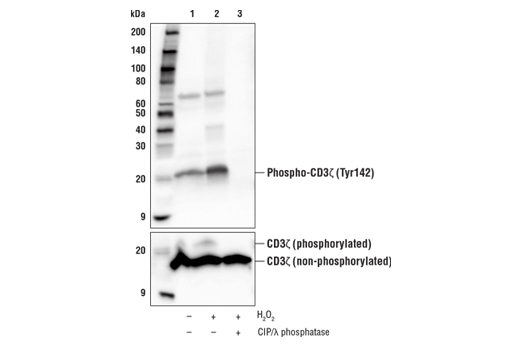
The CD3ζ invariant chain is a type-I transmembrane protein that exists in the TCR signaling complex as a disulfide-linked homodimer (6). The cytoplasmic tail of each CD3ζ monomer contains three distinct ITAM motifs, each containing two tyrosine residues. Phosphorylation of CD3ζ ITAM tyrosine residues, including Y142, is driven by recruitment of the Lck and Fyn tyrosine kinases to the TCR (7). Lck/Fyn-mediated ITAM phosphorylation creates docking sites that promote the SH2 domain-dependent recruitment and activation of Zap-70 (8-10), which drives amplification of signaling events downstream of the TCR that facilitate T cell activation (10). Phosphorylation of a pool of p16 CD3ζ leads to the generation of p21 and p23 species, which differ in the degree of ITAM phosphorylation. It has been proposed that the ratio of p21/p23 contributes to regulating the amplitude of T cell activation (11). CD3ζ plays an important role in the assembly and surface expression of the TCR complex. Indeed, research studies have demonstrated that CD3ζ is degraded in response to Ag-dependent TCR stimulation as a mechanism to tightly control T cell activation (12).
Background References
- Kuhns, M.S. et al. (2006) Immunity 24, 133-139.
- Pitcher, L.A. and van Oers, N.S. (2003) Trends Immunol. 24, 554-560.
- Osman, N. et al. (1996) Eur. J. Immunol. 26, 1063-1068.
- Hatada, M.H. et al. (1995) Nature 377, 32-38.
- Gil, D. et al. (2002) Cell 109, 901-912.
- Call, M.E. et al. (2006) Cell 127, 355-68.
- Housden, H.R. et al. (2003) Eur J Biochem 270, 2369-76.
- Hatada, M.H. et al. (1995) Nature 377, 32-8.
- Visco, C. et al. (2000) Biochemistry 39, 2784-91.
- Iwashima, M. et al. (1994) Science 263, 1136-9.
- Pitcher, L.A. et al. (2003) Immunol Rev 191, 47-61.
- Dumont, C. et al. (2002) J Immunol 169, 1705-12.
Species Reactivity
Species reactivity is determined by testing in at least one approved application (e.g., western blot).
Western Blot Buffer
IMPORTANT: For western blots, incubate membrane with diluted primary antibody in 5% w/v BSA, 1X TBS, 0.1% Tween® 20 at 4°C with gentle shaking, overnight.
Applications Key
W: Western Blotting
Cross-Reactivity Key
H: Human
Trademarks and Patents
Cell Signaling Technology is a trademark of Cell Signaling Technology, Inc.
All other trademarks are the property of their respective owners. Visit cellsignal.com/trademarks for more information.
Limited Uses
Except as otherwise expressly agreed in a writing signed by a legally authorized representative of CST, the following terms apply to Products provided by CST, its affiliates or its distributors. Any Customer's terms and conditions that are in addition to, or different from, those contained herein, unless separately accepted in writing by a legally authorized representative of CST, are rejected and are of no force or effect.
Products are labeled with For Research Use Only or a similar labeling statement and have not been approved, cleared, or licensed by the FDA or other regulatory foreign or domestic entity, for any purpose. Customer shall not use any Product for any diagnostic or therapeutic purpose, or otherwise in any manner that conflicts with its labeling statement. Products sold or licensed by CST are provided for Customer as the end-user and solely for research and development uses. Any use of Product for diagnostic, prophylactic or therapeutic purposes, or any purchase of Product for resale (alone or as a component) or other commercial purpose, requires a separate license from CST. Customer shall (a) not sell, license, loan, donate or otherwise transfer or make available any Product to any third party, whether alone or in combination with other materials, or use the Products to manufacture any commercial products, (b) not copy, modify, reverse engineer, decompile, disassemble or otherwise attempt to discover the underlying structure or technology of the Products, or use the Products for the purpose of developing any products or services that would compete with CST products or services, (c) not alter or remove from the Products any trademarks, trade names, logos, patent or copyright notices or markings, (d) use the Products solely in accordance with CST Product Terms of Sale and any applicable documentation, and (e) comply with any license, terms of service or similar agreement with respect to any third party products or services used by Customer in connection with the Products.
Revision 1