Revision 1
#2600
Store at -20C
877-616-CELL (2355)
877-678-TECH (8324)
3 Trask Lane | Danvers | Massachusetts | 01923 | USA
For Research Use Only. Not for Use in Diagnostic Procedures.
Applications:
W, IP, IF-IC
Reactivity:
H M R Mk
Sensitivity:
Endogenous
MW (kDa):
22
Source/Isotype:
Rabbit
UniProt ID:
#Q13185
Entrez-Gene Id:
11335
Product Usage Information
| Application | Dilution |
|---|---|
| Western Blotting | 1:1000 |
| Immunoprecipitation | 1:25 |
| Immunofluorescence (Immunocytochemistry) | 1:200 |
Storage
Specificity/Sensitivity
Species predicted to react based on 100% sequence homology
Source / Purification
Background
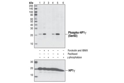
HP1γ is phosphorylated on Ser83 by protein kinase A (PKA) in vitro, and activation of PKA by forskolin and IBMX treatment leads to increased phosphorylation in vivo (14). Phosphorylation of HP1γ on Ser83 also increases during mitosis as demonstrated by the Phospho-HP1γ (Ser83) Antibody, which shows increased immunofluorescent staining in untreated mitotic cells and increased Western blot signal in lysates from cells arrested in mitosis by treatment with paclitaxel. Phosphorylation of Ser83 only occurs on a subpopulation of HP1γ found associated with euchromatin, specifically HP1γ bound to coding regions of active genes (14). This phosphorylation impairs the ability of HP1γ to silence transcription and may be a marker for transcription elongation (14).
Background References
- Maison, C. and Almouzni, G. (2004) Nat. Rev. Mol. Cell Biol. 5, 296-304.
- Minc, E. et al. (2000) Cytogenet. Cell Genet. 90, 279-284.
- Nielsen, A.L. et al. (2001) Mol. Cell 7, 729-739.
- Lachner, M. et al. (2001) Nature 410, 116-120.
- Bannister, A.J. et al. (2001) Nature 410, 120-124.
- Muchardt, C. et al. (2002) EMBO Rep. 3, 975-981.
- Yamamoto, K. and Sonoda, M. (2003) Biochem. Biophys. Res. Commun. 301, 287-292.
- Fuks, F. et al. (2003) Nucleic Acids Res. 31, 2305-2312.
- Murzina, N. et al. (1999) Mol. Cell 4, 529-540.
- Nielsen, S.J. et al. (2001) Nature 412, 561-565.
- Ogawa, H. et al. (2002) Science 296, 1132-1136.
- Minc, E. et al. (1999) Chromosoma 108, 220-234.
- Zhao, T. et al. (2001) J. Biol. Chem. 276, 9512-9518.
- Lomberk, G. et al. (2006) Nat. Cell Biol. 8, 407-415.
Species Reactivity
Species reactivity is determined by testing in at least one approved application (e.g., western blot).
Western Blot Buffer
IMPORTANT: For western blots, incubate membrane with diluted primary antibody in 5% w/v BSA, 1X TBS, 0.1% Tween® 20 at 4°C with gentle shaking, overnight.
Applications Key
W: Western Blotting IP: Immunoprecipitation IF-IC: Immunofluorescence (Immunocytochemistry)
Cross-Reactivity Key
H: Human M: Mouse R: Rat Mk: Monkey
Trademarks and Patents
Cell Signaling Technology is a trademark of Cell Signaling Technology, Inc.
Alexa Fluor is a registered trademark of Life Technologies Corporation.
All other trademarks are the property of their respective owners. Visit cellsignal.com/trademarks for more information.
Limited Uses
Except as otherwise expressly agreed in a writing signed by a legally authorized representative of CST, the following terms apply to Products provided by CST, its affiliates or its distributors. Any Customer's terms and conditions that are in addition to, or different from, those contained herein, unless separately accepted in writing by a legally authorized representative of CST, are rejected and are of no force or effect.
Products are labeled with For Research Use Only or a similar labeling statement and have not been approved, cleared, or licensed by the FDA or other regulatory foreign or domestic entity, for any purpose. Customer shall not use any Product for any diagnostic or therapeutic purpose, or otherwise in any manner that conflicts with its labeling statement. Products sold or licensed by CST are provided for Customer as the end-user and solely for research and development uses. Any use of Product for diagnostic, prophylactic or therapeutic purposes, or any purchase of Product for resale (alone or as a component) or other commercial purpose, requires a separate license from CST. Customer shall (a) not sell, license, loan, donate or otherwise transfer or make available any Product to any third party, whether alone or in combination with other materials, or use the Products to manufacture any commercial products, (b) not copy, modify, reverse engineer, decompile, disassemble or otherwise attempt to discover the underlying structure or technology of the Products, or use the Products for the purpose of developing any products or services that would compete with CST products or services, (c) not alter or remove from the Products any trademarks, trade names, logos, patent or copyright notices or markings, (d) use the Products solely in accordance with CST Product Terms of Sale and any applicable documentation, and (e) comply with any license, terms of service or similar agreement with respect to any third party products or services used by Customer in connection with the Products.
Revision 1