Revision 6
#9913
Store at -20C
Phospho-p38 MAPK Pathway Antibody Sampler Kit
1 Kit
(6 x 20 microliters)
877-616-CELL (2355)
877-678-TECH (8324)
3 Trask Lane | Danvers | Massachusetts | 01923 | USA
For Research Use Only. Not for Use in Diagnostic Procedures.
| Product Includes | Product # | Quantity | Mol. Wt | Isotype/Source |
|---|---|---|---|---|
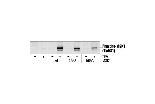
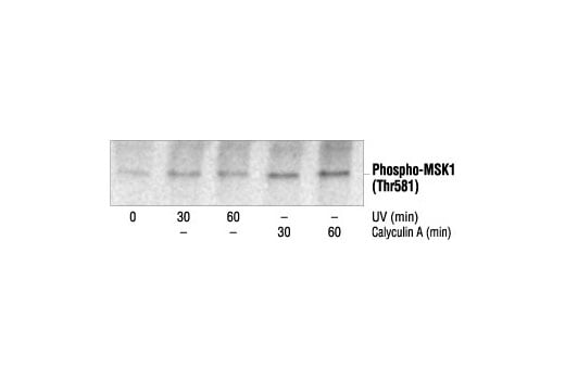
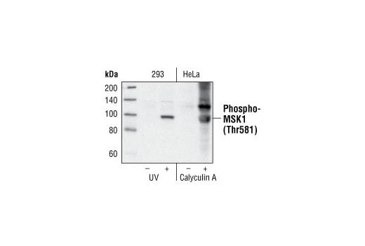
| Phospho-MSK1 (Thr581) Antibody | 9595 | 20 µl | 90 kDa | Rabbit |
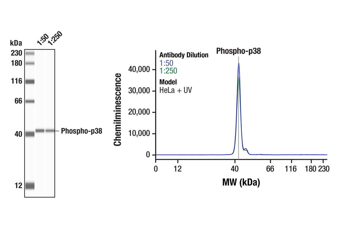
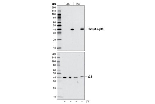
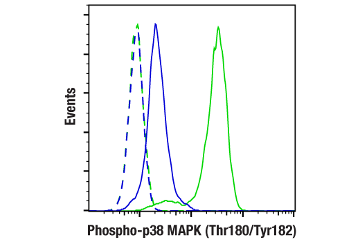
| Phospho-p38 MAPK (Thr180/Tyr182) (D3F9) XP® Rabbit mAb | 4511 | 20 µl | 43 kDa | Rabbit IgG |
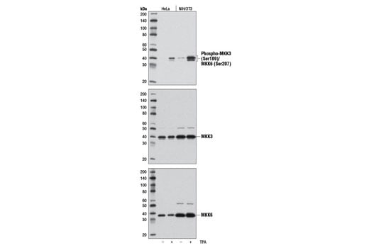
| Phospho-MKK3 (Ser189)/MKK6 (Ser207) (D8E9) Rabbit mAb | 12280 | 20 µl | 38 MKK6, 40 MKK3 kDa | Rabbit IgG |
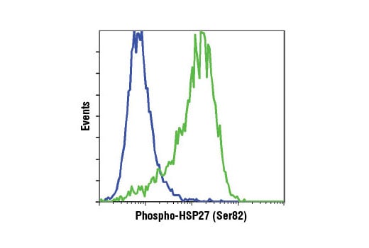
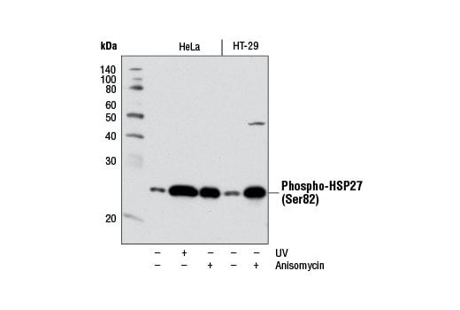
| Phospho-HSP27 (Ser82) (D1H2F6) XP® Rabbit mAb | 9709 | 20 µl | 27 kDa | Rabbit IgG |
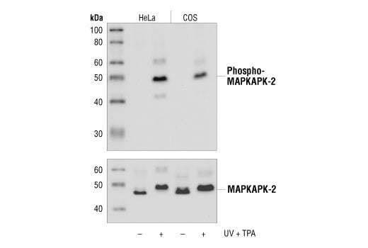
| Phospho-MAPKAPK-2 (Thr334) (27B7) Rabbit mAb | 3007 | 20 µl | 49 kDa | Rabbit IgG |
| Anti-rabbit IgG, HRP-linked Antibody | 7074 | 100 µl | Goat | |
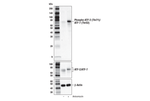
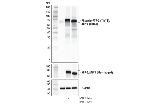
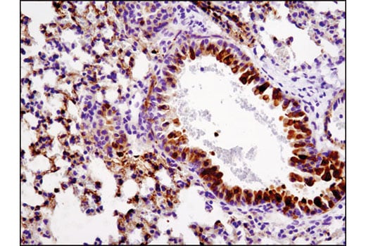
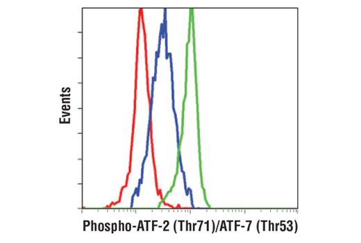
| Phospho-ATF-2 (Thr71)/ATF-7 (Thr53) (A8J7P) Rabbit mAb | 15411 | 20 µl | 65,75 kDa | Rabbit IgG |
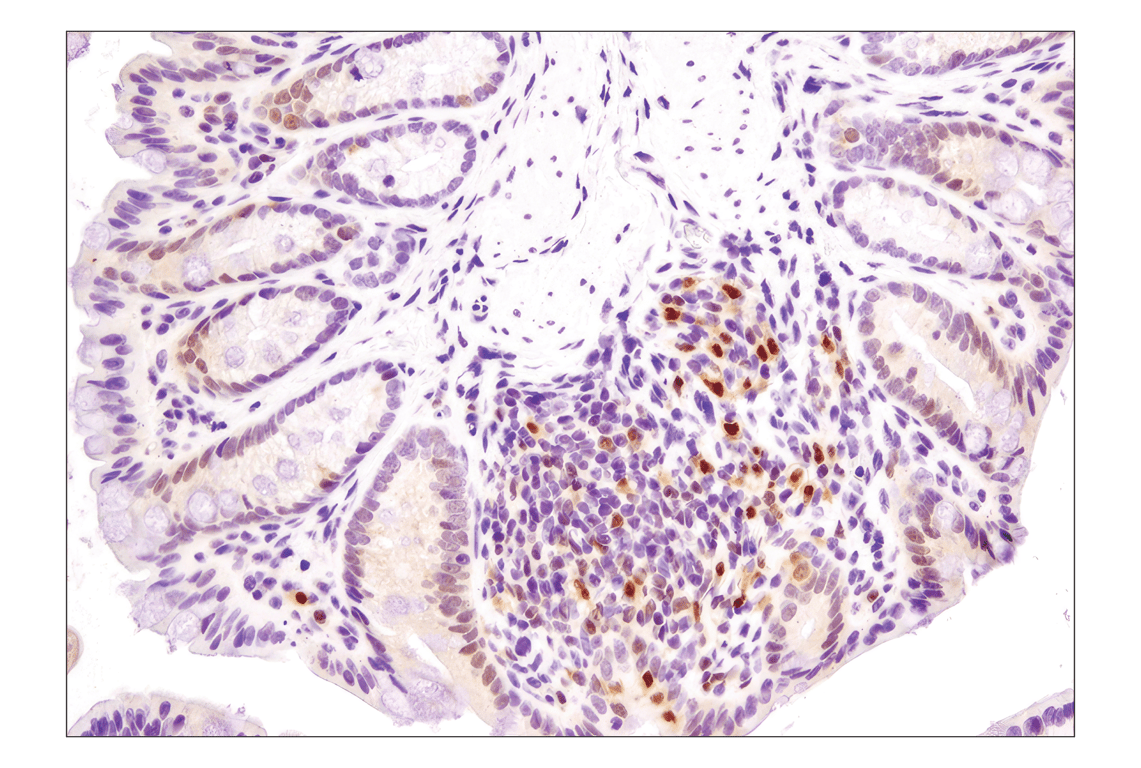
Please visit cellsignal.com for individual component applications, species cross-reactivity, dilutions, protocols, and additional product information.
Description
Storage
Background
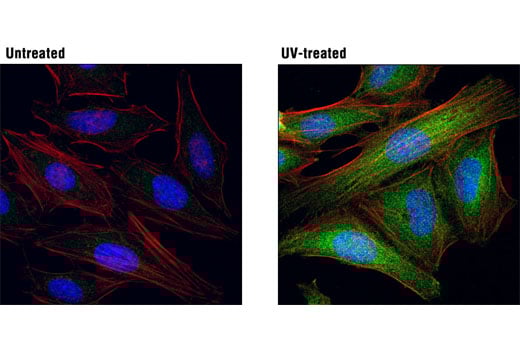
Four residues (Thr25, Thr222, Ser272 and Thr334) of MAPKAPK-2 are phosphorylated by p38 in an in vitro kinase assay (3). Phosphorylation at Thr222, Ser272 and Thr334 seems to be essential for the activity of MAPKAPK-2 (3). Activated MAPKAPK-2 can in return phosphorylate HSP27 at serines 15, 78 and 82 (3,9). Phosphorylation of HSP27 causes a change in the tertiary structure of HSP27, which shifts from large homotypic multimers to dimmers and monomers (10). It has been illustrated that phosphorylation and increased concentration of HSP27 modulate actin polymerization and reorganization (11,12).
Background References
- Rouse, J. et al. (1994) Cell 78, 1027-37.
- Han, J. et al. (1994) Science 265, 808-11.
- Lee, J.C. et al. (1994) Nature 372, 739-46.
- Freshney, N.W. et al. (1994) Cell 78, 1039-49.
- Raingeaud, J. et al. (1995) J Biol Chem 270, 7420-6.
- Zervos, A.S. et al. (1995) Proc Natl Acad Sci U S A 92, 10531-4.
- Zhao, M. et al. (1999) Mol Cell Biol 19, 21-30.
- Yang, S.H. et al. (1999) Mol Cell Biol 19, 4028-38.
- Cuenda, A. et al. (1995) FEBS Lett 364, 229-33.
- Kumar, S. et al. (1999) Biochem Biophys Res Commun 263, 825-31.
- Landry, J. et al. (1992) J Biol Chem 267, 794-803.
- Rogalla, T. et al. (1999) J Biol Chem 274, 18947-56.
- Lavoie, J.N. et al. (1993) J Biol Chem 268, 24210-4.
- Rousseau, S. et al. (1997) Oncogene 15, 2169-77.
Trademarks and Patents
Cell Signaling Technology is a trademark of Cell Signaling Technology, Inc.
All other trademarks are the property of their respective owners. Visit cellsignal.com/trademarks for more information.
Limited Uses
Except as otherwise expressly agreed in a writing signed by a legally authorized representative of CST, the following terms apply to Products provided by CST, its affiliates or its distributors. Any Customer's terms and conditions that are in addition to, or different from, those contained herein, unless separately accepted in writing by a legally authorized representative of CST, are rejected and are of no force or effect.
Products are labeled with For Research Use Only or a similar labeling statement and have not been approved, cleared, or licensed by the FDA or other regulatory foreign or domestic entity, for any purpose. Customer shall not use any Product for any diagnostic or therapeutic purpose, or otherwise in any manner that conflicts with its labeling statement. Products sold or licensed by CST are provided for Customer as the end-user and solely for research and development uses. Any use of Product for diagnostic, prophylactic or therapeutic purposes, or any purchase of Product for resale (alone or as a component) or other commercial purpose, requires a separate license from CST. Customer shall (a) not sell, license, loan, donate or otherwise transfer or make available any Product to any third party, whether alone or in combination with other materials, or use the Products to manufacture any commercial products, (b) not copy, modify, reverse engineer, decompile, disassemble or otherwise attempt to discover the underlying structure or technology of the Products, or use the Products for the purpose of developing any products or services that would compete with CST products or services, (c) not alter or remove from the Products any trademarks, trade names, logos, patent or copyright notices or markings, (d) use the Products solely in accordance with CST Product Terms of Sale and any applicable documentation, and (e) comply with any license, terms of service or similar agreement with respect to any third party products or services used by Customer in connection with the Products.
Revision 6
Revision 6

Revision 6
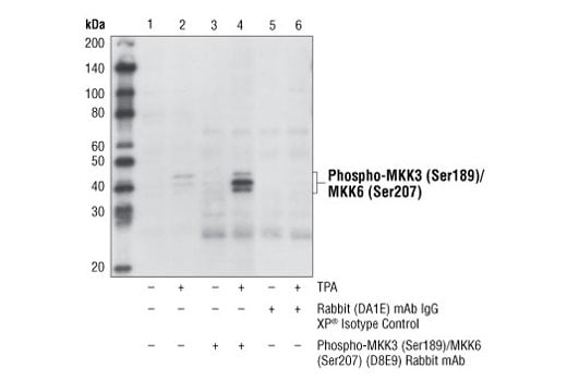
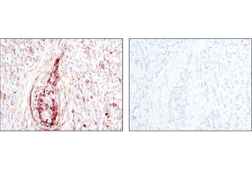
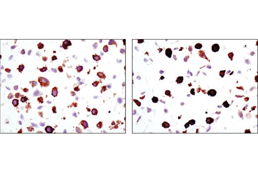
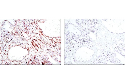
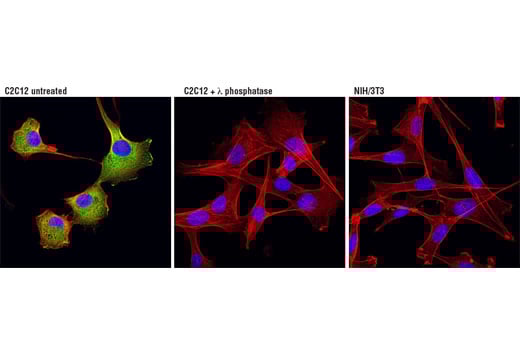
Phospho-MKK3 (Ser189)/MKK6 (Ser207) Antibody #9231. Mouse Anti-rabbit IgG (Conformation Specific) (L27A9) mAb #3678 was used as a secondary antibody.
Revision 6
Revision 6
Revision 6

Revision 6
Revision 6
Revision 6