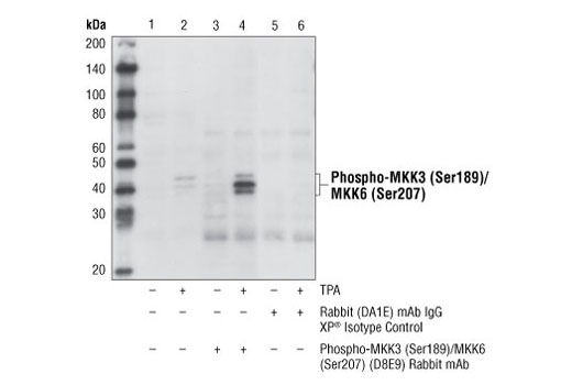
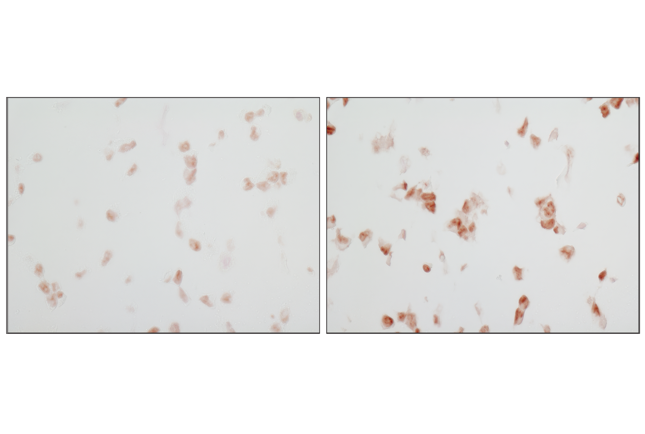
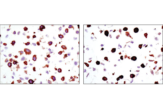
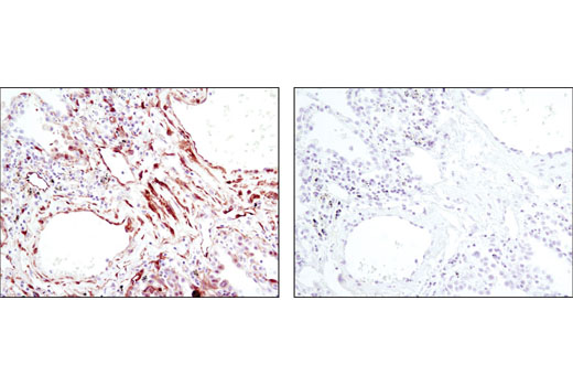
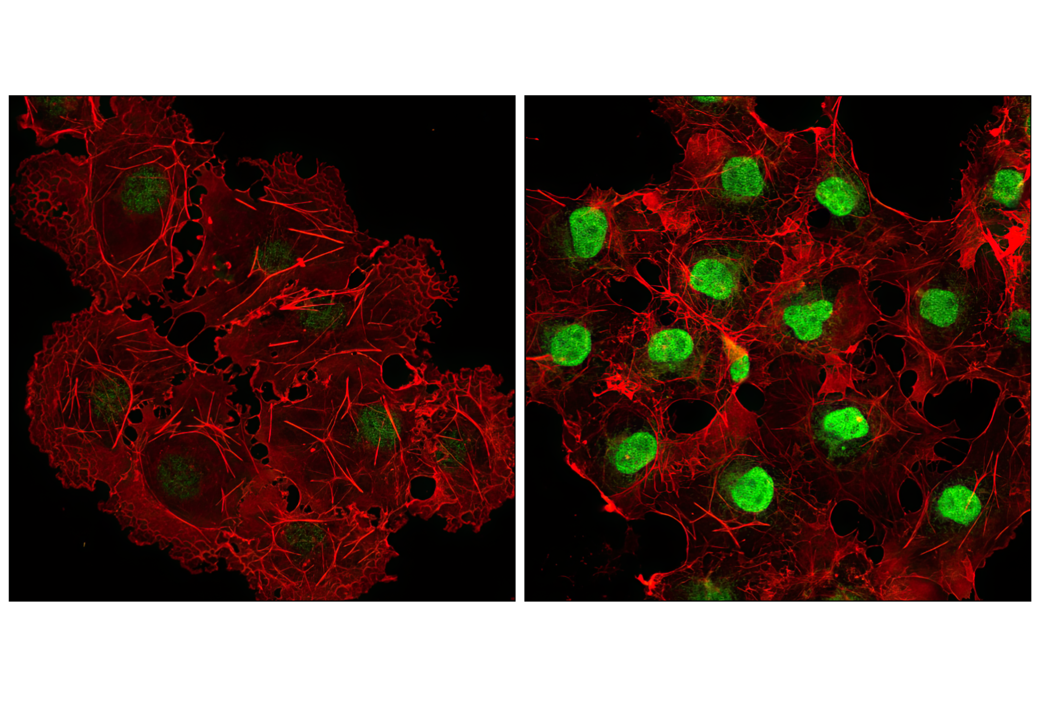
Phospho-MKK3 (Ser189)/MKK6 (Ser207) Antibody #9231. Mouse Anti-rabbit IgG (Conformation Specific) (L27A9) mAb #3678 was used as a secondary antibody.
| Cat. # | Size | Qty. | Price |
|---|---|---|---|
| 9913T | 1 Kit (6 x 20 microliters) |
|
| Product Includes | Quantity | Applications | Reactivity | MW(kDa) | Isotype |
|---|---|---|---|---|---|
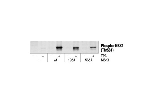
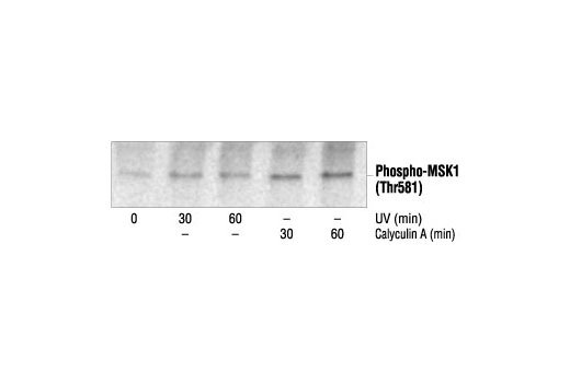
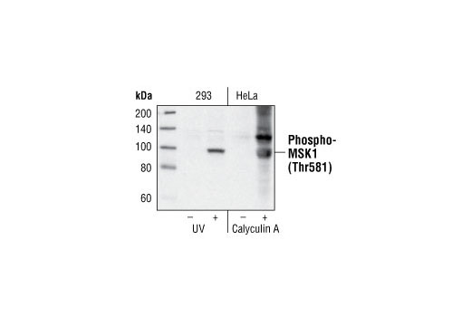
| Phospho-MSK1 (Thr581) Antibody 9595 | 20 µl |
|
H M | 90 | Rabbit |
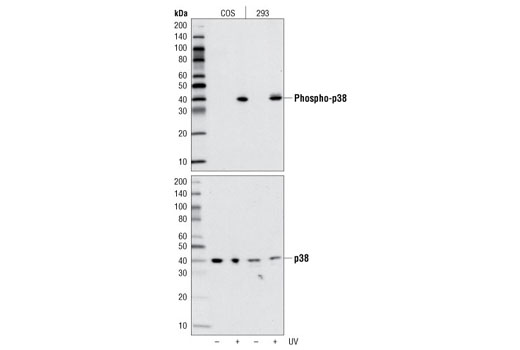
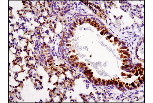
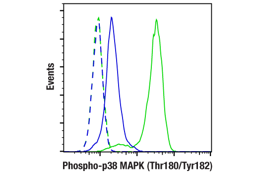
| Phospho-p38 MAPK (Thr180/Tyr182) (D3F9) XP® Rabbit mAb 4511 | 20 µl |
|
H M R Mk Mi Pg Sc | 43 | Rabbit IgG |
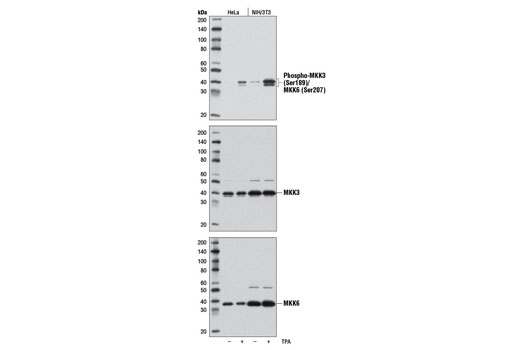
| Phospho-MKK3 (Ser189)/MKK6 (Ser207) (D8E9) Rabbit mAb 12280 | 20 µl |
|
H M R Mk | 38 MKK6, 40 MKK3 | Rabbit IgG |
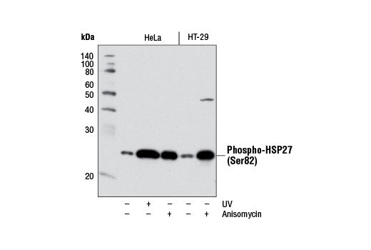
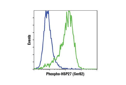
| Phospho-HSP27 (Ser82) (D1H2F6) XP® Rabbit mAb 9709 | 20 µl |
|
H M | 27 | Rabbit IgG |
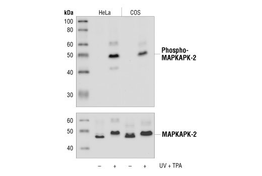
| Phospho-MAPKAPK-2 (Thr334) (27B7) Rabbit mAb 3007 | 20 µl |
|
H M R Mk | 49 | Rabbit IgG |
| Anti-rabbit IgG, HRP-linked Antibody 7074 | 100 µl |
|
Rab | Goat | |
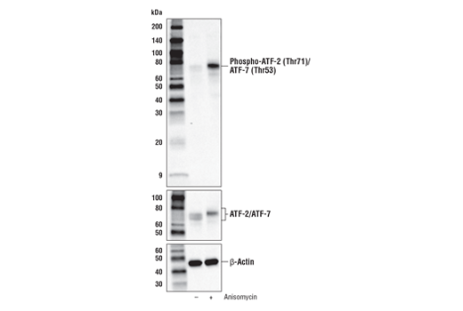
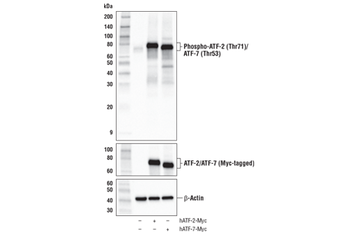
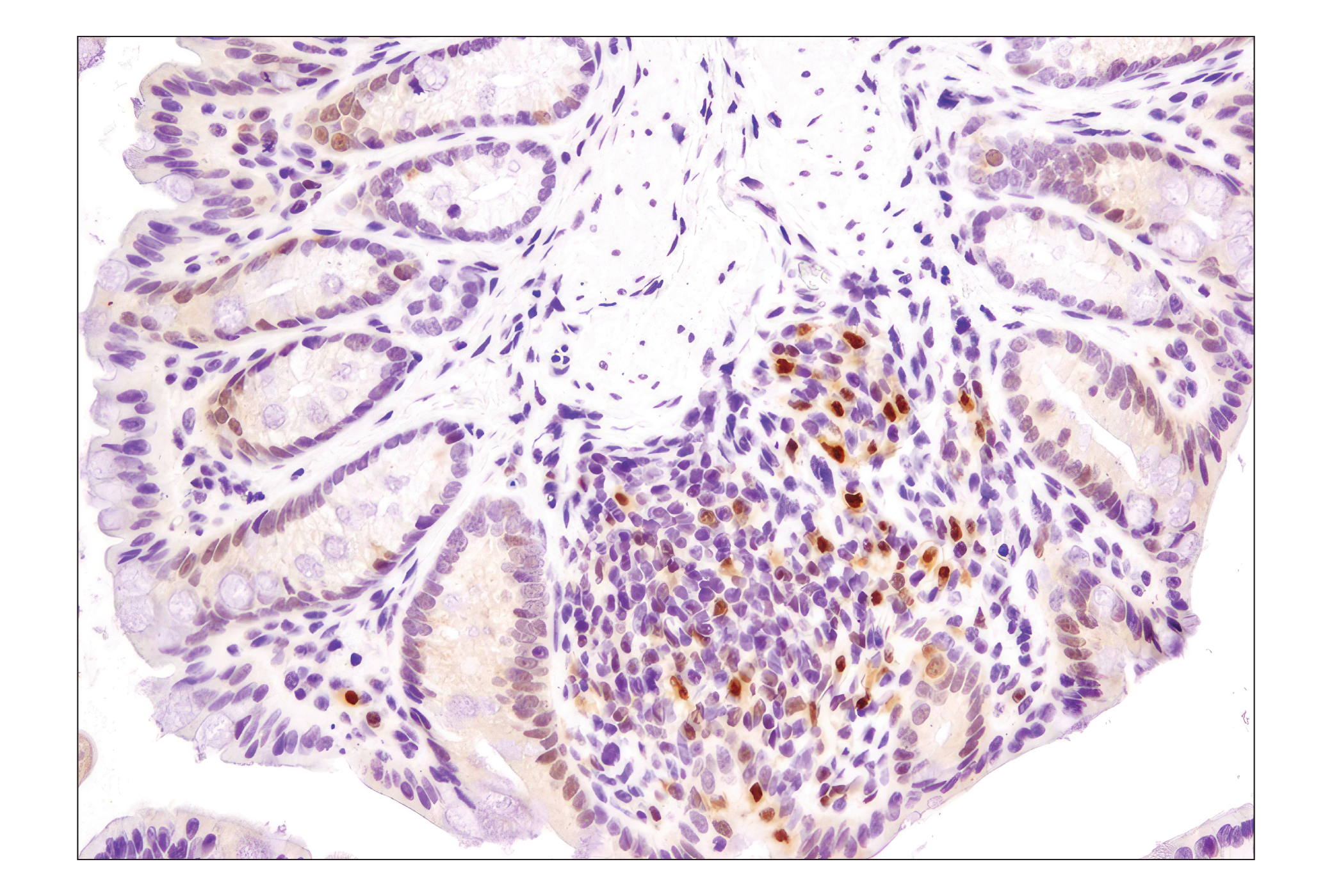
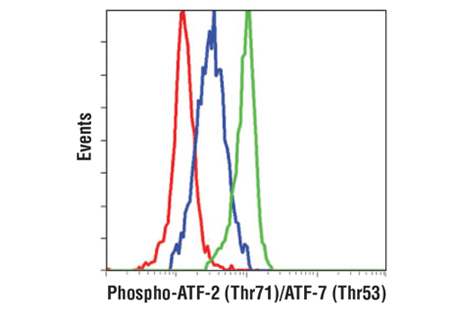
| Phospho-ATF-2 (Thr71)/ATF-7 (Thr53) (A8J7P) Rabbit mAb 15411 | 20 µl |
|
H M R Mk | 65,75 | Rabbit IgG |
Product Information
Monoclonal antibodies are produced by immunizing animals with synthetic phosphopeptides corresponding to residues surrounding Thr581 of human MSK1, Thr180/Tyr182 of human p38 MAPK, Ser189 of human MKK3, Thr71 of human ATF-2, Ser82 of human HSP27 or Thr334 of human MAPKAPK-2.
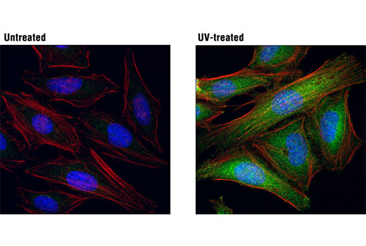
p38 MAP kinase (MAPK), also called RK (1) or CSBP (2), is the mammalian orthologue of the yeast HOG kinase that participates in a signaling cascade controlling cellular responses to cytokines and stress (1-4). Four isoforms of p38 MAPK, p38α, β, γ (also known as Erk6 or SAPK3), and δ (also known as SAPK4) have been identified. Similar to the SAPK/JNK pathway, p38 MAPK is activated by a variety of cellular stresses, including osmotic shock, inflammatory cytokines, lipopolysaccharide (LPS), UV light, and growth factors (1-5). MKK3, MKK6, and SEK activate p38 MAPK by phosphorylation at Thr180 and Tyr182. Activated p38 MAPK has been shown to phosphorylate and activate MAPKAP kinase 2 (3) and to phosphorylate the transcription factors ATF-2 (5), Max (6), and MEF2 (5-8). SB203580 (4-(4-fluorophenyl)-2-(4-methylsulfinylphenyl)-5-(4-pyridyl)-imidazole) is a selective inhibitor of p38 MAPK. This compound inhibits the activation of MAPKAPK-2 by p38 MAPK and subsequent phosphorylation of HSP27 (9). SB203580 inhibits p38 MAPK catalytic activity by binding to the ATP-binding pocket, but does not inhibit phosphorylation of p38 MAPK by upstream kinases (10).
Four residues (Thr25, Thr222, Ser272 and Thr334) of MAPKAPK-2 are phosphorylated by p38 in an in vitro kinase assay (3). Phosphorylation at Thr222, Ser272 and Thr334 seems to be essential for the activity of MAPKAPK-2 (3). Activated MAPKAPK-2 can in return phosphorylate HSP27 at serines 15, 78 and 82 (3,9). Phosphorylation of HSP27 causes a change in the tertiary structure of HSP27, which shifts from large homotypic multimers to dimmers and monomers (10). It has been illustrated that phosphorylation and increased concentration of HSP27 modulate actin polymerization and reorganization (11,12).
Except as otherwise expressly agreed in a writing signed by a legally authorized representative of CST, the following terms apply to Products provided by CST, its affiliates or its distributors. Any Customer's terms and conditions that are in addition to, or different from, those contained herein, unless separately accepted in writing by a legally authorized representative of CST, are rejected and are of no force or effect.
Products are labeled with For Research Use Only or a similar labeling statement and have not been approved, cleared, or licensed by the FDA or other regulatory foreign or domestic entity, for any purpose. Customer shall not use any Product for any diagnostic or therapeutic purpose, or otherwise in any manner that conflicts with its labeling statement. Products sold or licensed by CST are provided for Customer as the end-user and solely for research and development uses. Any use of Product for diagnostic, prophylactic or therapeutic purposes, or any purchase of Product for resale (alone or as a component) or other commercial purpose, requires a separate license from CST. Customer shall (a) not sell, license, loan, donate or otherwise transfer or make available any Product to any third party, whether alone or in combination with other materials, or use the Products to manufacture any commercial products, (b) not copy, modify, reverse engineer, decompile, disassemble or otherwise attempt to discover the underlying structure or technology of the Products, or use the Products for the purpose of developing any products or services that would compete with CST products or services, (c) not alter or remove from the Products any trademarks, trade names, logos, patent or copyright notices or markings, (d) use the Products solely in accordance with CST Product Terms of Sale and any applicable documentation, and (e) comply with any license, terms of service or similar agreement with respect to any third party products or services used by Customer in connection with the Products.