Revision 1
#54442
Store at -20C
877-616-CELL (2355)
877-678-TECH (8324)
3 Trask Lane | Danvers | Massachusetts | 01923 | USA
For Research Use Only. Not for Use in Diagnostic Procedures.
Applications:
W, IP, FC-FP
Reactivity:
H
Sensitivity:
Endogenous
MW (kDa):
150
Source/Isotype:
Rabbit IgG
UniProt ID:
#P16885
Entrez-Gene Id:
5336
Product Usage Information
| Application | Dilution |
|---|---|
| Western Blotting | 1:1000 |
| Immunoprecipitation | 1:50 |
| Flow Cytometry (Fixed/Permeabilized) | 1:100 - 1:400 |
Storage
Specificity/Sensitivity
Source / Purification
Background
Phosphorylation is one of the key mechanisms that regulates the activity of PLC. Phosphorylation of Ser1105 by PKA or PKC inhibits PLCβ3 activity (4,5). Ser537 of PLCβ3 is phosphorylated by CaMKII, and this phosphorylation may contribute to the basal activity of PLCβ3. PLCγ is activated by both receptor and nonreceptor tyrosine kinases (6).
PLCγ forms a complex with EGF and PDGF receptors, which leads to the phosphorylation of PLCγ at Tyr771, 783 and 1248 (7). Phosphorylation by Syk at Tyr783 activates the enzymatic activity of PLCγ1 (8).
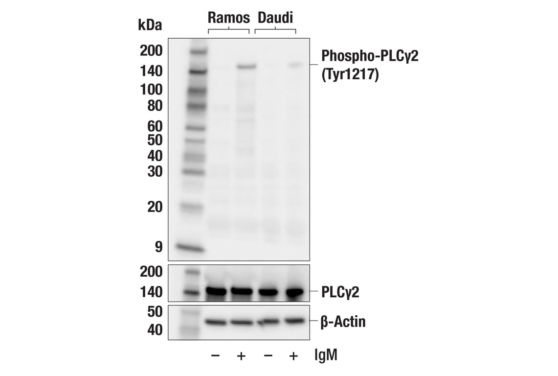
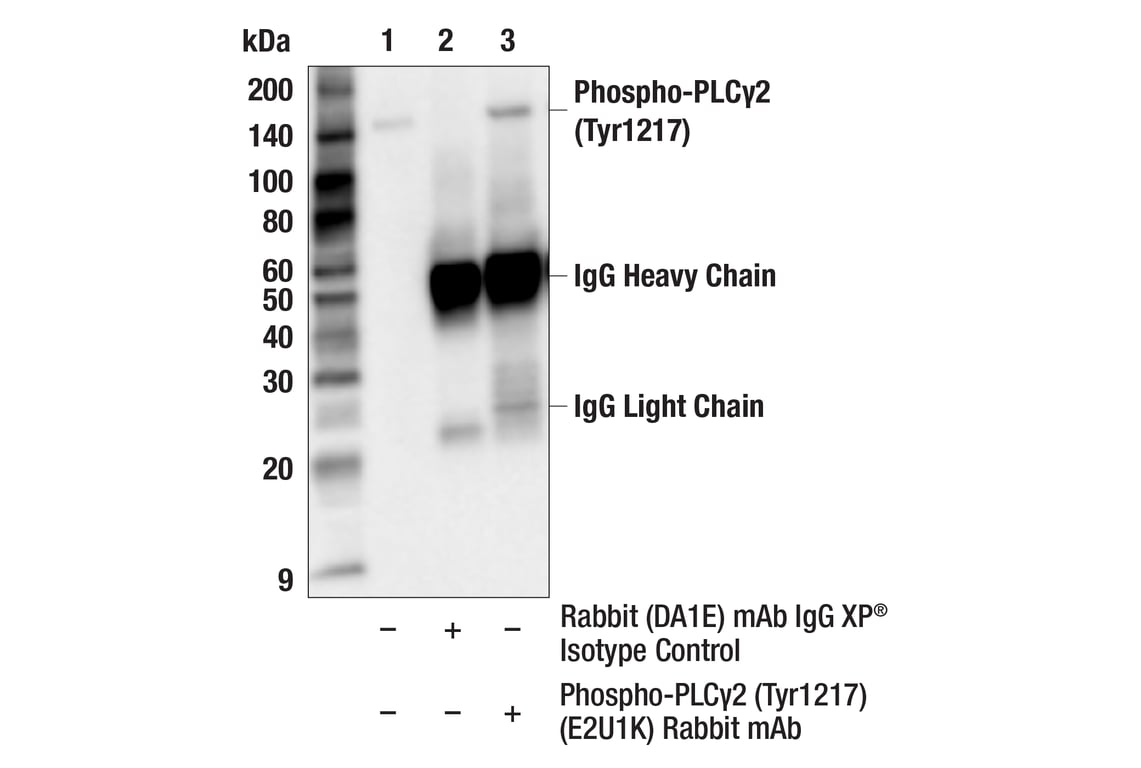
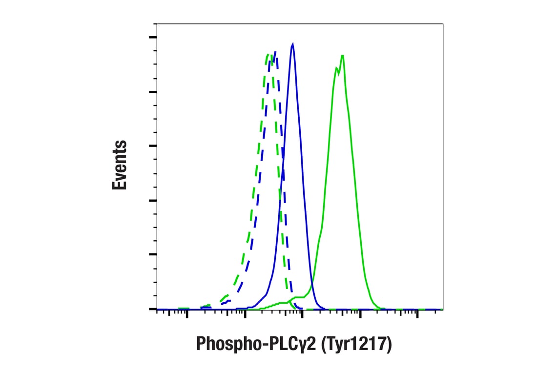
PLCgamma2 is engaged in antigen-dependent signaling in B cell and collagen-dependent signaling in platelets. Phosphorylation by Btk or Lck at tyrosines 753, 759, 1197 and 1217 is correlated with PLCgamma2 activity (9,10).
Background References
- Singer, W.D. et al. (1997) Annu Rev Biochem 66, 475-509.
- Smrcka, A.V. et al. (1991) Science 251, 804-7.
- Taylor, S.J. et al. (1991) Nature 350, 516-8.
- Yue, C. et al. (1998) J Biol Chem 273, 18023-7.
- Yue, C. et al. (2000) J Biol Chem 275, 30220-5.
- Margolis, B. et al. (1989) Cell 57, 1101-7.
- Kim, H.K. et al. (1991) Cell 65, 435-41.
- Wang, Z. et al. (1998) Mol Cell Biol 18, 590-7.
- Watanabe, D. et al. (2001) J Biol Chem 276, 38595-601.
- Ozdener, F. et al. (2002) Mol Pharmacol 62, 672-9.
Species Reactivity
Species reactivity is determined by testing in at least one approved application (e.g., western blot).
Western Blot Buffer
IMPORTANT: For western blots, incubate membrane with diluted primary antibody in 5% w/v BSA, 1X TBS, 0.1% Tween® 20 at 4°C with gentle shaking, overnight.
Applications Key
W: Western Blotting IP: Immunoprecipitation FC-FP: Flow Cytometry (Fixed/Permeabilized)
Cross-Reactivity Key
H: Human
Trademarks and Patents
Cell Signaling Technology is a trademark of Cell Signaling Technology, Inc.
All other trademarks are the property of their respective owners. Visit cellsignal.com/trademarks for more information.
Limited Uses
Except as otherwise expressly agreed in a writing signed by a legally authorized representative of CST, the following terms apply to Products provided by CST, its affiliates or its distributors. Any Customer's terms and conditions that are in addition to, or different from, those contained herein, unless separately accepted in writing by a legally authorized representative of CST, are rejected and are of no force or effect.
Products are labeled with For Research Use Only or a similar labeling statement and have not been approved, cleared, or licensed by the FDA or other regulatory foreign or domestic entity, for any purpose. Customer shall not use any Product for any diagnostic or therapeutic purpose, or otherwise in any manner that conflicts with its labeling statement. Products sold or licensed by CST are provided for Customer as the end-user and solely for research and development uses. Any use of Product for diagnostic, prophylactic or therapeutic purposes, or any purchase of Product for resale (alone or as a component) or other commercial purpose, requires a separate license from CST. Customer shall (a) not sell, license, loan, donate or otherwise transfer or make available any Product to any third party, whether alone or in combination with other materials, or use the Products to manufacture any commercial products, (b) not copy, modify, reverse engineer, decompile, disassemble or otherwise attempt to discover the underlying structure or technology of the Products, or use the Products for the purpose of developing any products or services that would compete with CST products or services, (c) not alter or remove from the Products any trademarks, trade names, logos, patent or copyright notices or markings, (d) use the Products solely in accordance with CST Product Terms of Sale and any applicable documentation, and (e) comply with any license, terms of service or similar agreement with respect to any third party products or services used by Customer in connection with the Products.
Revision 1