Revision 7
#8713
Store at -20C
877-616-CELL (2355)
877-678-TECH (8324)
3 Trask Lane | Danvers | Massachusetts | 01923 | USA
For Research Use Only. Not for Use in Diagnostic Procedures.
Applications:
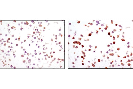
W, W-S, IP, IHC-P, IF-IC, FC-FP
Reactivity:
H M Mk
Sensitivity:
Endogenous
MW (kDa):
150
Source/Isotype:
Rabbit IgG
UniProt ID:
#P19174
Entrez-Gene Id:
5335
Product Usage Information
| Application | Dilution |
|---|---|
| Western Blotting | 1:1000 |
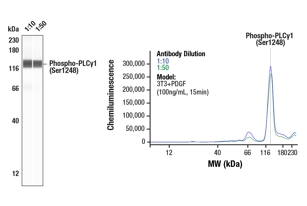
| Simple Western™ | 1:10 - 1:50 |
| Immunoprecipitation | 1:50 |
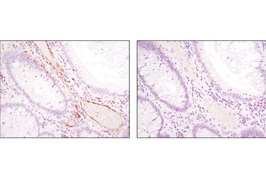
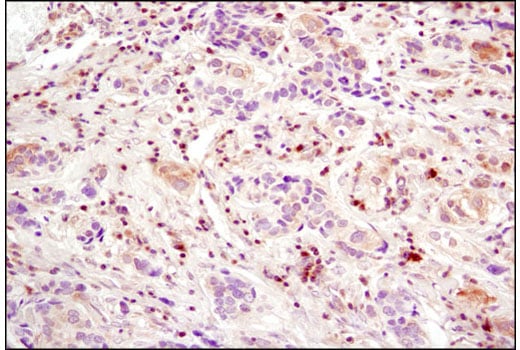
| Immunohistochemistry (Paraffin) | 1:100 - 1:400 |
| Immunofluorescence (Immunocytochemistry) | 1:100 - 1:200 |
| Flow Cytometry (Fixed/Permeabilized) | 1:800 |
Storage
For a carrier free (BSA and azide free) version of this product see product #16685.
Specificity/Sensitivity
Species predicted to react based on 100% sequence homology
Source / Purification
Background
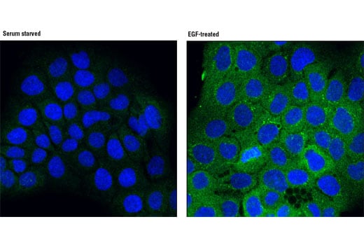
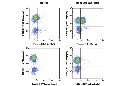
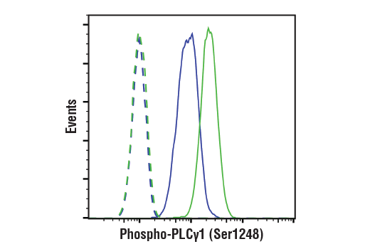
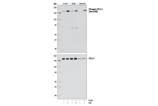
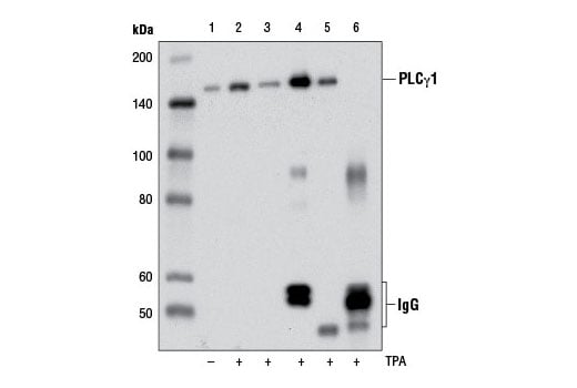
Two mammalian PLCγ isoforms (γ1 and γ2) have been cloned and characterized (7,8). Like other PLC-family members, PLCγ1 and PLCγ2 contain calcium-binding (EF-hand, C2) and lipid-interacting (PH, EF-hand) domains necessary for their enzymatic activity and substrate recognition. Uniquely, PLCγ isoforms have additional, conserved SH2 and SH3 domains critical for their functions as signaling molecules and scaffolding proteins. Upon growth factor stimulation, PLCγ1 is recruited (via SH2 domains) to phosphotyrosine residues within the cytoplasmic tail of many RTKs where it serves as a substrate for the RTK and provides docking sites for additional proteins involved in RTK signaling (4-6,9-12). PLCγ1 and γ2 can also be activated downstream of receptors lacking intrinsic tyrosine kinase activity. This has been reported downstream of multiple G protein-coupled receptors and the T cell receptor in which tyrosine kinases of the Src, Syk, and Tec families serve to bind, phosphorylate, and activate PLCγ (reviewed in 13-15). Phosphorylation at tyrosine residues by both receptor and non-receptor tyrosine kinases results in robust activation of PLCγ1 activity, leading to generation of second messengers. In response to agonists, PLCγ1 is phosphorylated on Tyr783, Tyr711, and Tyr1253 (Tyr753, Tyr759, and Tyr1217 in PLCγ2) resulting in robust PI-4,5-P2 hydrolysis (4-6,9-12). Interestingly recent evidence suggests a role for tyrosine kinase-independent regulation of PLCγ in some systems. For example, in response to EGF, proline-rich regions of Akt interact with the SH3 domain of PLCγ1 resulting in association of the two enzymes, phosphorylation of PLCγ1 at Ser1248, and enhanced cellular motility (16). This finding demonstrates that PLCγ1 can function as a "scaffold" between RTKs and Akt, thereby establishing a mechanism by which the Akt signaling pathway cross-talks with tyrosine kinases. However, the mechanism and functional significance of phosphorylation at Ser1248 remains to be fully clarified, as it has also been shown that PKA-mediated phosphorylation at this site is inhibitory to PLCγ1 tyrosine phosphorylation and phospholipase activity in CD3-treated Jurkat cells (17), suggesting that Ser1248 may be an allosteric regulator of PLCγ1 activity.
Background References
- Singer, W.D. et al. (1997) Annu Rev Biochem 66, 475-509.
- Margolis, B. et al. (1989) Cell 57, 1101-7.
- Kim, H.K. et al. (1991) Cell 65, 435-41.
- Wang, Z. et al. (1998) Mol Cell Biol 18, 590-7.
- Watanabe, D. et al. (2001) J Biol Chem 276, 38595-601.
- Ozdener, F. et al. (2002) Mol Pharmacol 62, 672-9.
- Burgess, W.H. et al. (1990) Mol Cell Biol 10, 4770-7.
- Ohta, S. et al. (1988) FEBS Lett 242, 31-5.
- Rodriguez, R. et al. (2001) J Biol Chem 276, 47982-92.
- Humphries, L.A. et al. (2004) J Biol Chem 279, 37651-61.
- Kim, Y.J. et al. (2004) Mol Cell Biol 24, 9986-99.
- Sekiya, F. et al. (2004) J Biol Chem 279, 32181-90.
- Carpenter, G. and Ji, Q. (1999) Exp Cell Res 253, 15-24.
- Rebecchi, M.J. and Pentyala, S.N. (2000) Physiol Rev 80, 1291-335.
- Rhee, S.G. (2001) Annu Rev Biochem 70, 281-312.
- Wang, Y. et al. (2006) Mol Biol Cell 17, 2267-77.
- Park, D.J. et al. (1992) J Biol Chem 267, 1496-501.
Species Reactivity
Species reactivity is determined by testing in at least one approved application (e.g., western blot).
Western Blot Buffer
IMPORTANT: For western blots, incubate membrane with diluted primary antibody in 5% w/v BSA, 1X TBS, 0.1% Tween® 20 at 4°C with gentle shaking, overnight.
Applications Key
W: Western Blotting W-S: Simple Western™ IP: Immunoprecipitation IHC-P: Immunohistochemistry (Paraffin) IF-IC: Immunofluorescence (Immunocytochemistry) FC-FP: Flow Cytometry (Fixed/Permeabilized)
Cross-Reactivity Key
H: Human M: Mouse Mk: Monkey
Trademarks and Patents
Cell Signaling Technology is a trademark of Cell Signaling Technology, Inc.
All other trademarks are the property of their respective owners. Visit cellsignal.com/trademarks for more information.
Limited Uses
Except as otherwise expressly agreed in a writing signed by a legally authorized representative of CST, the following terms apply to Products provided by CST, its affiliates or its distributors. Any Customer's terms and conditions that are in addition to, or different from, those contained herein, unless separately accepted in writing by a legally authorized representative of CST, are rejected and are of no force or effect.
Products are labeled with For Research Use Only or a similar labeling statement and have not been approved, cleared, or licensed by the FDA or other regulatory foreign or domestic entity, for any purpose. Customer shall not use any Product for any diagnostic or therapeutic purpose, or otherwise in any manner that conflicts with its labeling statement. Products sold or licensed by CST are provided for Customer as the end-user and solely for research and development uses. Any use of Product for diagnostic, prophylactic or therapeutic purposes, or any purchase of Product for resale (alone or as a component) or other commercial purpose, requires a separate license from CST. Customer shall (a) not sell, license, loan, donate or otherwise transfer or make available any Product to any third party, whether alone or in combination with other materials, or use the Products to manufacture any commercial products, (b) not copy, modify, reverse engineer, decompile, disassemble or otherwise attempt to discover the underlying structure or technology of the Products, or use the Products for the purpose of developing any products or services that would compete with CST products or services, (c) not alter or remove from the Products any trademarks, trade names, logos, patent or copyright notices or markings, (d) use the Products solely in accordance with CST Product Terms of Sale and any applicable documentation, and (e) comply with any license, terms of service or similar agreement with respect to any third party products or services used by Customer in connection with the Products.
Revision 7
Revision 7
Revision 7