Revision 1
#9920
Store at -20C
Phospho-(Ser/Thr) Kinase Substrate Antibody Sampler Kit
1 Kit
(6 x 20 microliters)
877-616-CELL (2355)
877-678-TECH (8324)
3 Trask Lane | Danvers | Massachusetts | 01923 | USA
For Research Use Only. Not for Use in Diagnostic Procedures.
| Product Includes | Product # | Quantity | Mol. Wt | Isotype/Source |
|---|---|---|---|---|
| Phospho-AMPK Substrate Motif [LXRXX(pS/pT) MultiMab® Rabbit Monoclonal Antibody mix | 5759 | 20 µl | Rabbit | |
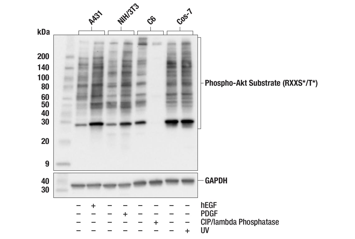
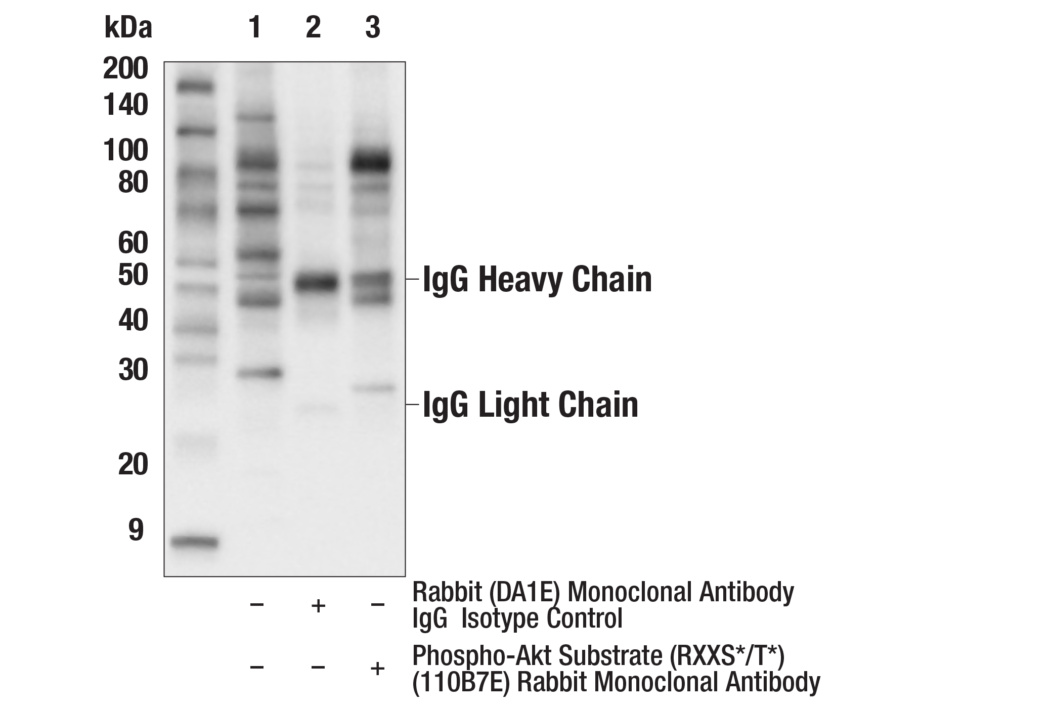
| Phospho-Akt Substrate (RXXS*/T*) (110B7E) Rabbit Monoclonal Antibody | 9614 | 20 µl | Rabbit IgG | |
| Phospho-PKA Substrate (RRXS*/T*) (100G7E) Rabbit Monoclonal Antibody | 9624 | 20 µl | Rabbit IgG | |
| Phospho-ATM/ATR Substrate Motif [(pS/pT) QG] MultiMab® Rabbit Monoclonal Antibody mix | 6966 | 20 µl | Rabbit IgG | |
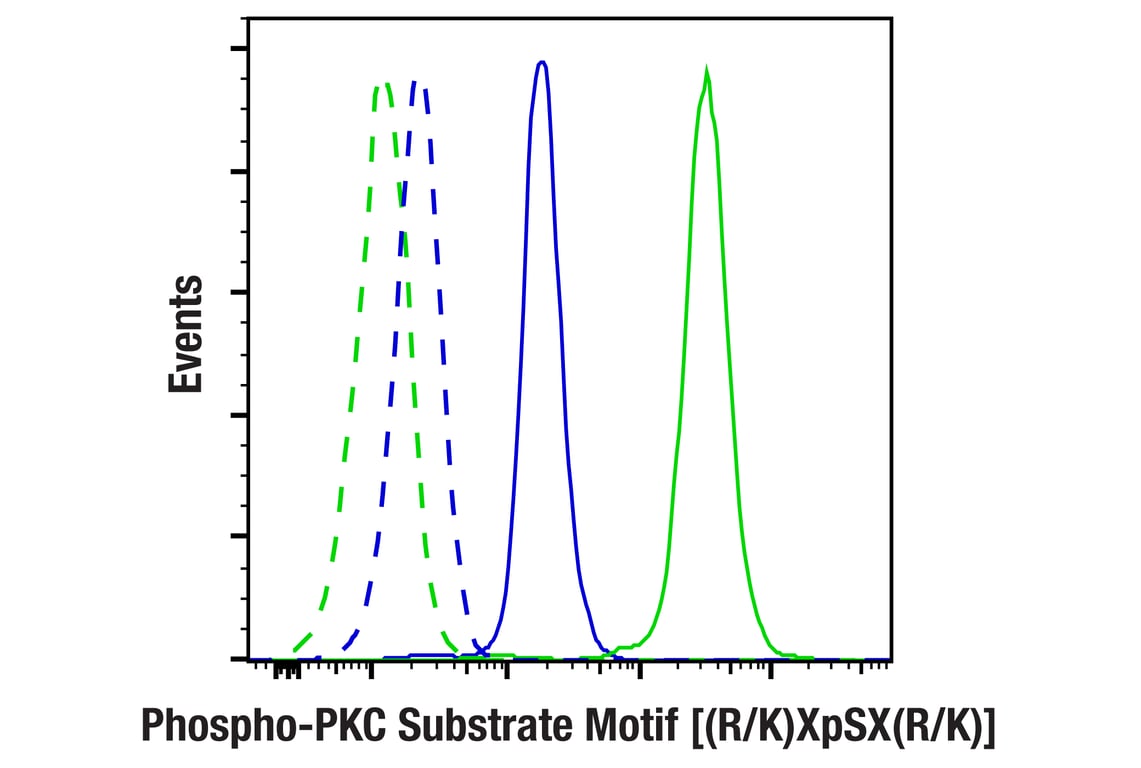
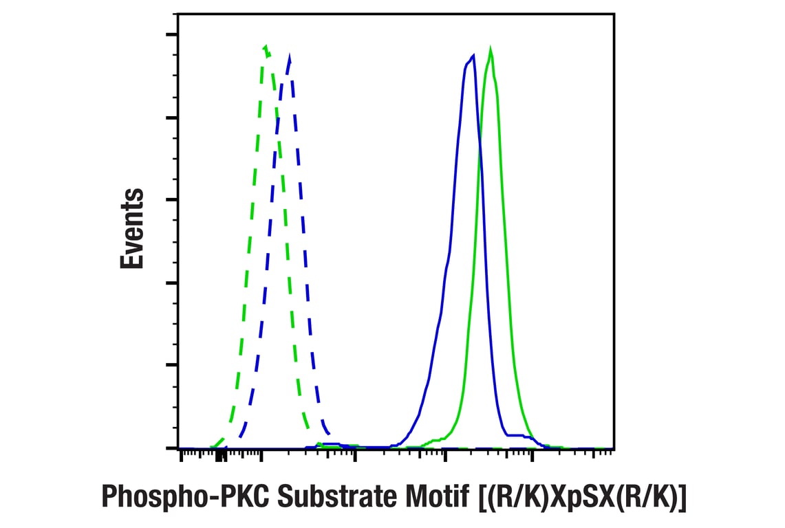
| Phospho-PKC Substrate Motif [(R/K)XpSX(R/K)] MultiMab® Rabbit Monoclonal Antibody mix | 6967 | 20 µl | Rabbit IgG | |
| Phospho-CDK Substrate Motif [(K/H)pSP] MultiMab® Rabbit Monoclonal Antibody mix | 9477 | 20 µl | Rabbit IgG | |
| Anti-rabbit IgG, HRP-linked Antibody | 7074 | 100 µl | Goat |
Please visit cellsignal.com for individual component applications, species cross-reactivity, dilutions, protocols, and additional product information.
Description
Storage
Background
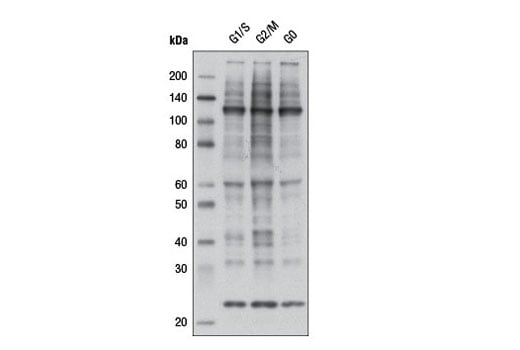
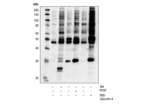
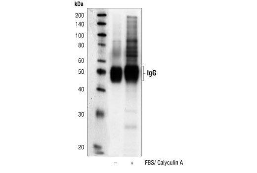
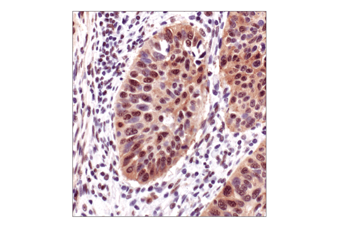
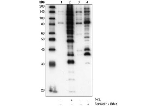
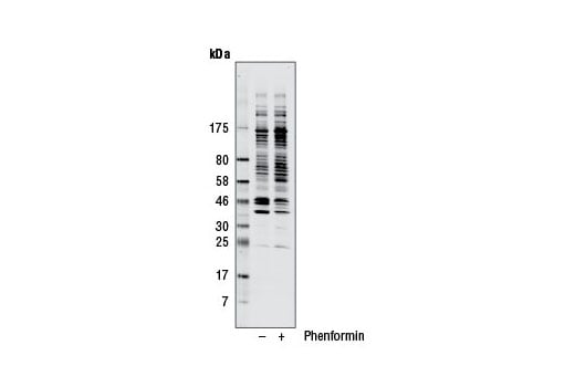
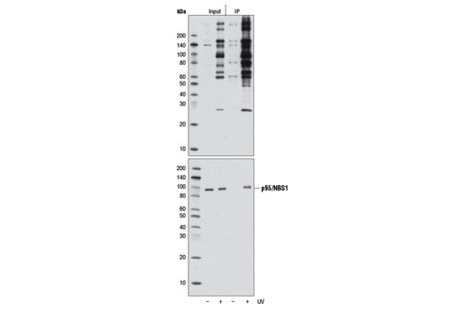
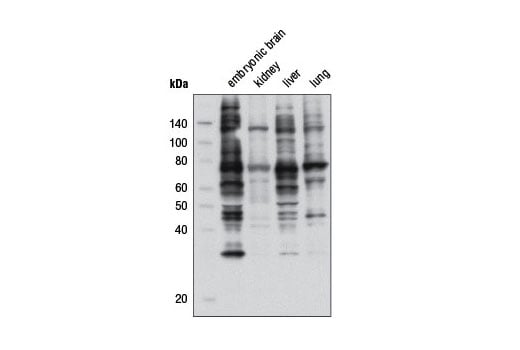
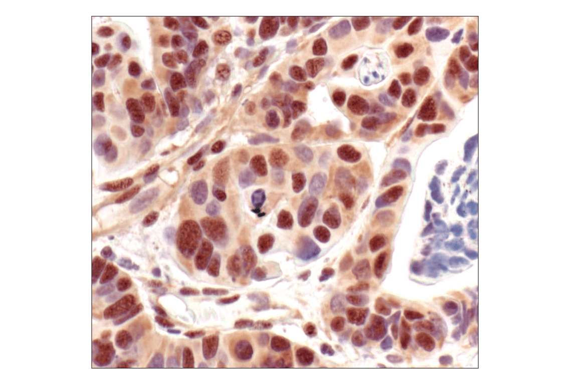
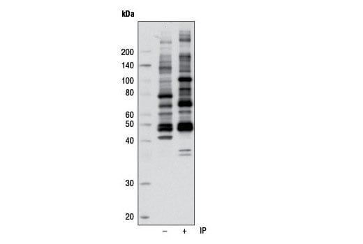
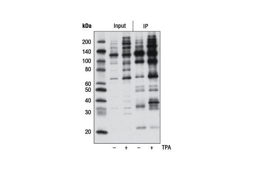
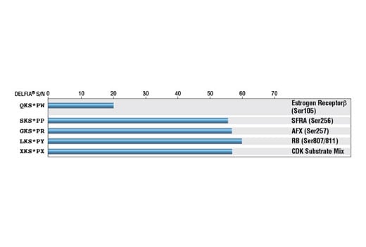
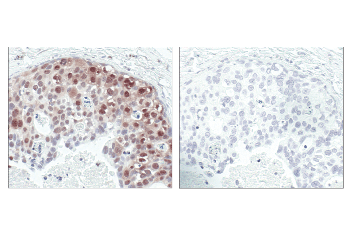
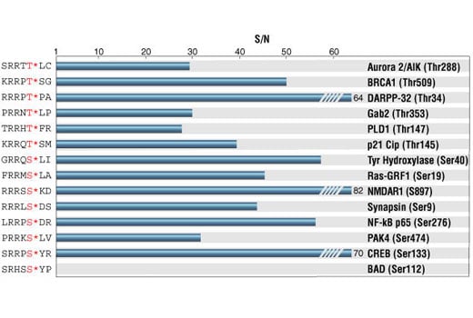
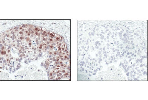
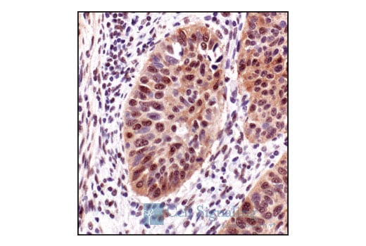
Antibodies specific to particular kinase substrates are invaluable reagents in determining kinase activity and identifying potential new kinase substrates. CST has developed antibodies that recognize phosphorylated serine or threonine within the context of a protein motif that is phosphorylated by Akt, PKC, PKA, MAPK/CDK, CDKs or ATM/ATR. As shown by peptide pairing ELISA, each phospho-(Ser/Thr) kinase substrate antibody in this sampler kit is specific to its kinase substrate motif.
Background References
- Alessi, D.R. et al. (1996) FEBS Lett 399, 333-8.
- Nishikawa, K. et al. (1997) J Biol Chem 272, 952-60.
- Pearson, R.B. and Kemp, B.E. (1991) Methods Enzymol 200, 62-81.
- Songyang, Z. et al. (1996) Mol Cell Biol 16, 6486-93.
- Songyang, Z. (1999) Prog Biophys Mol Biol 71, 359-72.
- Holmes, J.K. and Solomon, M.J. (1996) J Biol Chem 271, 25240-6.
- Kastan, M.B. and Lim, D.S. (2000) Nat Rev Mol Cell Biol 1, 179-86.
- Zhao, H. and Piwnica-Worms, H. (2001) Mol Cell Biol 21, 4129-39.
Trademarks and Patents
Cell Signaling Technology is a trademark of Cell Signaling Technology, Inc.
All other trademarks are the property of their respective owners. Visit cellsignal.com/trademarks for more information.
Limited Uses
Except as otherwise expressly agreed in a writing signed by a legally authorized representative of CST, the following terms apply to Products provided by CST, its affiliates or its distributors. Any Customer's terms and conditions that are in addition to, or different from, those contained herein, unless separately accepted in writing by a legally authorized representative of CST, are rejected and are of no force or effect.
Products are labeled with For Research Use Only or a similar labeling statement and have not been approved, cleared, or licensed by the FDA or other regulatory foreign or domestic entity, for any purpose. Customer shall not use any Product for any diagnostic or therapeutic purpose, or otherwise in any manner that conflicts with its labeling statement. Products sold or licensed by CST are provided for Customer as the end-user and solely for research and development uses. Any use of Product for diagnostic, prophylactic or therapeutic purposes, or any purchase of Product for resale (alone or as a component) or other commercial purpose, requires a separate license from CST. Customer shall (a) not sell, license, loan, donate or otherwise transfer or make available any Product to any third party, whether alone or in combination with other materials, or use the Products to manufacture any commercial products, (b) not copy, modify, reverse engineer, decompile, disassemble or otherwise attempt to discover the underlying structure or technology of the Products, or use the Products for the purpose of developing any products or services that would compete with CST products or services, (c) not alter or remove from the Products any trademarks, trade names, logos, patent or copyright notices or markings, (d) use the Products solely in accordance with CST Product Terms of Sale and any applicable documentation, and (e) comply with any license, terms of service or similar agreement with respect to any third party products or services used by Customer in connection with the Products.
Revision 1
Revision 1

Revision 1
Revision 1
Revision 1
Revision 1
Revision 1
Revision 1
Revision 1