Revision 6
#61407
Store at -20C
877-616-CELL (2355)
877-678-TECH (8324)
3 Trask Lane | Danvers | Massachusetts | 01923 | USA
For Research Use Only. Not for Use in Diagnostic Procedures.
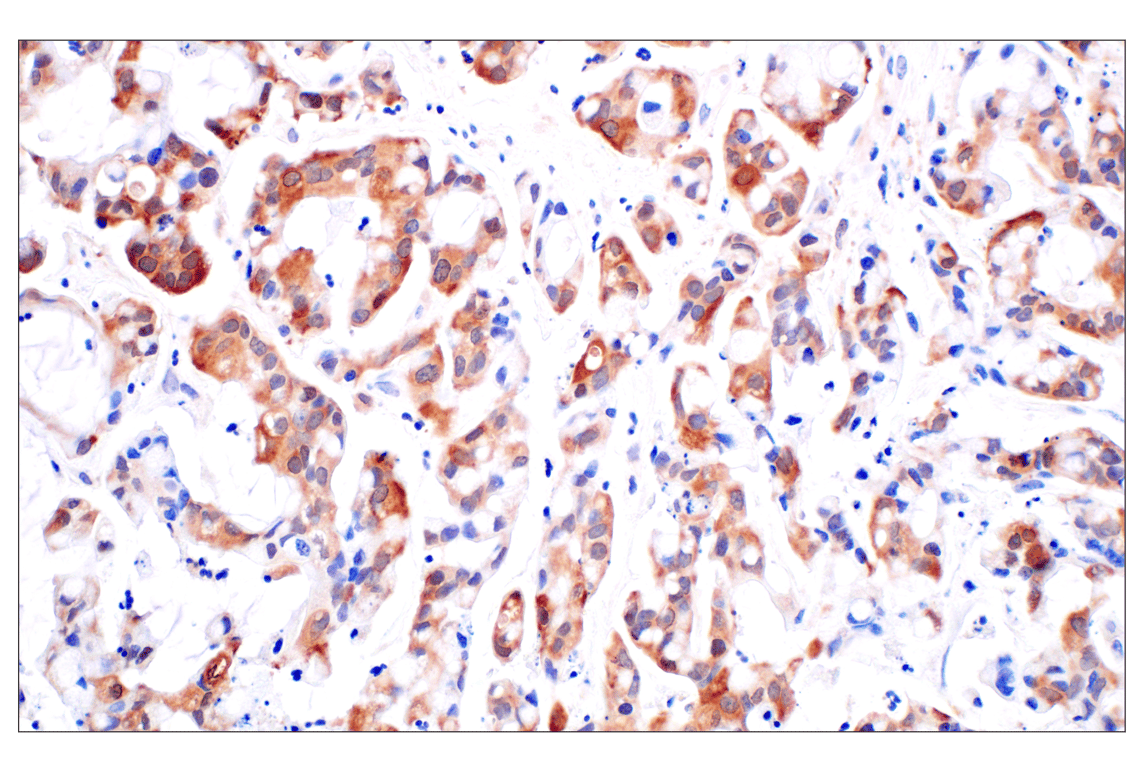
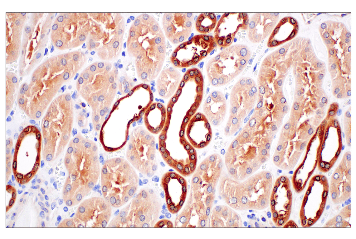
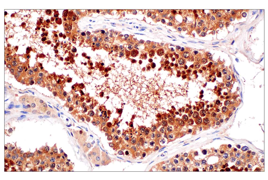
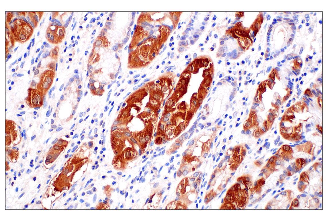
Applications:
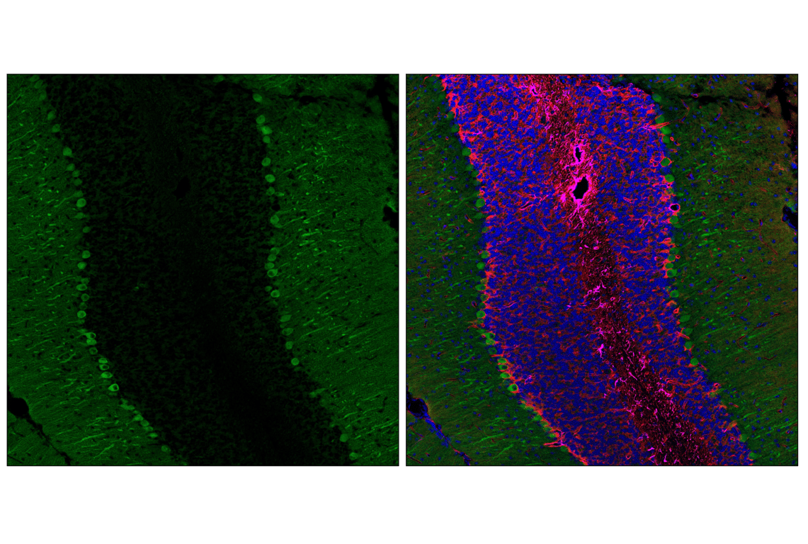
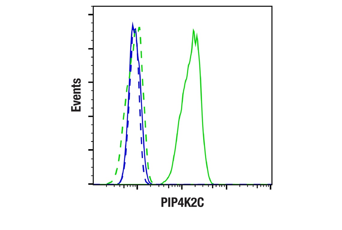
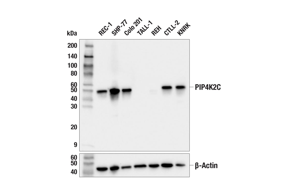
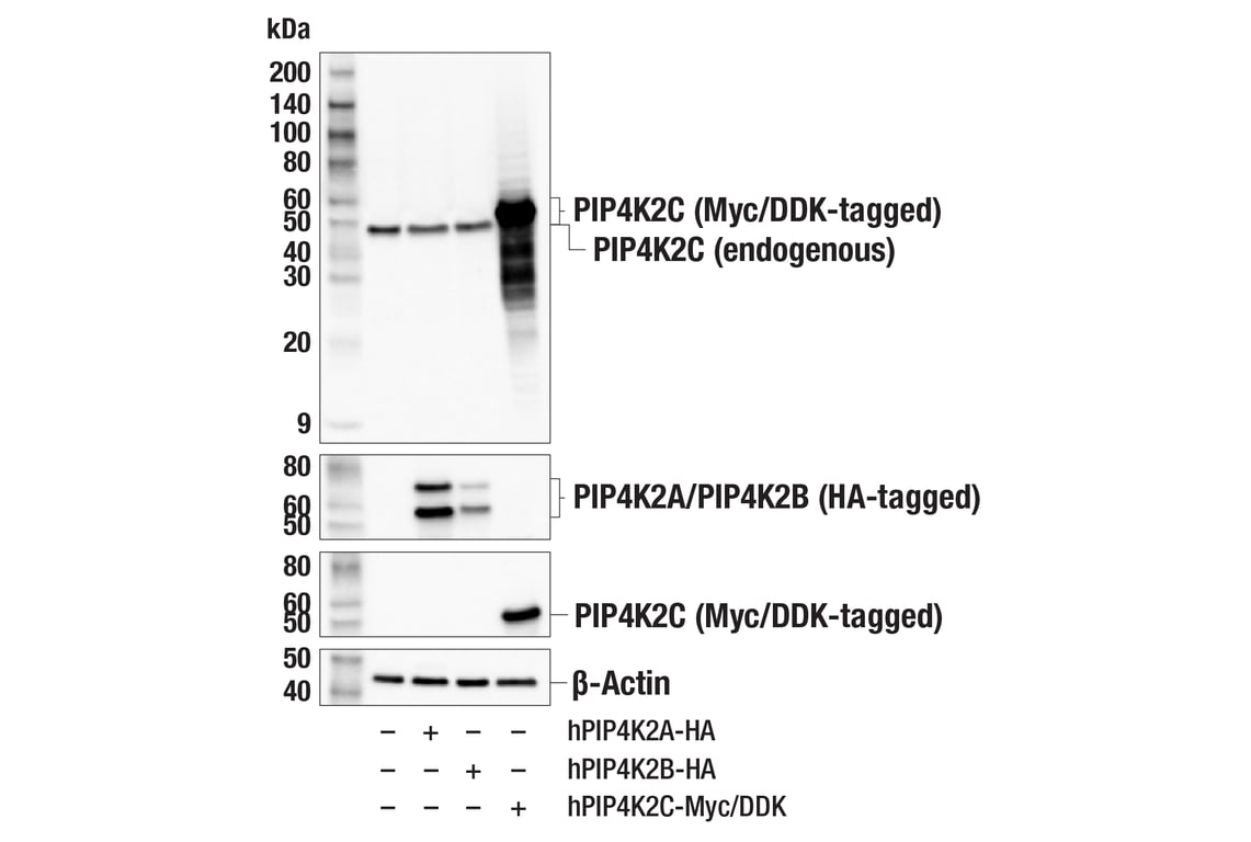
W, W-S, IP, IHC-P, IF-F, IF-IC, FC-FP
Reactivity:
H M R
Sensitivity:
Endogenous
MW (kDa):
50
Source/Isotype:
Rabbit IgG
UniProt ID:
#Q8TBX8
Entrez-Gene Id:
79837
Product Usage Information
| Application | Dilution |
|---|---|
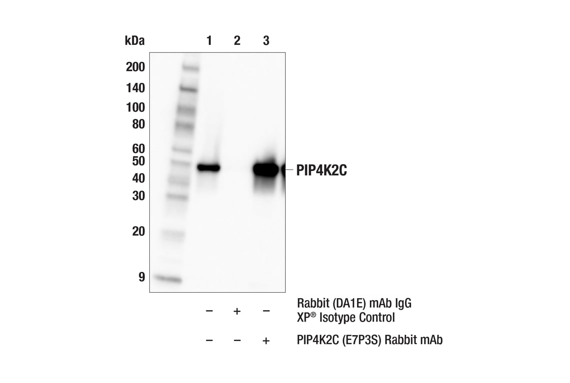
| Western Blotting | 1:1000 |
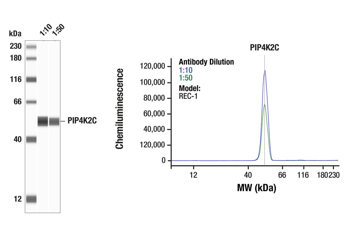
| Simple Western™ | 1:10 - 1:50 |
| Immunoprecipitation | 1:50 |
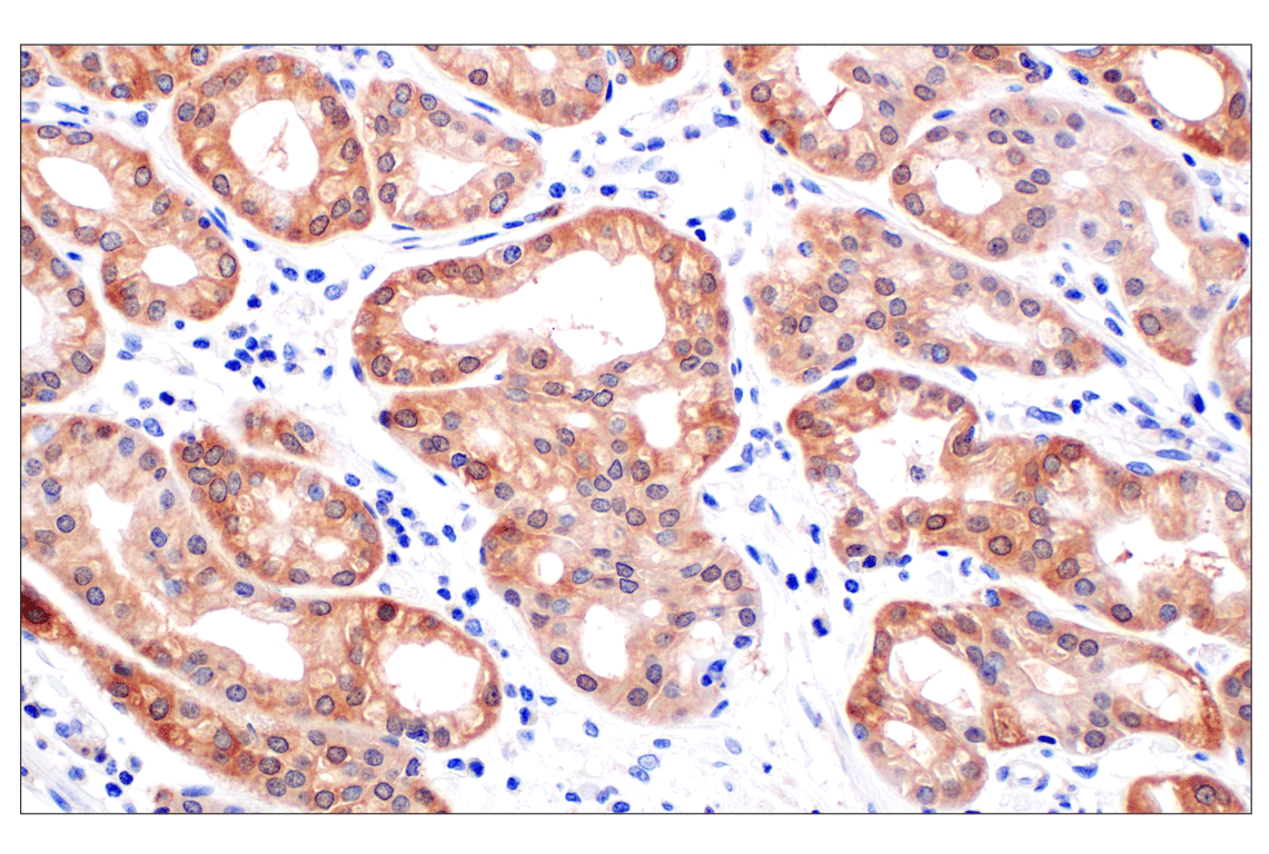
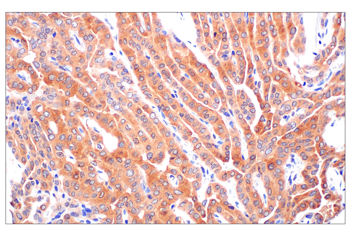
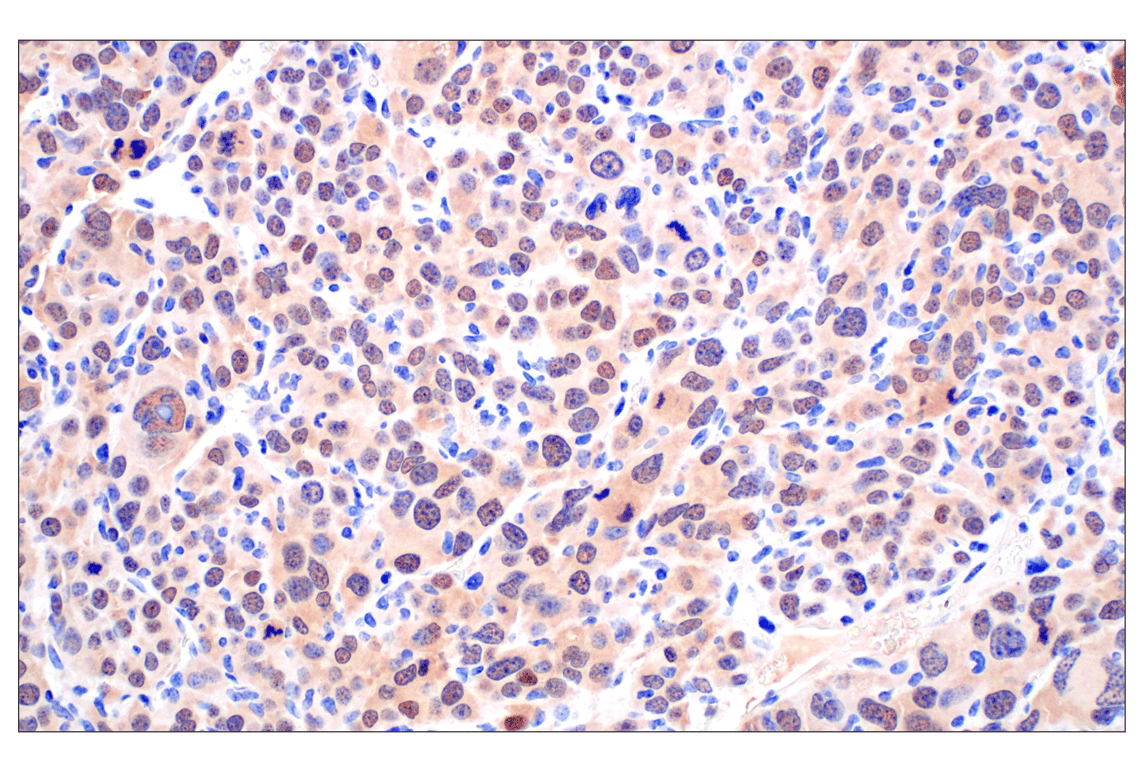
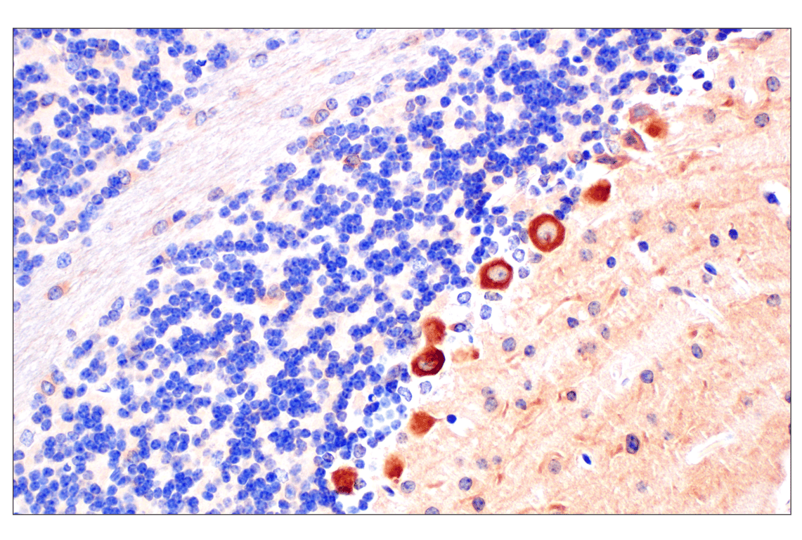
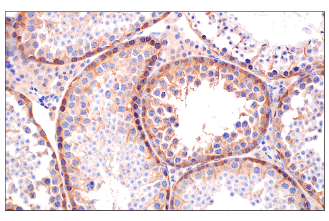
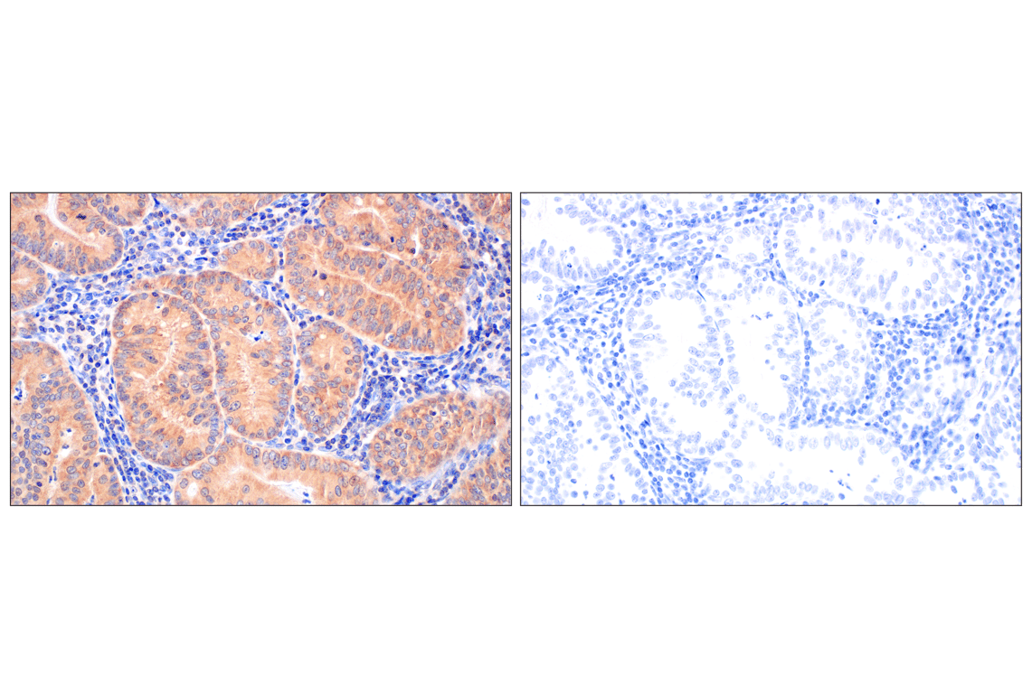
| Immunohistochemistry (Paraffin) | 1:800 - 1:3200 |
| Immunofluorescence (Frozen) | 1:50 - 1:200 |
| Immunofluorescence (Immunocytochemistry) | 1:200 - 1:800 |
| Flow Cytometry (Fixed/Permeabilized) | 1:50 - 1:200 |
Storage
Specificity/Sensitivity
Source / Purification
Background
PIP4K2C, also known as phosphatidylinositol-5-phosphate 4-kinase type-2 gamma (PI5P4Kγ), is a member of the type II PIP kinase family, and is broadly expressed with the highest levels in kidney and brain tissue (9). PIP4K2C can form a homodimer or heterodimer with its two other known family members, PIP4K2A and PIP4K2B (10). PIP4K2C is a regulator of mammalian target of rapamycin complex 1 (mTORC1) signaling, and Pip4k2c KO mice demonstrate enhanced immune responses and develop autoimmunity (11,12).
Background References
- Rameh, L.E. et al. (1997) Nature 390, 192-6.
- Zhang, X. et al. (1997) J Biol Chem 272, 17756-61.
- Ishihara, H. et al. (1996) J Biol Chem 271, 23611-4.
- Loijens, J.C. and Anderson, R.A. (1996) J Biol Chem 271, 32937-43.
- Ishihara, H. et al. (1998) J Biol Chem 273, 8741-8.
- Itoh, T. et al (1998) J. Biol. Chem. 273, 20292-20299
- Boronenkov, I.V. et al. (1998) Mol Biol Cell 9, 3547-60.
- Oude Weernink, P.A. et al. (2004) Eur J Pharmacol 500, 87-99.
- Clarke, J.H. et al. (2008) Am J Physiol Renal Physiol 295, F1422-30.
- Ciruela, A. et al. (2000) Biochem J 346 Pt 3, 587-91.
- Mackey, A.M. et al. (2014) Sci Signal 7, ra104.
- Shim, H. et al. (2016) Proc Natl Acad Sci USA 113, 7596-601.
Species Reactivity
Species reactivity is determined by testing in at least one approved application (e.g., western blot).
Western Blot Buffer
IMPORTANT: For western blots, incubate membrane with diluted primary antibody in 5% w/v BSA, 1X TBS, 0.1% Tween® 20 at 4°C with gentle shaking, overnight.
Applications Key
W: Western Blotting W-S: Simple Western™ IP: Immunoprecipitation IHC-P: Immunohistochemistry (Paraffin) IF-F: Immunofluorescence (Frozen) IF-IC: Immunofluorescence (Immunocytochemistry) FC-FP: Flow Cytometry (Fixed/Permeabilized)
Cross-Reactivity Key
H: Human M: Mouse R: Rat
Trademarks and Patents
Cell Signaling Technology is a trademark of Cell Signaling Technology, Inc.
U.S. Patent No. 7,429,487, foreign equivalents, and child patents deriving therefrom.
All other trademarks are the property of their respective owners. Visit cellsignal.com/trademarks for more information.
Limited Uses
Except as otherwise expressly agreed in a writing signed by a legally authorized representative of CST, the following terms apply to Products provided by CST, its affiliates or its distributors. Any Customer's terms and conditions that are in addition to, or different from, those contained herein, unless separately accepted in writing by a legally authorized representative of CST, are rejected and are of no force or effect.
Products are labeled with For Research Use Only or a similar labeling statement and have not been approved, cleared, or licensed by the FDA or other regulatory foreign or domestic entity, for any purpose. Customer shall not use any Product for any diagnostic or therapeutic purpose, or otherwise in any manner that conflicts with its labeling statement. Products sold or licensed by CST are provided for Customer as the end-user and solely for research and development uses. Any use of Product for diagnostic, prophylactic or therapeutic purposes, or any purchase of Product for resale (alone or as a component) or other commercial purpose, requires a separate license from CST. Customer shall (a) not sell, license, loan, donate or otherwise transfer or make available any Product to any third party, whether alone or in combination with other materials, or use the Products to manufacture any commercial products, (b) not copy, modify, reverse engineer, decompile, disassemble or otherwise attempt to discover the underlying structure or technology of the Products, or use the Products for the purpose of developing any products or services that would compete with CST products or services, (c) not alter or remove from the Products any trademarks, trade names, logos, patent or copyright notices or markings, (d) use the Products solely in accordance with CST Product Terms of Sale and any applicable documentation, and (e) comply with any license, terms of service or similar agreement with respect to any third party products or services used by Customer in connection with the Products.
Revision 6
Revision 6
Revision 6
Revision 6
Revision 6

Revision 6