Revision 23
#83732
Store at -20C
877-616-CELL (2355)
877-678-TECH (8324)
3 Trask Lane | Danvers | Massachusetts | 01923 | USA
For Research Use Only. Not for Use in Diagnostic Procedures.
Applications:
W, IF-IC
Reactivity:
All
Sensitivity:
Endogenous
Source/Isotype:
Rabbit IgG
Product Usage Information
| Application | Dilution |
|---|---|
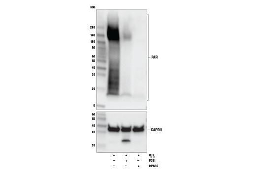
| Western Blotting | 1:1000 |
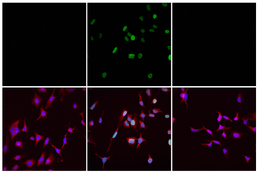
| Immunofluorescence (Immunocytochemistry) | 1:12000 - 1:48000 |
Storage
For a carrier free (BSA and azide free) version of this product see product #71630.
Specificity/Sensitivity
Source / Purification
Background
ADP-ribosylation is involved in a variety of cellular processes, including mitotic spindle formation, chromatin decondensation, cell stress response, retroviral silencing, RNA biology, and transcription, but the most well-known function of ADP-ribose chains is to serve as a scaffold for recruiting DNA repair proteins that contain PAR-binding modules to sites of DNA damage (6). X-ray repair cross-complementing protein 1 (XRCC1), histone macroH2A1, RNF146 (Iduna) an E3 ubiquitin ligase, and many of the PARPs themselves, among others, contain PAR-binding motifs (PBMs) or domains: WWE, PAR-binding zinc-finger (PBZ), or macrodomains (7). PARylation has a central role in cell survival, and is tightly regulated. PARP deficiency can leave a cell vulnerable to DNA damage-induced apoptosis, while hyper PARylation can lead to parthanatos, a unique form of cell death (8). The role of PARylation in DNA repair has inspired great interest in developing candidate drug inhibitors for PARP, in particular to treat breast, prostate and small cell lung cancers with mutations in DNA repair genes like BRCA1/2, CHK2 or ATM. Stat1, PERK, p53, G-actin and Ras are just a few examples of proteins that are functionally modulated by ADP-ribosylation (6,7). Modification by ADP-ribose can block protein interactions or, in the case of P2X7, cause a conformational change that in the presence of ART2 expression sensitizes naive murine T-cells to extracellular NAD+ leading to apoptosis (9).
Background References
- Koch-Nolte, F. et al. (2008) Front Biosci 13, 6716-29.
- Leung, A.K. (2014) J Cell Biol 205, 613-9.
- Laing, S. et al. (2011) Amino Acids 41, 257-69.
- Vyas, S. et al. (2014) Nat Commun 5, 4426.
- Vivelo, C.A. and Leung, A.K. (2015) Proteomics 15, 203-17.
- Gupte, R. et al. (2017) Genes Dev 31, 101-126.
- Wei, H. and Yu, X. (2016) Genomics Proteomics Bioinformatics 14, 131-139.
- David, K.K. et al. (2009) Front Biosci (Landmark Ed) 14, 1116-28.
- Seman, M. et al. (2003) Immunity 19, 571-82.
Species Reactivity
Species reactivity is determined by testing in at least one approved application (e.g., western blot).
Western Blot Buffer
IMPORTANT: For western blots, incubate membrane with diluted primary antibody in 5% w/v BSA, 1X TBS, 0.1% Tween® 20 at 4°C with gentle shaking, overnight.
Applications Key
W: Western Blotting IF-IC: Immunofluorescence (Immunocytochemistry)
Cross-Reactivity Key
All: All Species Expected
Trademarks and Patents
Cell Signaling Technology is a trademark of Cell Signaling Technology, Inc.
All other trademarks are the property of their respective owners. Visit cellsignal.com/trademarks for more information.
Limited Uses
Except as otherwise expressly agreed in a writing signed by a legally authorized representative of CST, the following terms apply to Products provided by CST, its affiliates or its distributors. Any Customer's terms and conditions that are in addition to, or different from, those contained herein, unless separately accepted in writing by a legally authorized representative of CST, are rejected and are of no force or effect.
Products are labeled with For Research Use Only or a similar labeling statement and have not been approved, cleared, or licensed by the FDA or other regulatory foreign or domestic entity, for any purpose. Customer shall not use any Product for any diagnostic or therapeutic purpose, or otherwise in any manner that conflicts with its labeling statement. Products sold or licensed by CST are provided for Customer as the end-user and solely for research and development uses. Any use of Product for diagnostic, prophylactic or therapeutic purposes, or any purchase of Product for resale (alone or as a component) or other commercial purpose, requires a separate license from CST. Customer shall (a) not sell, license, loan, donate or otherwise transfer or make available any Product to any third party, whether alone or in combination with other materials, or use the Products to manufacture any commercial products, (b) not copy, modify, reverse engineer, decompile, disassemble or otherwise attempt to discover the underlying structure or technology of the Products, or use the Products for the purpose of developing any products or services that would compete with CST products or services, (c) not alter or remove from the Products any trademarks, trade names, logos, patent or copyright notices or markings, (d) use the Products solely in accordance with CST Product Terms of Sale and any applicable documentation, and (e) comply with any license, terms of service or similar agreement with respect to any third party products or services used by Customer in connection with the Products.
Revision 23