Revision 1
#9942
Store at -20C
Pro-Apoptosis Bcl-2 Family Antibody Sampler Kit
1 Kit
(8 x 20 microliters)
877-616-CELL (2355)
877-678-TECH (8324)
3 Trask Lane | Danvers | Massachusetts | 01923 | USA
For Research Use Only. Not for Use in Diagnostic Procedures.
| Product Includes | Product # | Quantity | Mol. Wt | Isotype/Source |
|---|---|---|---|---|
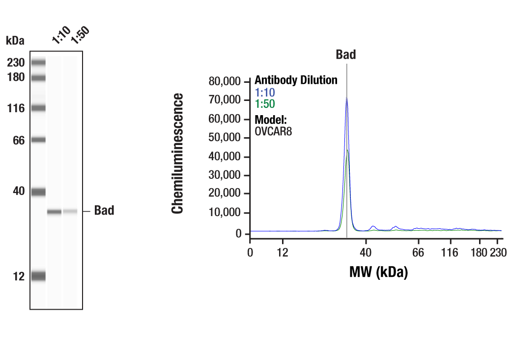
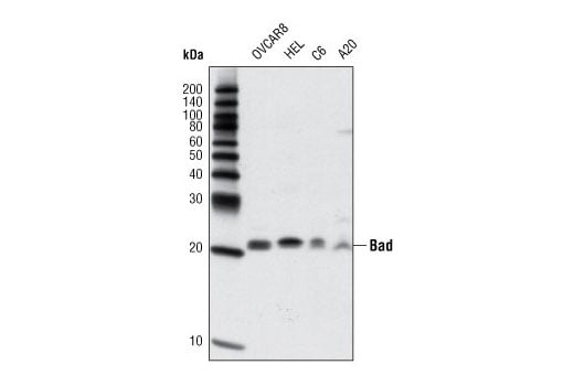
| Bad (D24A9) Rabbit Monoclonal Antibody | 9239 | 20 µl | 23 kDa | Rabbit IgG |
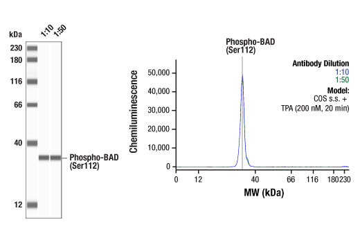
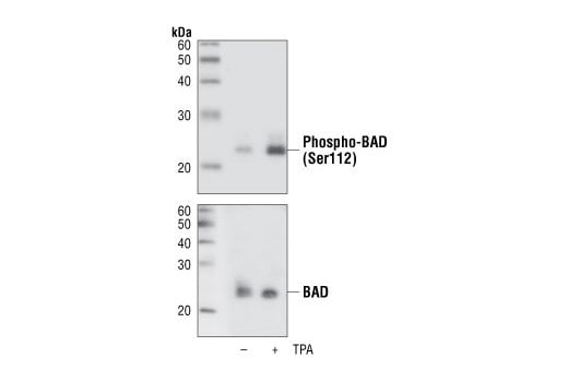
| Phospho-Bad (Ser112) (40A9) Rabbit Monoclonal Antibody | 5284 | 20 µl | 23 kDa | Rabbit IgG |
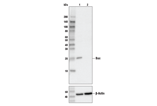
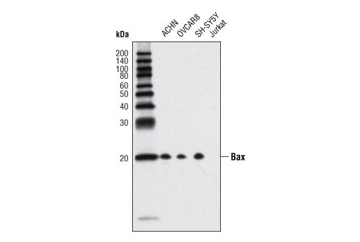
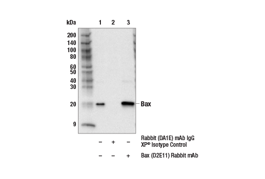
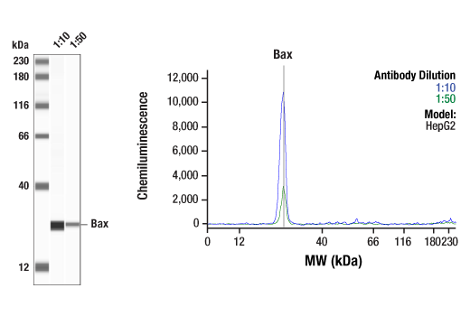
| Bax (D2E11) Rabbit Monoclonal Antibody | 5023 | 20 µl | 20 kDa | Rabbit IgG |
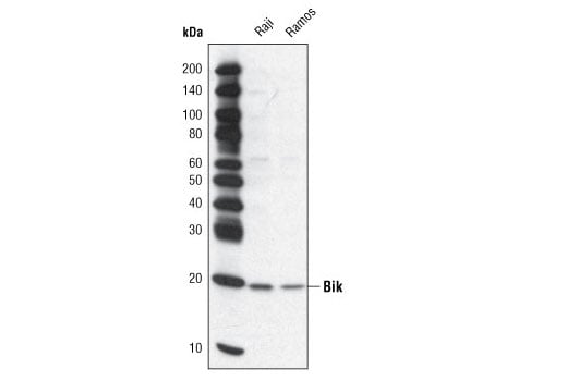
| Bik Antibody | 4592 | 20 µl | 20 kDa | Rabbit |
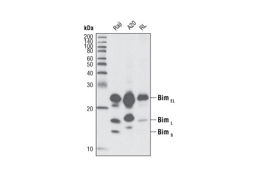
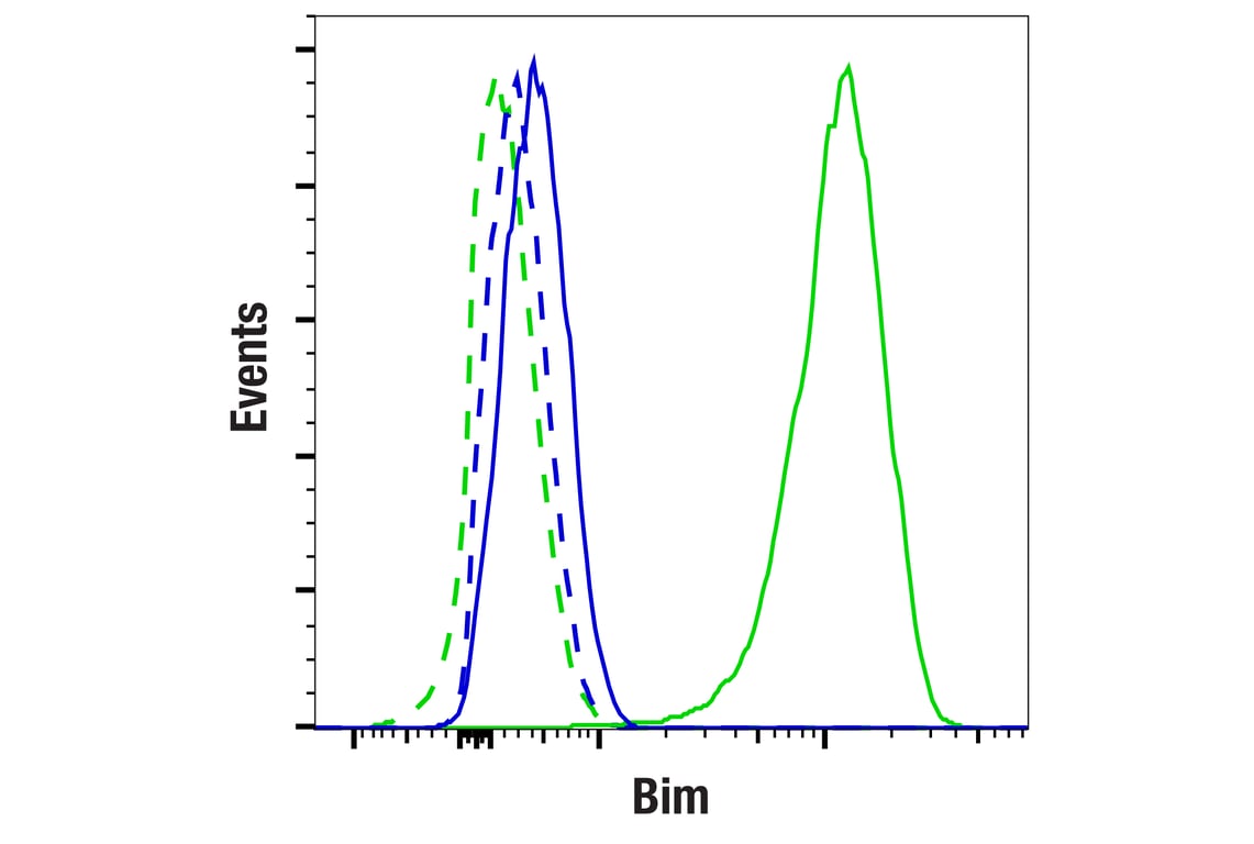
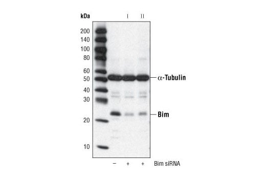
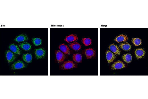
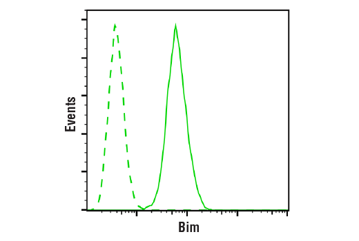
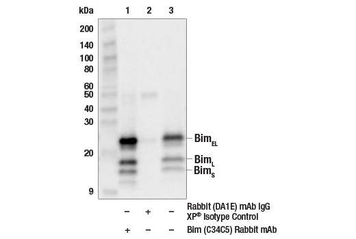
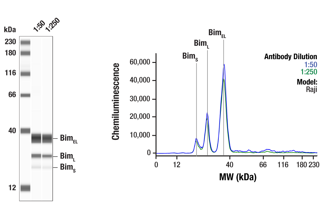
| Bim (C34C5) Rabbit Monoclonal Antibody | 2933 | 20 µl | 12, 15, 23 kDa | Rabbit IgG |
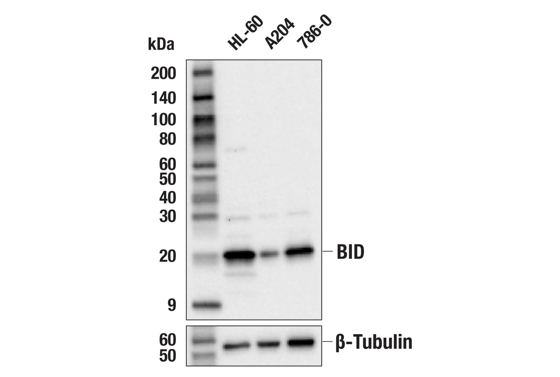
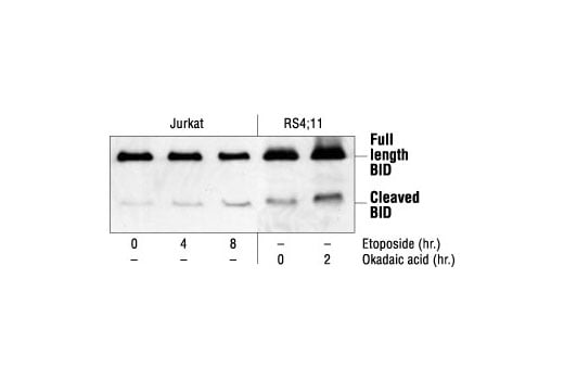
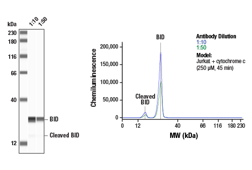
| BID Antibody | 2002 | 20 µl | 15, 22 kDa | Rabbit |
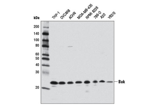
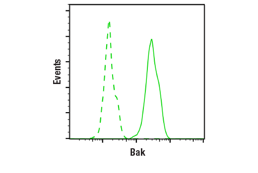
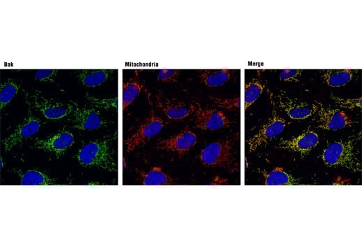
| Bak (D4E4) Rabbit Monoclonal Antibody | 12105 | 20 µl | 25 kDa | Rabbit IgG |
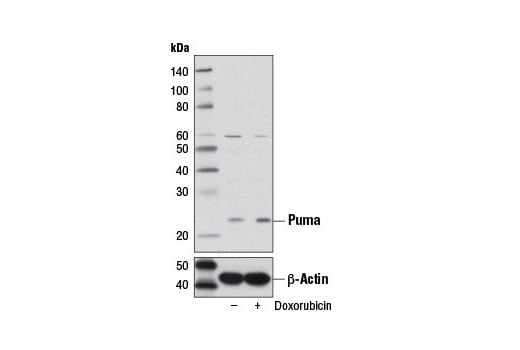
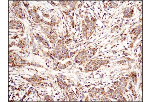
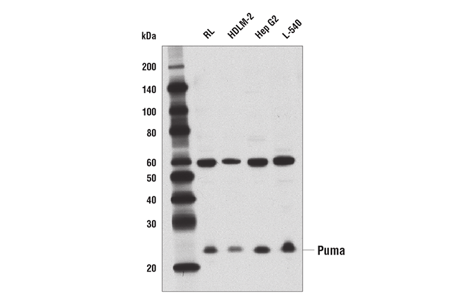
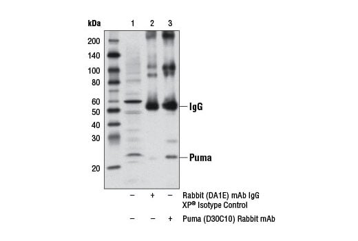
| Puma (D30C10) Rabbit Monoclonal Antibody | 12450 | 20 µl | 23 kDa | Rabbit IgG |
| Anti-rabbit IgG, HRP-linked Antibody | 7074 | 100 µl | Goat |
Please visit cellsignal.com for individual component applications, species cross-reactivity, dilutions, protocols, and additional product information.
Description
Storage
Background
Bad is a pro-apoptotic member of the Bcl-2 family that can displace Bax from binding to Bcl-2 and Bcl-xL, resulting in cell death (6,7). Survival factors such as IL-3 can inhibit the apoptotic activity of Bad by activating intracellular signaling pathways that result in the phosphorylation of Bad at Ser112 and Ser136 (7). Phosphorylation at these sites results in the binding of Bad to 14-3-3 proteins and the inhibition of Bad binding to Bcl-2 and Bcl-xL (7). Akt has been shown to promote cell survival via its ability to phosphorylate Bad at Ser136 (8,9). Ser112 has been shown to be the substrate in vivo and in vitro of p90RSK (10,11) and mitochondria-anchored PKA (12).
Background References
- Cory, S. et al. (2003) Oncogene 22, 8590-607.
- Antonsson, B. and Martinou, J.C. (2000) Exp Cell Res 256, 50-7.
- Sharpe, J.C. et al. (2004) Biochim Biophys Acta 1644, 107-13.
- Korsmeyer, S.J. et al. (1993) Semin Cancer Biol 4, 327-32.
- Bouillet, P. and Strasser, A. (2002) J Cell Sci 115, 1567-74.
- Yang, E. et al. (1995) Cell 80, 285-91.
- Zha, J. et al. (1996) Cell 87, 619-28.
- Datta, S.R. et al. (1997) Cell 91, 231-41.
- del Peso, L. et al. (1997) Science 278, 687-9.
- Bonni, A. et al. (1999) Science 286, 1358-62.
- Tan, Y. et al. (1999) J Biol Chem 274, 34859-67.
- Harada, H. et al. (1999) Mol Cell 3, 413-22.
Trademarks and Patents
Cell Signaling Technology is a trademark of Cell Signaling Technology, Inc.
All other trademarks are the property of their respective owners. Visit cellsignal.com/trademarks for more information.
Limited Uses
Except as otherwise expressly agreed in a writing signed by a legally authorized representative of CST, the following terms apply to Products provided by CST, its affiliates or its distributors. Any Customer's terms and conditions that are in addition to, or different from, those contained herein, unless separately accepted in writing by a legally authorized representative of CST, are rejected and are of no force or effect.
Products are labeled with For Research Use Only or a similar labeling statement and have not been approved, cleared, or licensed by the FDA or other regulatory foreign or domestic entity, for any purpose. Customer shall not use any Product for any diagnostic or therapeutic purpose, or otherwise in any manner that conflicts with its labeling statement. Products sold or licensed by CST are provided for Customer as the end-user and solely for research and development uses. Any use of Product for diagnostic, prophylactic or therapeutic purposes, or any purchase of Product for resale (alone or as a component) or other commercial purpose, requires a separate license from CST. Customer shall (a) not sell, license, loan, donate or otherwise transfer or make available any Product to any third party, whether alone or in combination with other materials, or use the Products to manufacture any commercial products, (b) not copy, modify, reverse engineer, decompile, disassemble or otherwise attempt to discover the underlying structure or technology of the Products, or use the Products for the purpose of developing any products or services that would compete with CST products or services, (c) not alter or remove from the Products any trademarks, trade names, logos, patent or copyright notices or markings, (d) use the Products solely in accordance with CST Product Terms of Sale and any applicable documentation, and (e) comply with any license, terms of service or similar agreement with respect to any third party products or services used by Customer in connection with the Products.
Revision 1
Revision 1
Revision 1
Revision 1

Revision 1
Revision 1
Revision 1
Revision 1
Revision 1

Revision 1
Revision 1

Revision 1
Revision 1
Revision 1