Revision 1
#4889
Store at -20C
Protein Folding and Stability Antibody Sampler Kit
1 Kit
(8 x 20 microliters)
877-616-CELL (2355)
877-678-TECH (8324)
3 Trask Lane | Danvers | Massachusetts | 01923 | USA
For Research Use Only. Not for Use in Diagnostic Procedures.
| Product Includes | Product # | Quantity | Mol. Wt | Isotype/Source |
|---|---|---|---|---|
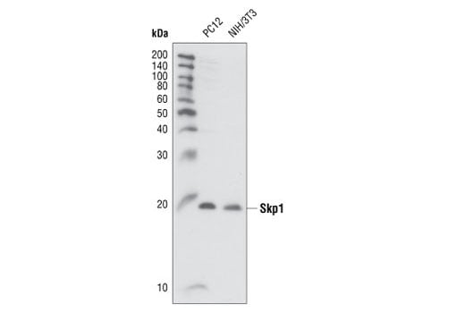
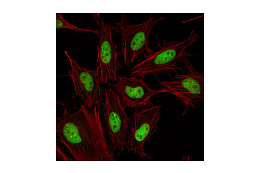
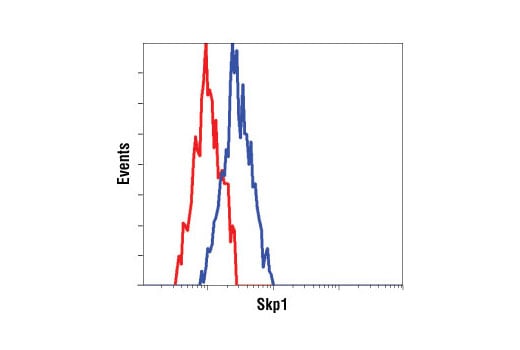
| Skp1 Antibody | 2156 | 20 µl | 19 kDa | Rabbit |
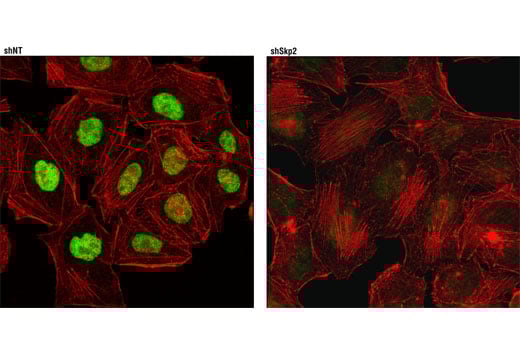
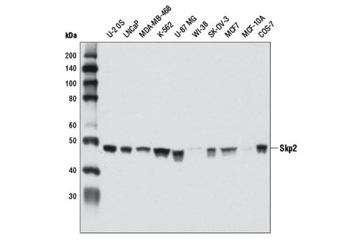
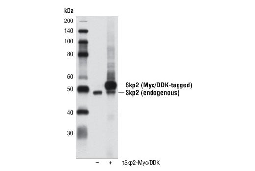
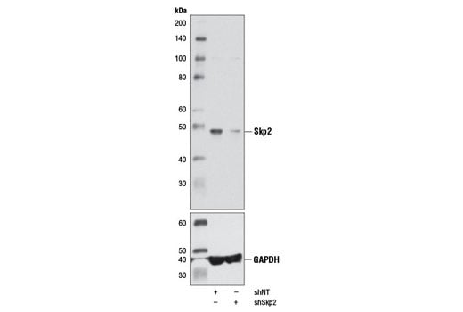
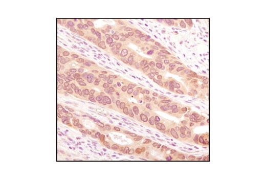
| Skp2 (D3G5) Rabbit Monoclonal Antibody | 2652 | 20 µl | 48 kDa | Rabbit IgG |
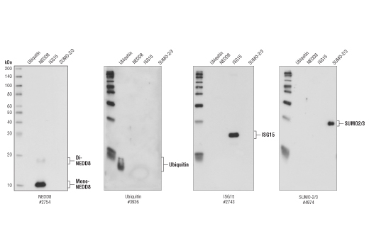
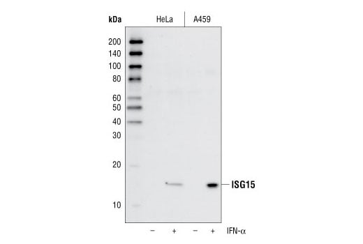
| ISG15 (22D2) Rabbit Monoclonal Antibody | 2758 | 20 µl | 15 kDa | Rabbit IgG |
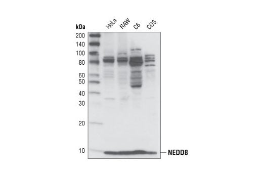
| NEDD8 (19E3) Rabbit Monoclonal Antibody | 2754 | 20 µl | 9 kDa | Rabbit IgG |
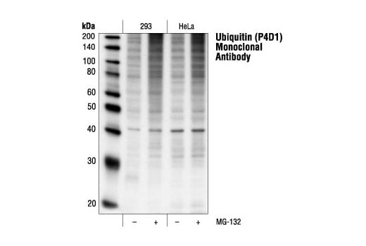
| Ubiquitin (P4D1) Mouse Monoclonal Antibody | 3936 | 20 µl | Mouse IgG1 | |
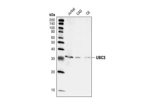
| UBC3 Antibody | 4997 | 20 µl | 32 kDa | Rabbit |
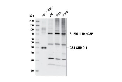
| SUMO-1 Antibody | 4930 | 20 µl | Rabbit | |
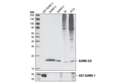
| SUMO-2/3 (18H8) Rabbit Monoclonal Antibody | 4971 | 20 µl | Rabbit IgG | |
| Anti-rabbit IgG, HRP-linked Antibody | 7074 | 100 µl | Goat | |
| Anti-mouse IgG, HRP-linked Antibody | 7076 | 100 µl | Horse |
Please visit cellsignal.com for individual component applications, species cross-reactivity, dilutions, protocols, and additional product information.
Description
Storage
Background
The ubiquitin-like protein family contains three small ubiquitin-related modifier proteins (SUMO-1, -2 and -3), neural precursor cell-expressed developmentally down-regulated protein 8 (NEDD8) and interferon-stimulated 15 kDa protein (ISG15) (4-6). Their covalent attachment to target proteins is a reversible, multi-step process that is analogous to protein ubiquitination. Mature molecules are linked to the activating enzyme E1, conjugated to E2 and ligated to the target proteins by E3 (7-10). Ubiquitin is the predominant regulator for the degradation of a wide range of target proteins (8) while SUMO, NEDD8 and ISG15 modify a limited set of substrates to regulate various other biological processes (4, 11-18).
During ubiquitination, the combinatorial interaction of different E2 and E3 proteins produces variable substrate specificity (4). UBC3 and UBC3B are E2 ubiquitin-carrier proteins (19, 20). The SCF (Skp1/CUL1/F-box) E3 ubiquitin ligase protein complex is composed of three protein components, including the S phase kinase associated protein 1 (Skp1), Cullin homolog 1 (CUL1) and the Skp2 F-box protein (21-23).
Background References
- Ciechanover, A. (1998) EMBO J. 17, 7151-7160.
- Bernardi, R. et al. (2000) Oncogene 19, 2447-2454.
- Jesenberger, V. and Jentsch, S. (2002) Nat. Rev. Mol. Cell Biol. 3, 112-121.
- Schwartz, D.C. and Hochstrasser, M. (2003) Trends Biochem. Sci. 28, 321-328.
- Chiba, T. and Tanaka, K. (2004) Curr. Protein Pept. Sci. 5, 177-184.
- Ritchie, K.J. and Zhang, D.E. (2004) Semin. Cell Dev. Biol. 15, 237-246.
- Kim, K.I. et al. (2002) J. Cell Physiol. 191, 257-268.
- Osaka, F. et al. (1998) Genes Dev. 12, 2263-2268.
- Loeb, K.R. and Haas, A.L. (1992) J. Biol. Chem. 267, 7806-7813.
- Zhao, C. et al. (2005) Proc. Natl. Acad. Sci. USA 102, 10200-10205.
- Matunis, M.J. et al. (1996) J. Cell Biol. 135, 1457-1470.
- Duprez, E. et al. (1999) J. Cell Sci. 112 ( Pt 3), 381-393.
- Gostissa, M. et al. (1999) EMBO J. 18, 6462-6471.
- Rodriguez, M.S. et al. (1999) EMBO J. 18, 6455-6461.
- Desterro, J.M. et al. (1998) Mol. Cell 2, 233-239.
- Stickle, N.H. et al. (2004) Mol. Cell. Biol. 24, 3251-3261.
- Xirodimas, D.P. et al. (2004) Cell 118, 83-97.
- Hamerman, J.A. et al. (2002) J. Immunol. 168, 2415-2423.
- Semplici, F. et al. (2002) Oncogene 21, 3978-3987.
- Pagano, M. et al. (1995) Science 269, 682-685.
- Yu, Z.K. et al. (1998) Proc. Natl. Acad. Sci. USA 95, 11324-11329.
- Pagano, M. (2004) Mol. Cell 14, 414-416.
- Reed, S.I. (2003) Nat. Rev. Mol. Cell Biol. 4, 855-864.
Trademarks and Patents
Cell Signaling Technology is a trademark of Cell Signaling Technology, Inc.
All other trademarks are the property of their respective owners. Visit cellsignal.com/trademarks for more information.
Limited Uses
Except as otherwise expressly agreed in a writing signed by a legally authorized representative of CST, the following terms apply to Products provided by CST, its affiliates or its distributors. Any Customer's terms and conditions that are in addition to, or different from, those contained herein, unless separately accepted in writing by a legally authorized representative of CST, are rejected and are of no force or effect.
Products are labeled with For Research Use Only or a similar labeling statement and have not been approved, cleared, or licensed by the FDA or other regulatory foreign or domestic entity, for any purpose. Customer shall not use any Product for any diagnostic or therapeutic purpose, or otherwise in any manner that conflicts with its labeling statement. Products sold or licensed by CST are provided for Customer as the end-user and solely for research and development uses. Any use of Product for diagnostic, prophylactic or therapeutic purposes, or any purchase of Product for resale (alone or as a component) or other commercial purpose, requires a separate license from CST. Customer shall (a) not sell, license, loan, donate or otherwise transfer or make available any Product to any third party, whether alone or in combination with other materials, or use the Products to manufacture any commercial products, (b) not copy, modify, reverse engineer, decompile, disassemble or otherwise attempt to discover the underlying structure or technology of the Products, or use the Products for the purpose of developing any products or services that would compete with CST products or services, (c) not alter or remove from the Products any trademarks, trade names, logos, patent or copyright notices or markings, (d) use the Products solely in accordance with CST Product Terms of Sale and any applicable documentation, and (e) comply with any license, terms of service or similar agreement with respect to any third party products or services used by Customer in connection with the Products.
Revision 1
Revision 1
Revision 1

Revision 1

Revision 1
Revision 1
Revision 1
Revision 1
Revision 1
Revision 1