Revision 1

#9385Store at -20C

1 Kit

(5 x 20 microliters)

Cell Signaling Technology

Orders: 877-616-CELL (2355) [email protected]

Support: 877-678-TECH (8324)

Web: [email protected] cellsignal.com

3 Trask LaneDanversMassachusetts01923USA
For Research Use Only. Not for Use in Diagnostic Procedures.
Product Includes Product # Quantity Mol. Wt Isotype/Source
Rab4 Antibody 2167 20 µl 25 kDa Rabbit 
Rab5A (E6N8S) Mouse mAb 46449 20 µl 25 kDa Mouse IgG1
Rab7 (D95F2) XP® Rabbit mAb 9367 20 µl 23 kDa Rabbit IgG
Rab9A (D52G8) XP® Rabbit mAb 5118 20 µl 23 kDa Rabbit IgG
Rab11 (D4F5) XP® Rabbit mAb 5589 20 µl 25 kDa Rabbit IgG
Anti-rabbit IgG, HRP-linked Antibody 7074 100 µl Goat 
Anti-mouse IgG, HRP-linked Antibody 7076 100 µl Horse 

Please visit cellsignal.com for individual component applications, species cross-reactivity, dilutions, protocols, and additional product information.

Description

The Rab Family Antibody Sampler Kit provides an economical means to evaluate the presence and status of Rab proteins in cells. This kit provides enough primary and secondary antibodies to perform two Western blot experiments per primary antibody.

Storage

Supplied in 10 mM sodium HEPES (pH 7.5), 150 mM NaCl, 100 µg/ml BSA, 50% glycerol and less than 0.02% sodium azide. Store at –20°C. Do not aliquot the antibody.

Background

Rab family proteins are GTPases and members of the Ras superfamily of monomeric G proteins. These membrane-associated proteins are involved in many aspects of vesicle-mediated transport, taking part in the initial vesicle formation, transport of vesicles along the cytoskeleton, and eventual fusion of vesicle and target membranes. Rab4 is localized at early endosomes/recycling endosomes and functions as a key regulator for sorting/recycling of membrane and proteins (1,2). Both Rab4A and Rab4B isoforms are localized to similar cellular compartments and are believed to have similar functions (4). Rab4 interacts with several Rab4 effectors in a complex on a special endosome site to promote membrane/protein recycling (1,3). Rab5 is localized to the plasma membrane and early endosome and functions as a key regulator of vesicle trafficking during early endocytosis (1). The conformational change between Rab5-GTP and Rab5-GDP is essential for its biological function as a rate-limiting regulator at multiple steps during endocytosis (1,5). Similar to Rab4, Rab5 also interacts with specific Rab5 effectors on a specialized endosomal Rab domain to promote recycling between endosome and the plasma membrane (1,5,6). Both Rab7 and Rab9 are located in late endosomes but exert different functions. Rab7 associates with the RIPL effector protein to control membrane trafficking from early to late endosome and to lysosomes (7,8). Rab7 also helps to regulate growth receptor endocytic trafficking and degradation, and maturation of phagosome and autophagic vacuoles (8-11). Rab9 interacts with its effector proteins p40 and TIP47 (12,13) to promote the MPR (mannose 6-phosphate receptor)-associated lysosomal enzyme transport between late endosomes and the trans Golgi network (14,15). Rab11 (isoforms Rab11a and Rab11b) functions as a key regulator in the recycling of perinuclear, plasma membrane and Golgi compartment endosomes (16,17). Despite some overlap, distinct differences exist between Rab11a and Rab11b in both their cellular distribution and functional roles. Rab11a is ubiquitously expressed while Rab11b is found mainly in the heart and brain (18,19). Like other Rab proteins, Rab11 functions when associated with Rab11 family interacting proteins (FIPs). The three distinct classes of Rab11 FIPs all share a conserved carboxy-terminal Rab-binding domain that allows Rab-FIP protein interaction. When bound together, these proteins are thought to regulate membrane-associated protein sorting (20,21).

  1. Zerial, M. and McBride, H. (2001) Nat Rev Mol Cell Biol 2, 107-17.
  2. van der Sluijs, P. et al. (1992) Cell 70, 729-40.
  3. Deneka, M. et al. (2003) EMBO J 22, 2645-57.
  4. Krawczyk, M. et al. (2007) Nucleic Acids Res 35, 595-605.
  5. van der Bliek, A.M. (2005) Nat Cell Biol 7, 548-50.
  6. Haas, A.K. et al. (2005) Nat Cell Biol 7, 887-93.
  7. Feng, Y. et al. (1995) J Cell Biol 131, 1435-52.
  8. Méresse, S. et al. (1995) J Cell Sci 108 ( Pt 11), 3349-58.
  9. Ceresa, B.P. and Bahr, S.J. (2006) J Biol Chem 281, 1099-106.
  10. Jäger, S. et al. (2004) J Cell Sci 117, 4837-48.
  11. Méresse, S. et al. (1999) EMBO J 18, 4394-403.
  12. Díaz, E. et al. (1997) J Cell Biol 138, 283-90.
  13. Barbero, P. et al. (2002) J Cell Biol 156, 511-8.
  14. Lombardi, D. et al. (1993) EMBO J 12, 677-82.
  15. Riederer, M.A. et al. (1994) J Cell Biol 125, 573-82.
  16. Ullrich, O. et al. (1996) J Cell Biol 135, 913-24.
  17. Chen, W. et al. (1998) Mol Biol Cell 9, 3241-57.
  18. Lapierre, L.A. et al. (2003) Exp Cell Res 290, 322-31.
  19. Khvotchev, M.V. et al. (2003) J Neurosci 23, 10531-9.
  20. Junutula, J.R. et al. (2004) J Biol Chem 279, 33430-7.
  21. Hales, C.M. et al. (2001) J Biol Chem 276, 39067-75.

Background References

    Trademarks and Patents

    Cell Signaling Technology is a trademark of Cell Signaling Technology, Inc.
    U.S. Patent No. 7,429,487, foreign equivalents, and child patents deriving therefrom.
    All other trademarks are the property of their respective owners. Visit cellsignal.com/trademarks for more information.

    Limited Uses

    Except as otherwise expressly agreed in a writing signed by a legally authorized representative of CST, the following terms apply to Products provided by CST, its affiliates or its distributors. Any Customer's terms and conditions that are in addition to, or different from, those contained herein, unless separately accepted in writing by a legally authorized representative of CST, are rejected and are of no force or effect.

    Products are labeled with For Research Use Only or a similar labeling statement and have not been approved, cleared, or licensed by the FDA or other regulatory foreign or domestic entity, for any purpose. Customer shall not use any Product for any diagnostic or therapeutic purpose, or otherwise in any manner that conflicts with its labeling statement. Products sold or licensed by CST are provided for Customer as the end-user and solely for research and development uses. Any use of Product for diagnostic, prophylactic or therapeutic purposes, or any purchase of Product for resale (alone or as a component) or other commercial purpose, requires a separate license from CST. Customer shall (a) not sell, license, loan, donate or otherwise transfer or make available any Product to any third party, whether alone or in combination with other materials, or use the Products to manufacture any commercial products, (b) not copy, modify, reverse engineer, decompile, disassemble or otherwise attempt to discover the underlying structure or technology of the Products, or use the Products for the purpose of developing any products or services that would compete with CST products or services, (c) not alter or remove from the Products any trademarks, trade names, logos, patent or copyright notices or markings, (d) use the Products solely in accordance with CST Product Terms of Sale and any applicable documentation, and (e) comply with any license, terms of service or similar agreement with respect to any third party products or services used by Customer in connection with the Products.

    Revision 1
    #9385

    Rab Family Antibody Sampler Kit

    Rab Family Antibody Sampler Kit: Image 1 Expand Image
    Western blot analysis of extracts from various cell types using Rab4 Antibody.
    Rab Family Antibody Sampler Kit: Image 2 Expand Image
    Western blot analysis of extracts from various cell lines using Rab5A (E6N8S) Mouse mAb.
    Rab Family Antibody Sampler Kit: Image 3 Expand Image
    Western blot analysis of extracts from various cell lines using Rab9A (D52G8) XP® Rabbit mAb.
    Rab Family Antibody Sampler Kit: Image 4 Expand Image
    Western blot analysis of extracts from various cell lines using Rab11 (D4F5) XP® Rabbit mAb.
    Rab Family Antibody Sampler Kit: Image 5 Expand Image
    After the primary antibody is bound to the target protein, a complex with HRP-linked secondary antibody is formed. The LumiGLO® is added and emits light during enzyme catalyzed decomposition.
    Rab Family Antibody Sampler Kit: Image 6 Expand Image
    After the primary antibody is bound to the target protein, a complex with HRP-linked secondary antibody is formed. The LumiGLO* is added and emits light during enzyme catalyzed decomposition.
    Rab Family Antibody Sampler Kit: Image 7 Expand Image
    Western blot analysis of extracts from various cell lines using Rab7 (D95F2) XP® Rabbit mAb.
    Rab Family Antibody Sampler Kit: Image 8 Expand Image
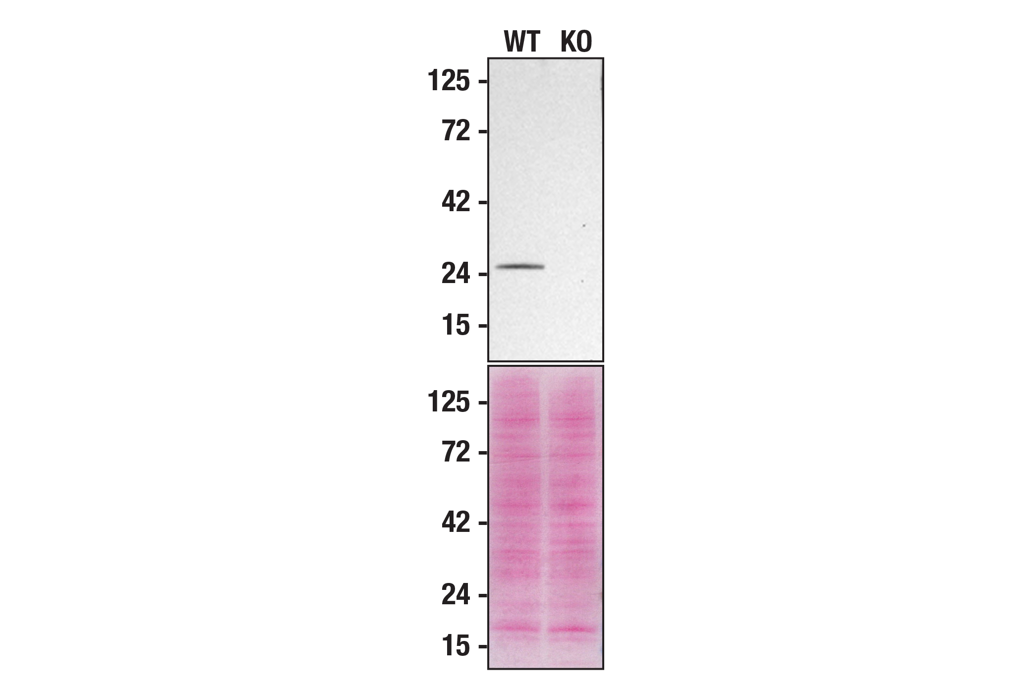
    Western blot analysis of HAP1 extracts from WT (left) or RAB5A KO (right) using Rab5A (E6N8S) Mouse mAb. Membranes stained with Ponceau S for total protein normalization (lower). These data were provided by YCharOS Inc., an open science company with the mission of characterizing commercially available antibodies, as a companion to validation data generated by CST scientists.
    Rab Family Antibody Sampler Kit: Image 9 Expand Image
    Confocal Immunofluorescent analysis of HeLa cells using Rab9A (D52G8) XP® Rabbit mAb (green). Actin filaments have been labeled with DY-554 phalloidin (red). Blue pseudocolor = DRAQ5® #4084 (fluorescent DNA dye).
    Rab Family Antibody Sampler Kit: Image 10 Expand Image
    Confocal immunofluorescent analysis of A549 cells using Rab11 (D4F5) XP® Rabbit mAb (green). Actin filaments were labeled wtih DY-554 phalloidin (red). Blue pseudocolor = DRAQ5® #4084 (fluorescent DNA dye).
    Rab Family Antibody Sampler Kit: Image 11 Expand Image
    Confocal immunofluorescent analysis of SK-MEL-28 cells using Rab7 (D95F2) XP® Rabbit mAb (green). Actin filaments were labeled using DY-554 phalloidin (red). Blue pseudocolor = DRAQ5® #4084 (fluorescent DNA dye).
    Rab Family Antibody Sampler Kit: Image 12 Expand Image
    Immunoprecipitation of Rab5A protein from HeLa cell extracts. Lane 1 is 10% input, lane 2 is Mouse (G3A1) mAb IgG1 Isotype Control #5415, lane 3 is Rab5A (E6N8S) Mouse mAb without HeLa cell extracts, and lane 4 is Rab5A (E6N8S) Mouse mAb. Western blot analysis was performed using Rab5A (E6N8S) Mouse mAb.
    Rab Family Antibody Sampler Kit: Image 13 Expand Image
    Immunoprecipitation of Rab5A protein from HeLa cell extracts. Lane 1 is 10% input, lane 2 is Mouse (G3A1) mAb IgG1 Isotype Control #5415, and lane 3 is Rab5A (E6N8S) Mouse mAb. Western blot analysis was performed using Rab5 (C8B1) Rabbit mAb #3547.
    Rab Family Antibody Sampler Kit: Image 14 Expand Image
    Immunohistochemical analysis of paraffin-embedded human ductal carcinoma of the breast using Rab5A (E6N8S) Mouse mAb.
    Rab Family Antibody Sampler Kit: Image 15 Expand Image
    Immunohistochemical analysis of paraffin-embedded human serous papillary carcinoma of the ovary using Rab5A (E6N8S) Mouse mAb.
    Rab Family Antibody Sampler Kit: Image 16 Expand Image
    Immunohistochemical analysis of paraffin-embedded human colon adenocarcinoma using Rab5A (E6N8S) Mouse mAb (left) compared to concentration matched Mouse (G3A1) IgG1 Isotype Control #5415 (right).
    Rab Family Antibody Sampler Kit: Image 17 Expand Image
    Immunohistochemical analysis of paraffin-embedded human prostate adenocarcinoma using Rab5A (E6N8S) Mouse mAb.
    Rab Family Antibody Sampler Kit: Image 18 Expand Image
    Confocal immunofluorescent analysis of HeLa cells using Rab5A (E6N8S) Mouse mAb (green). Actin filaments were labeled with DyLight 554 Phalloidin #13054 (red). Samples were mounted in ProLong® Gold Antifade Reagent with DAPI #8961 (blue).