Revision 5
#8965
Store at -20C
877-616-CELL (2355)
877-678-TECH (8324)
3 Trask Lane | Danvers | Massachusetts | 01923 | USA
For Research Use Only. Not for Use in Diagnostic Procedures.
Applications:
W, IP, IHC-P, IF-IC, FC-FP
Reactivity:
H M
Sensitivity:
Endogenous
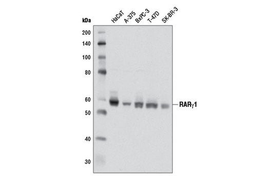
MW (kDa):
58
Source/Isotype:
Rabbit IgG
UniProt ID:
#P13631
Entrez-Gene Id:
5916
Product Usage Information
| Application | Dilution |
|---|---|
| Western Blotting | 1:1000 |
| Immunoprecipitation | 1:100 |
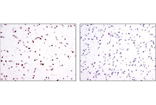
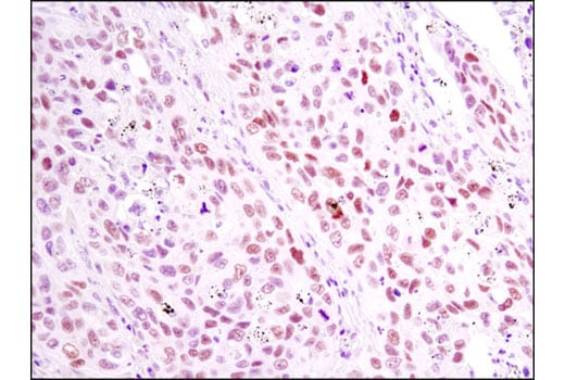
| Immunohistochemistry (Paraffin) | 1:200 - 1:800 |
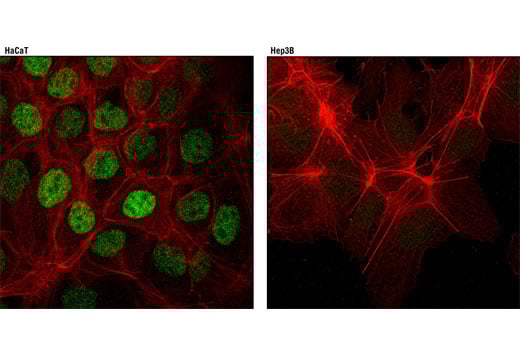
| Immunofluorescence (Immunocytochemistry) | 1:400 - 1:800 |
| Flow Cytometry (Fixed/Permeabilized) | 1:400 - 1:1600 |
Storage
For a carrier free (BSA and azide free) version of this product see product #82517.
Specificity/Sensitivity
Species predicted to react based on 100% sequence homology
Source / Purification
Background
In contrast to the ubiquitously expressed RARα subtype, RARγ displays a complex tissue-specific expression pattern (7). The hematopoietic system expresses significant levels of RARγ, and a recent study identified a role for RARγ in hematopoietic stem cell maintenance (8). RARγ is the predominant subtype in human and mouse epidermis, representing 90% of the RARs in this tissue (9-11). Given the high level of RARγ expression in the skin, it has been suggested that this nuclear receptor participates in a transcriptional program that governs maintenance and differentiation of normal epidermis and skin appendages. The transcriptional activity of RARγ is under stringent control, in part, through retinoic acid-induced phosphorylation and proteasomal degradation (12).
Background References
- Rochette-Egly, C. and Germain, P. (2009) Nucl Recept Signal 7, e005.
- Delacroix, L. et al. (2010) Mol Cell Biol 30, 231-44.
- Eifert, C. et al. (2006) Mol Reprod Dev 73, 796-824.
- Mark, M. et al. (2006) Annu Rev Pharmacol Toxicol 46, 451-80.
- Niederreither, K. and Dollé, P. (2008) Nat Rev Genet 9, 541-53.
- Mark, M. et al. (2009) Nucl Recept Signal 7, e002.
- Dollé, P. (2009) Nucl Recept Signal 7, e006.
- Purton, L.E. et al. (2006) J Exp Med 203, 1283-93.
- Fisher, G.J. et al. (1994) J Biol Chem 269, 20629-35.
- Zelent, A. et al. (1989) Nature 339, 714-7.
- Elder, J.T. et al. (1991) J Invest Dermatol 96, 425-33.
- Giannì, M. et al. (2002) EMBO J 21, 3760-9.
Species Reactivity
Species reactivity is determined by testing in at least one approved application (e.g., western blot).
Western Blot Buffer
IMPORTANT: For western blots, incubate membrane with diluted primary antibody in 5% w/v nonfat dry milk, 1X TBS, 0.1% Tween® 20 at 4°C with gentle shaking, overnight.
Applications Key
W: Western Blotting IP: Immunoprecipitation IHC-P: Immunohistochemistry (Paraffin) IF-IC: Immunofluorescence (Immunocytochemistry) FC-FP: Flow Cytometry (Fixed/Permeabilized)
Cross-Reactivity Key
H: Human M: Mouse
Trademarks and Patents
Cell Signaling Technology is a trademark of Cell Signaling Technology, Inc.
All other trademarks are the property of their respective owners. Visit cellsignal.com/trademarks for more information.
Limited Uses
Except as otherwise expressly agreed in a writing signed by a legally authorized representative of CST, the following terms apply to Products provided by CST, its affiliates or its distributors. Any Customer's terms and conditions that are in addition to, or different from, those contained herein, unless separately accepted in writing by a legally authorized representative of CST, are rejected and are of no force or effect.
Products are labeled with For Research Use Only or a similar labeling statement and have not been approved, cleared, or licensed by the FDA or other regulatory foreign or domestic entity, for any purpose. Customer shall not use any Product for any diagnostic or therapeutic purpose, or otherwise in any manner that conflicts with its labeling statement. Products sold or licensed by CST are provided for Customer as the end-user and solely for research and development uses. Any use of Product for diagnostic, prophylactic or therapeutic purposes, or any purchase of Product for resale (alone or as a component) or other commercial purpose, requires a separate license from CST. Customer shall (a) not sell, license, loan, donate or otherwise transfer or make available any Product to any third party, whether alone or in combination with other materials, or use the Products to manufacture any commercial products, (b) not copy, modify, reverse engineer, decompile, disassemble or otherwise attempt to discover the underlying structure or technology of the Products, or use the Products for the purpose of developing any products or services that would compete with CST products or services, (c) not alter or remove from the Products any trademarks, trade names, logos, patent or copyright notices or markings, (d) use the Products solely in accordance with CST Product Terms of Sale and any applicable documentation, and (e) comply with any license, terms of service or similar agreement with respect to any third party products or services used by Customer in connection with the Products.
Revision 5

Revision 5

Revision 5
