Revision 1
#42344
Store at -20C
Receptor Tyrosine Kinase Antibody Sampler Kit
1 Kit
(8 x 20 microliters)
877-616-CELL (2355)
877-678-TECH (8324)
3 Trask Lane | Danvers | Massachusetts | 01923 | USA
For Research Use Only. Not for Use in Diagnostic Procedures.
| Product Includes | Product # | Quantity | Mol. Wt | Isotype/Source |
|---|---|---|---|---|
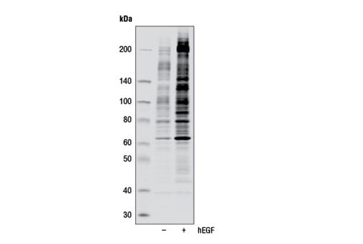
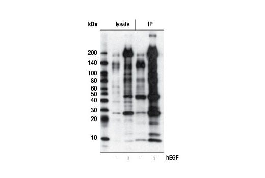
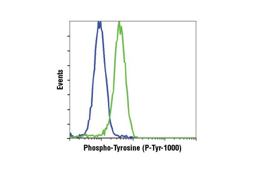
| Phospho-Tyrosine (P-Tyr-1000) MultiMab® Rabbit Monoclonal Antibody mix | 8954 | 20 µl | N/A kDa | Rabbit IgG |
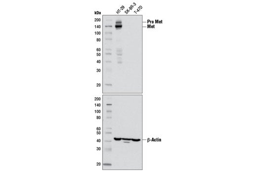
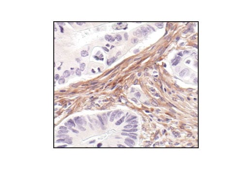
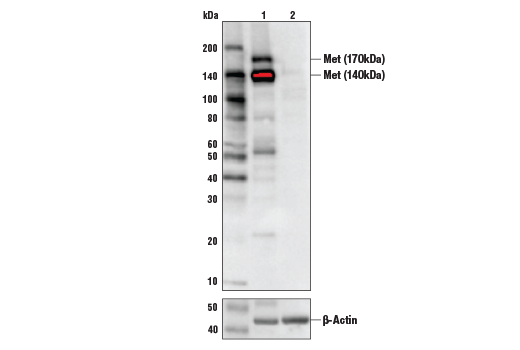
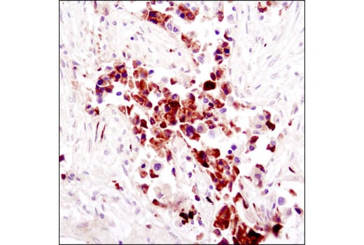
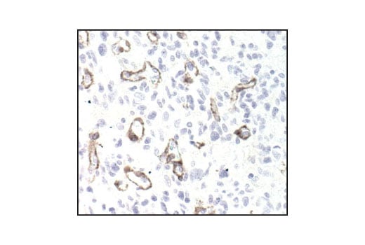
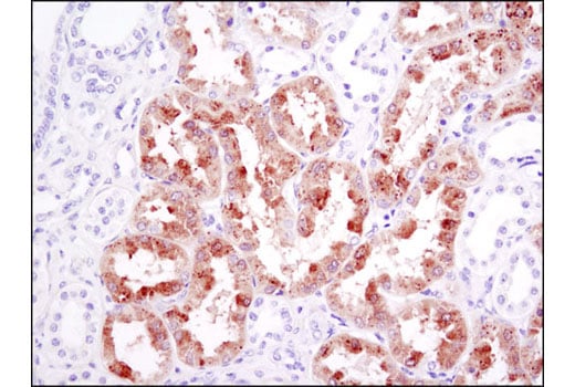
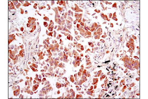
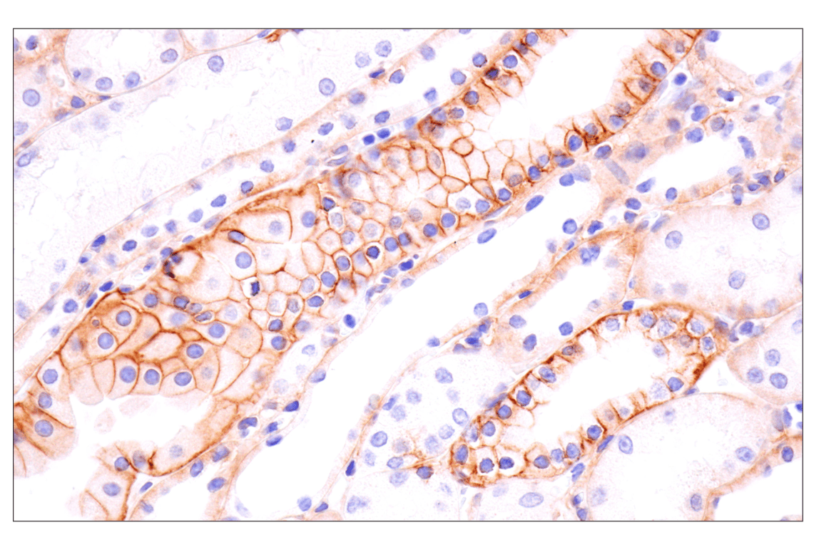
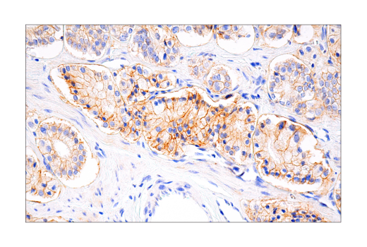
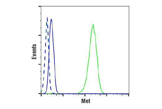
| Met (D1C2) Rabbit Monoclonal Antibody | 8198 | 20 µl | 140, 170 kDa | Rabbit IgG |
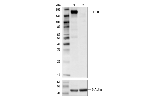
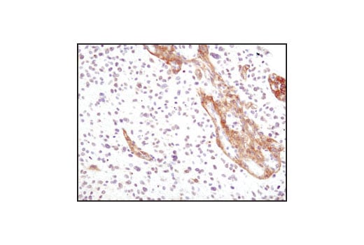
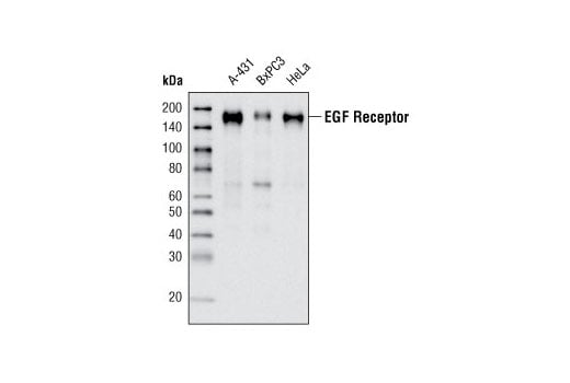
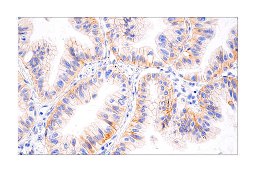
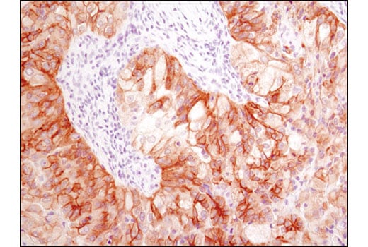
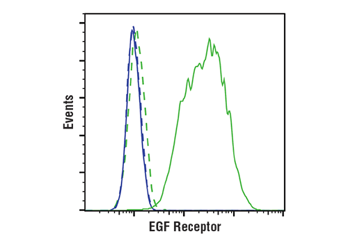
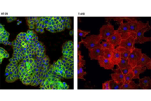
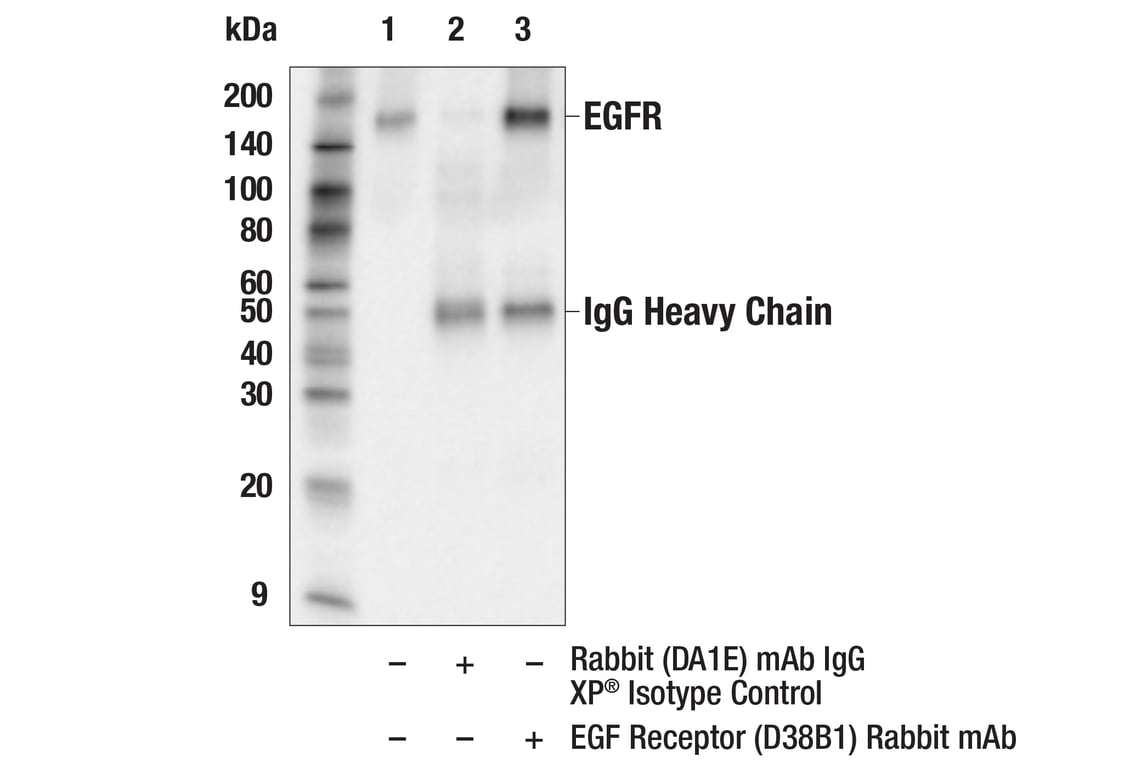
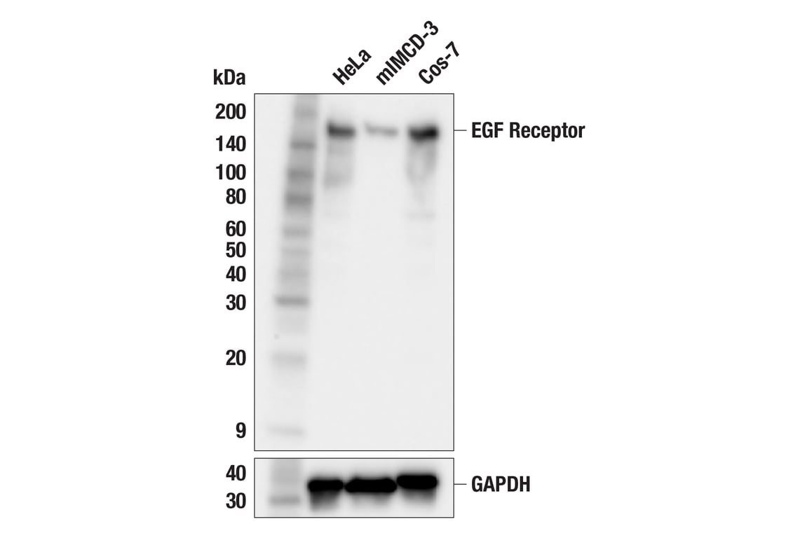
| EGF Receptor (D38B1) Rabbit Monoclonal Antibody | 4267 | 20 µl | 175 kDa | Rabbit IgG |
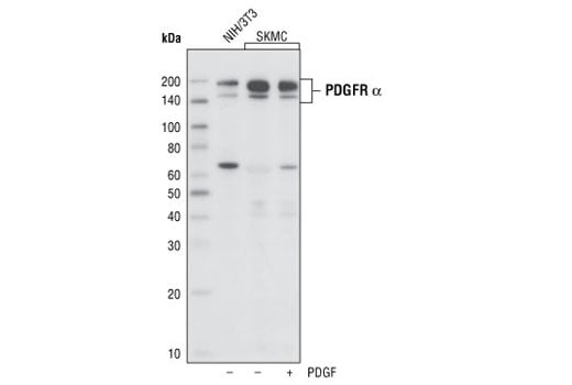
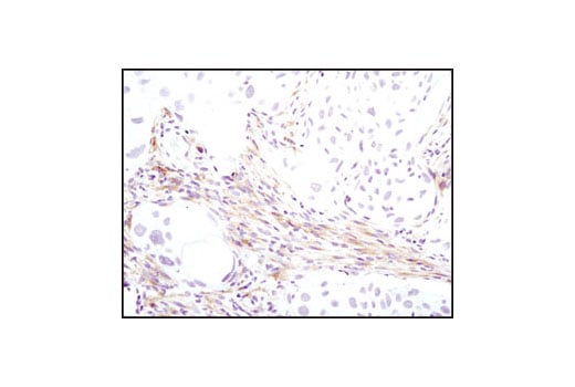
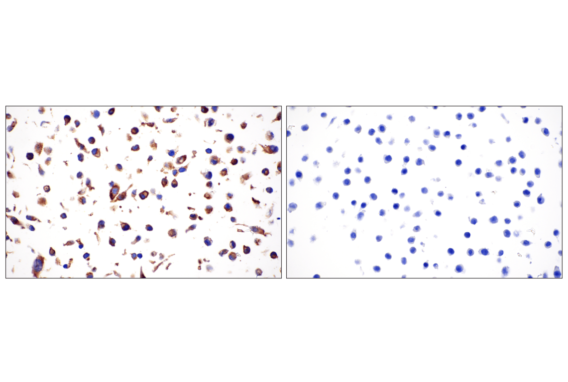
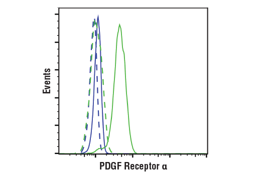
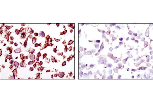
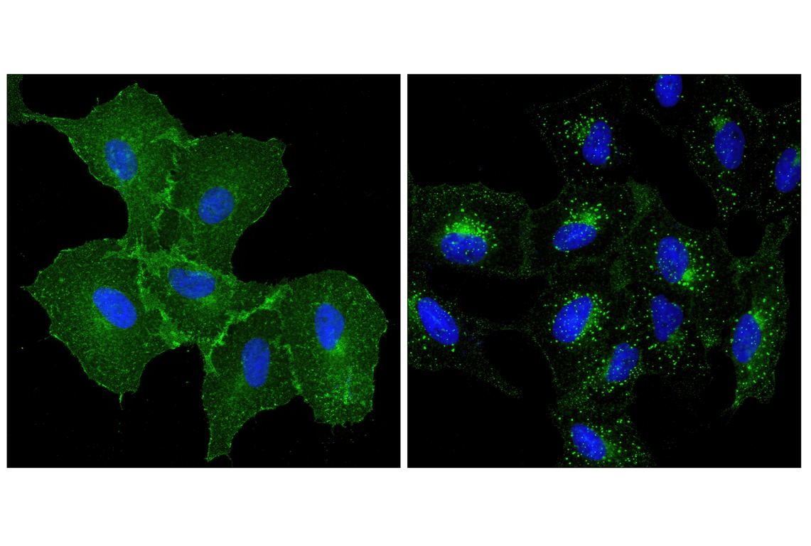
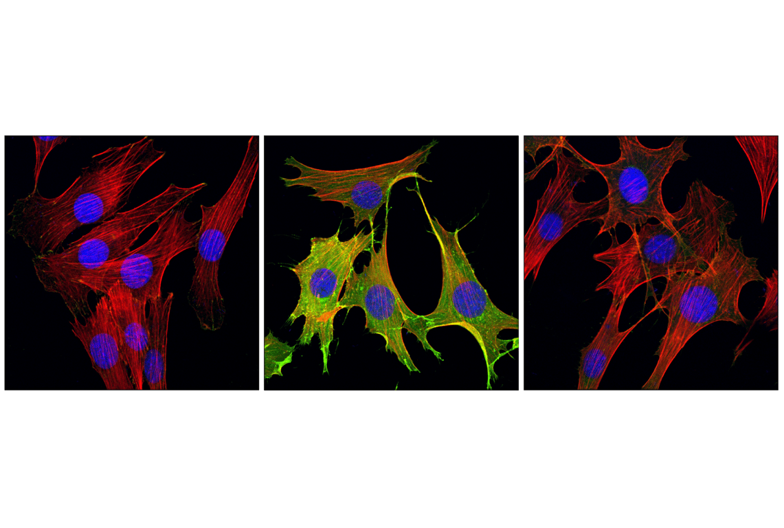
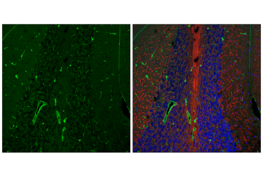
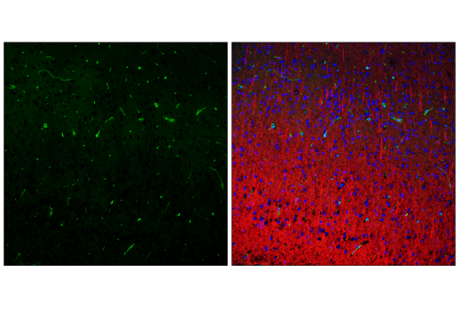
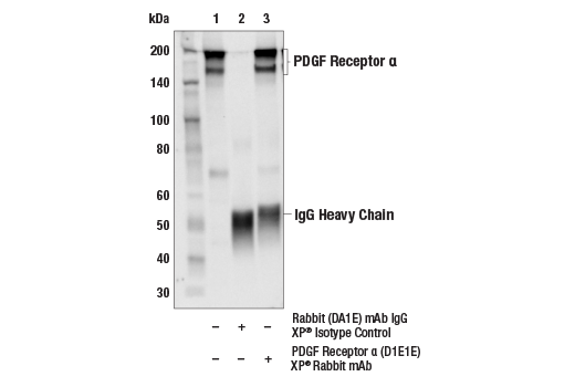
| PDGF Receptor alpha (D1E1E) Rabbit Monoclonal Antibody | 3174 | 20 µl | 190 kDa | Rabbit IgG |
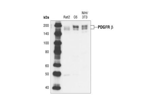
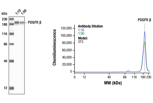
| PDGF Receptor beta (28E1) Rabbit Monoclonal Antibody | 3169 | 20 µl | 190 kDa | Rabbit IgG |
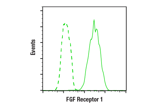
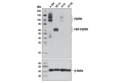
| FGF Receptor 1 (D8E4) Rabbit Monoclonal Antibody | 9740 | 20 µl | 92 , 120, 145 kDa | Rabbit IgG |
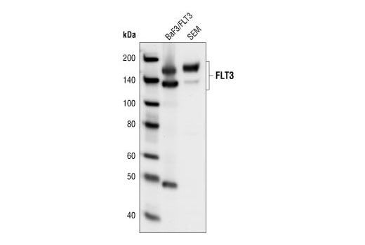
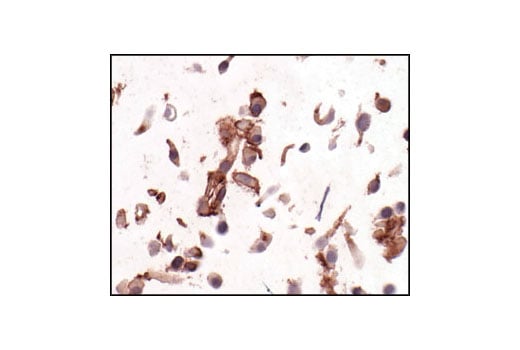
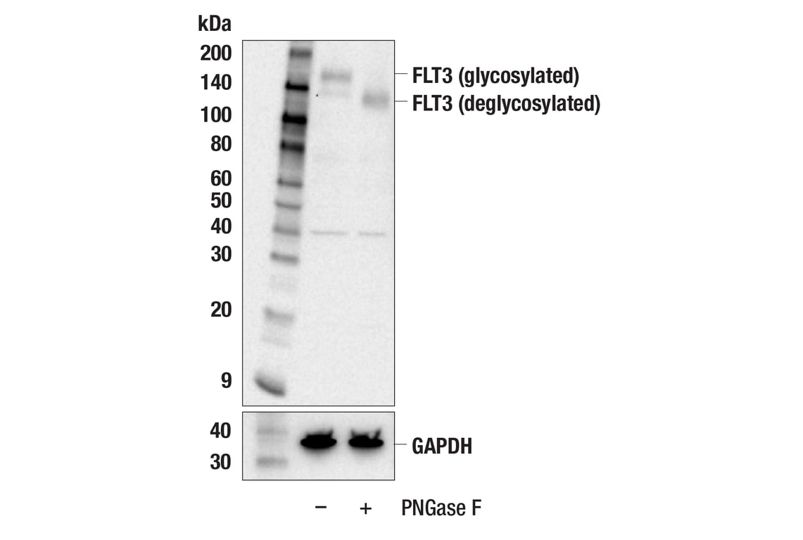
| FLT3 (8F2) Rabbit Monoclonal Antibody | 3462 | 20 µl | 130 nonglycosylated form;160 glycosylated mature form kDa | Rabbit IgG |
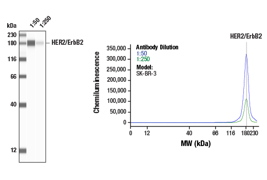
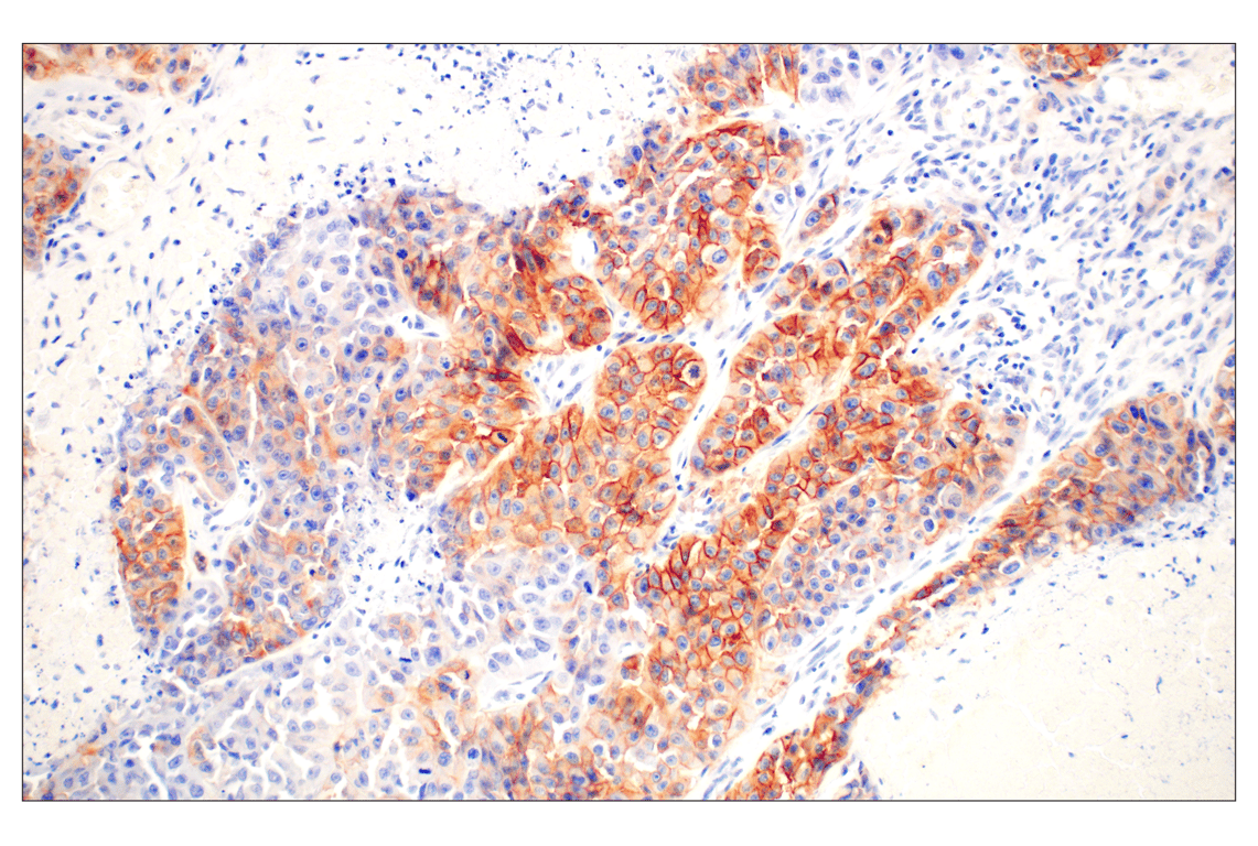
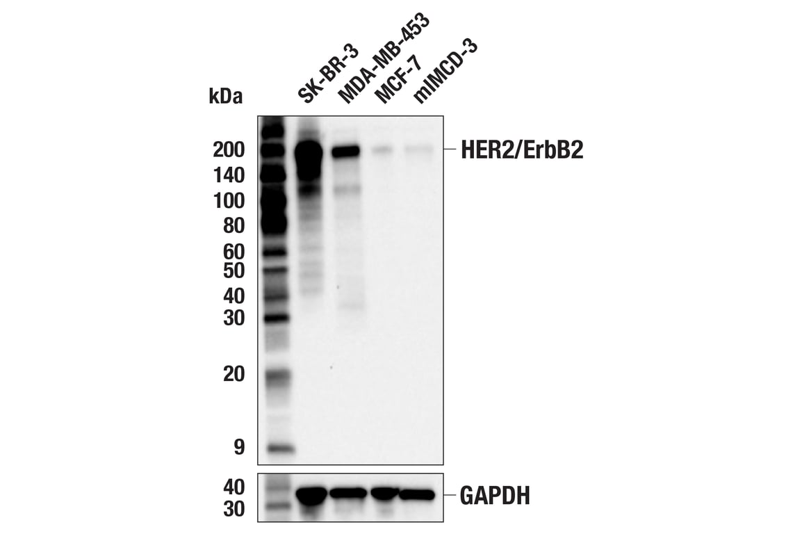
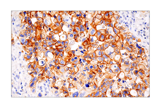
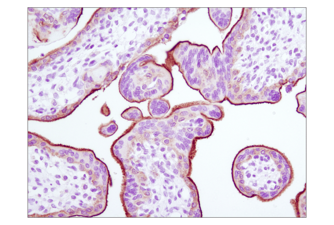
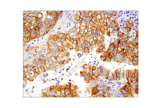
| HER2/ErbB2 (D8F12) Rabbit Monoclonal Antibody | 4290 | 20 µl | 185 kDa | Rabbit IgG |
| Anti-rabbit IgG, HRP-linked Antibody | 7074 | 100 µl | Goat |
Please visit cellsignal.com for individual component applications, species cross-reactivity, dilutions, protocols, and additional product information.
Description
Storage
Background
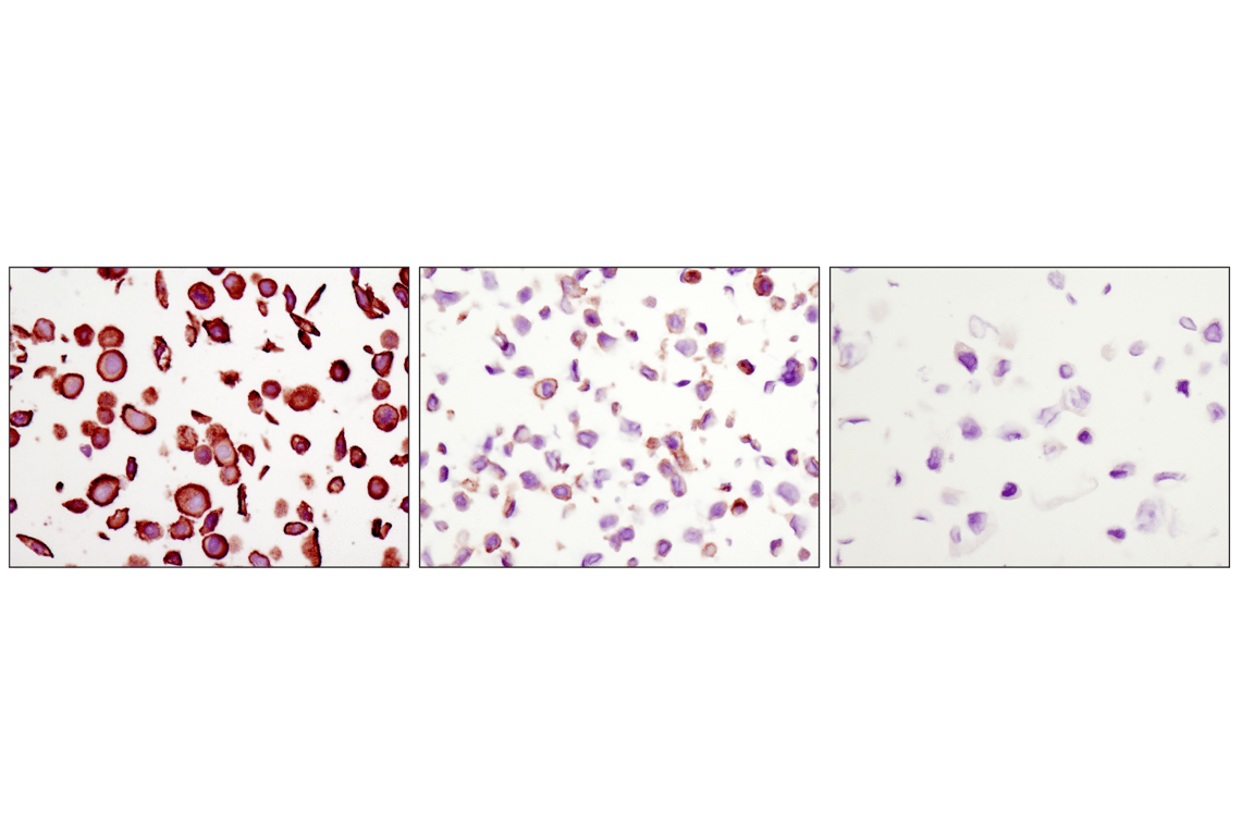
Met, a tyrosine kinase receptor for hepatocyte growth factor (HGF), is a heterodimer made of α- and β-subunits (5,6). The cytoplasmic region of the β-chain is essential for tyrosine kinase activity. Interaction of Met with HGF results in autophosphorylation at multiple tyrosines (Tyr1003, 1234/1235, 1349) which recruit downstream signaling components, including Gab1, c-Cbl, and PI3 kinase (7-9). Altered Met levels and/or tyrosine kinase activities are found in several types of tumors, including renal, colon, and breast (10,11).
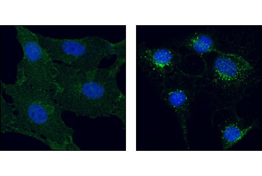
The epidermal growth factor (EGF) receptor is a transmembrane tyrosine kinase that belongs to the HER/ErbB protein family. Ligand binding results in receptor dimerization, autophosphorylation, activation of downstream signaling, internalization, and lysosomal degradation (12,13). c-Src mediated phosphorylation of EGF receptor (EGFR) at Tyr845 provides a binding surface for substrate proteins (14-16). The SH2 domain of PLCγ binds at phospho-Tyr992, activating PLCγ-mediated downstream signaling (17). Adaptor protein c-Cbl binds at phospho-Tyr1045, leading to receptor ubiquitination and degradation (18,19). The GRB2 adaptor protein binds activated EGFR at phospho-Tyr1068 (20), while phospho-Tyr1148 and -Tyr1173 provide a docking site for the Shc scaffold protein, playing a role in MAP kinase signaling (13).
Platelet derived growth factor (PDGF) family proteins bind to two closely related receptor tyrosine kinases, PDGF receptor α (PDGFRα) and PDGF receptor β (PDGFRβ) (21). PDGFRα and PDGFRβ can each form heterodimers with EGFR, which is also activated by PDGF (22). Ligand binding induces receptor dimerization and autophosphorylation, followed by binding and activation of signal transduction molecules such as GRB2, Src, GAP, PI3 kinase, PLCγ, and NCK. Signaling pathways initiated by activated PDGF receptors lead to control of cell growth, actin reorganization, migration, and differentiation (23). Tyr751 and Tyr740 of PDGFRβ regulate binding and activation of PI3 kinase (24,25).
Fibroblast growth factors (FGFs) produce mitogenic and angiogenic effects in target cells by signaling through cell surface receptor tyrosine kinases, after ligand binding and dimerization (26,27). Tyr653 and Tyr654 are important for catalytic activity of activated FGFR and are essential for signaling (28). The other phosphorylated tyrosine residues (Tyr463, 583, 585, 730, and 766) may provide docking sites for downstream signaling components such as Crk and PLCγ (29,30).
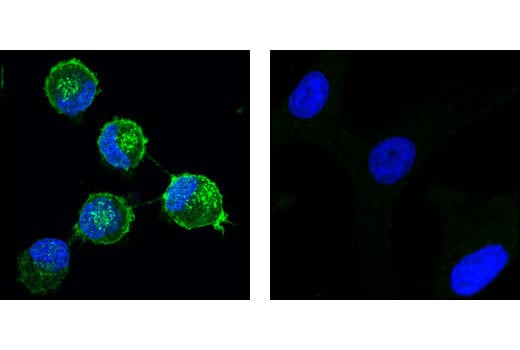
FMS-related tyrosine kinase 3 (FLT3), a member of the type III receptor tyrosine kinase family, is expressed on early hematopoietic progenitor cells and supports growth and differentiation within the hematopoietic system (31,32). FLT3 is activated after binding with its ligand FL, which results in a cascade of tyrosine autophosphorylation and tyrosine phosphorylation of downstream targets (33). The p85 subunit of PI3 kinase, SHP2, GRB2 and Shc are associated with FLT3 after FL stimulation (34-36). Tyr589/591 may play an important role in regulation of FLT3 tyrosine kinase activity (37).
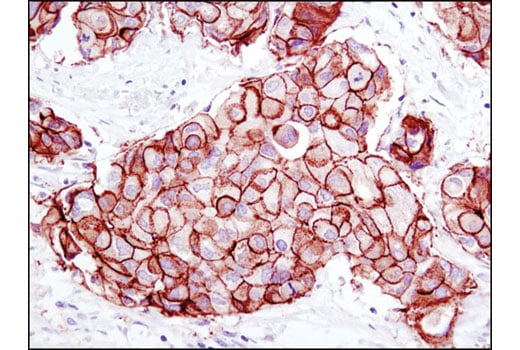
The ErbB2 (HER2) proto-oncogene encodes a transmembrane, receptor-like glycoprotein with tyrosine kinase activity (38). ErbB2 kinase activity can be activated in the absence of a ligand when overexpressed and through associations with other ErbB family members (39). Phosphorylation at Tyr877 may be involved in regulating ErbB2 activity. Autophosphorylation of ErbB2 at Tyr1248 and Tyr1221/1222 couples ErbB2 to the Ras-Raf-MAP kinase signal transduction pathway (38,40).
Background References
- Schlessinger, J. (2000) Cell 103, 211-25
- Blume-Jensen, P. and Hunter, T. (2001) Nature 411, 355-65
- Ward, S.G. et al. (1992) J Biol Chem 267, 23862-9
- Glenney, J.R. et al. (1988) J Immunol Methods 109, 277-85
- Cooper, C.S. et al. Nature 311, 29-33.
- Bottaro, D.P. et al. (1991) Science 251, 802-4.
- Bardelli, A. et al. (1997) Oncogene 15, 3103-11.
- Taher, T.E. et al. (2002) J Immunol 169, 3793-800.
- Schaeper, U. et al. (2000) J Cell Biol 149, 1419-32.
- Eder, J.P. et al. (2009) Clin Cancer Res 15, 2207-14.
- Sattler, M. and Salgia, R. (2009) Update Cancer Ther 3, 109-118.
- Hackel, P.O. et al. (1999) Curr Opin Cell Biol 11, 184-9.
- Zwick, E. et al. (1999) Trends Pharmacol Sci 20, 408-12.
- Cooper, J.A. and Howell, B. (1993) Cell 73, 1051-4.
- Hubbard, S.R. et al. Nature 372, 746-54.
- Biscardi, J.S. et al. (1999) J Biol Chem 274, 8335-43.
- Emlet, D.R. et al. (1997) J Biol Chem 272, 4079-86.
- Levkowitz, G. et al. (1999) Mol Cell 4, 1029-40.
- Ettenberg, S.A. et al. (1999) Oncogene 18, 1855-66.
- Rojas, M. et al. (1996) J Biol Chem 271, 27456-61.
- Deuel, T.F. et al. (1988) Biofactors 1, 213-7.
- Betsholtz, C. et al. (2001) Bioessays 23, 494-507.
- Ostman, A. and Heldin, C.H. (2001) Adv Cancer Res 80, 1-38.
- Panayotou, G. et al. (1992) EMBO J 11, 4261-72.
- Kashishian, A. et al. (1992) EMBO J 11, 1373-82.
- Powers, C.J. et al. (2000) Endocr Relat Cancer 7, 165-97.
- Reilly, J.F. et al. (2000) J Biol Chem 275, 7771-8.
- Mohammadi, M. et al. (1996) Mol Cell Biol 16, 977-89.
- Mohammadi, M. et al. (1991) Mol Cell Biol 11, 5068-78.
- Larsson, H. et al. (1999) J Biol Chem 274, 25726-34.
- Shurin, M.R. et al. (1998) Cytokine Growth Factor Rev 9, 37-48.
- Naoe, T. et al. (2001) Cancer Chemother Pharmacol 48 Suppl 1, S27-30.
- Namikawa, R. et al. (1996) Stem Cells 14, 388-95.
- Beslu, N. et al. (1996) J Biol Chem 271, 20075-81.
- Zhang, S. and Broxmeyer, H.E. (2000) Biochem Biophys Res Commun 277, 195-9.
- Zhang, S. et al. (1999) J Leukoc Biol 65, 372-80.
- Mizuki, M. et al. (2000) Blood 96, 3907-14.
- Muthuswamy, S.K. et al. (1999) Mol Cell Biol 19, 6845-57.
- Qian, X. et al. (1994) Proc Natl Acad Sci U S A 91, 1500-4.
- Kwon, Y.K. et al. (1997) J Neurosci 17, 8293-9.
Trademarks and Patents
Cell Signaling Technology is a trademark of Cell Signaling Technology, Inc.
MultiMab is a registered trademark of Cell Signaling Technology, Inc.
All other trademarks are the property of their respective owners. Visit cellsignal.com/trademarks for more information.
Limited Uses
Except as otherwise expressly agreed in a writing signed by a legally authorized representative of CST, the following terms apply to Products provided by CST, its affiliates or its distributors. Any Customer's terms and conditions that are in addition to, or different from, those contained herein, unless separately accepted in writing by a legally authorized representative of CST, are rejected and are of no force or effect.
Products are labeled with For Research Use Only or a similar labeling statement and have not been approved, cleared, or licensed by the FDA or other regulatory foreign or domestic entity, for any purpose. Customer shall not use any Product for any diagnostic or therapeutic purpose, or otherwise in any manner that conflicts with its labeling statement. Products sold or licensed by CST are provided for Customer as the end-user and solely for research and development uses. Any use of Product for diagnostic, prophylactic or therapeutic purposes, or any purchase of Product for resale (alone or as a component) or other commercial purpose, requires a separate license from CST. Customer shall (a) not sell, license, loan, donate or otherwise transfer or make available any Product to any third party, whether alone or in combination with other materials, or use the Products to manufacture any commercial products, (b) not copy, modify, reverse engineer, decompile, disassemble or otherwise attempt to discover the underlying structure or technology of the Products, or use the Products for the purpose of developing any products or services that would compete with CST products or services, (c) not alter or remove from the Products any trademarks, trade names, logos, patent or copyright notices or markings, (d) use the Products solely in accordance with CST Product Terms of Sale and any applicable documentation, and (e) comply with any license, terms of service or similar agreement with respect to any third party products or services used by Customer in connection with the Products.
Revision 1
Revision 1
Revision 1

Revision 1
Revision 1
Revision 1
Revision 1
Revision 1
Revision 1

Revision 1
Revision 1
Revision 1
Revision 1
Revision 1
Revision 1
Revision 1

Revision 1
Revision 1
Revision 1
Revision 1

Revision 1