| Cat. # | Size | Qty. | Price |
|---|---|---|---|
| 4102S | 100 µl |
|
| REACTIVITY | H M |
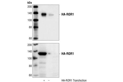
| SENSITIVITY | Transfected Only |
| MW (kDa) | 135 |
| SOURCE | Rabbit |
Product Information
| Application | Dilution |
|---|---|
| Western Blotting | 1:1000 |
For western blots, incubate membrane with diluted primary antibody in 5% w/v BSA, 1X TBS, 0.1% Tween® 20 at 4°C with gentle shaking, overnight.
NOTE: Please refer to primary antibody product webpage for recommended antibody dilution.
From sample preparation to detection, the reagents you need for your Western Blot are now in one convenient kit: #12957 Western Blotting Application Solutions Kit
NOTE: Prepare solutions with reverse osmosis deionized (RODI) or equivalent grade water.
Load 20 µl onto SDS-PAGE gel (10 cm x 10 cm).
NOTE: Loading of prestained molecular weight markers (#59329, 10 µl/lane) to verify electrotransfer and biotinylated protein ladder (#7727, 10 µl/lane) to determine molecular weights are recommended.
NOTE: Volumes are for 10 cm x 10 cm (100 cm2) of membrane; for different sized membranes, adjust volumes accordingly.
* Avoid repeated exposure to skin.
posted June 2005
revised June 2020
Protocol Id: 10
Human, Mouse
Human
Polyclonal antibodies are produced by immunizing animals with a synthetic peptide corresponding to the residues surrounding Pro87 of human ROR1 protein. The antibodies are purified by protein A and peptide affinity chromatography.
ROR1 and ROR2 are orphan receptor tyrosine kinases that are most closely related to MuSK and the Trk family of neurotrophin receptors. They are characterized by the presence of extracellular frizzled-like cysteine-rich domains and membrane-proximal kringle domains, both of which are assumed to mediate protein-protein interactions (1-3). The ROR family RTKs are evolutionarily conserved among Caenorhabditis elegans, Drosophila, mice, and humans (1,4). Although the functions of ROR kinases are unknown, similarities between ROR and MuSK and Trk kinases have led to speculation that ROR kinases regulate synaptic development. CAM-1, a C. elegans ortholog of the ROR family RTKs, plays several important roles in regulating cellular migration, polarity of asymmetric cell divisions, and axonal outgrowth of neurons during nematode development (4). mROR1 and mROR2 may play differential roles during the development of the nervous system (5).
Explore pathways related to this product.
STRING - Known and Predicted Protein-Protein Interactions.
Except as otherwise expressly agreed in a writing signed by a legally authorized representative of CST, the following terms apply to Products provided by CST, its affiliates or its distributors. Any Customer's terms and conditions that are in addition to, or different from, those contained herein, unless separately accepted in writing by a legally authorized representative of CST, are rejected and are of no force or effect.
Products are labeled with For Research Use Only or a similar labeling statement and have not been approved, cleared, or licensed by the FDA or other regulatory foreign or domestic entity, for any purpose. Customer shall not use any Product for any diagnostic or therapeutic purpose, or otherwise in any manner that conflicts with its labeling statement. Products sold or licensed by CST are provided for Customer as the end-user and solely for research and development uses. Any use of Product for diagnostic, prophylactic or therapeutic purposes, or any purchase of Product for resale (alone or as a component) or other commercial purpose, requires a separate license from CST. Customer shall (a) not sell, license, loan, donate or otherwise transfer or make available any Product to any third party, whether alone or in combination with other materials, or use the Products to manufacture any commercial products, (b) not copy, modify, reverse engineer, decompile, disassemble or otherwise attempt to discover the underlying structure or technology of the Products, or use the Products for the purpose of developing any products or services that would compete with CST products or services, (c) not alter or remove from the Products any trademarks, trade names, logos, patent or copyright notices or markings, (d) use the Products solely in accordance with CST Product Terms of Sale and any applicable documentation, and (e) comply with any license, terms of service or similar agreement with respect to any third party products or services used by Customer in connection with the Products.