Revision 1
#54020
Store at -20C
Rpb1 CTD Antibody Sampler Kit
1 Kit
(5 x 20 microliters)
877-616-CELL (2355)
877-678-TECH (8324)
3 Trask Lane | Danvers | Massachusetts | 01923 | USA
For Research Use Only. Not for Use in Diagnostic Procedures.
| Product Includes | Product # | Quantity | Mol. Wt | Isotype/Source |
|---|---|---|---|---|
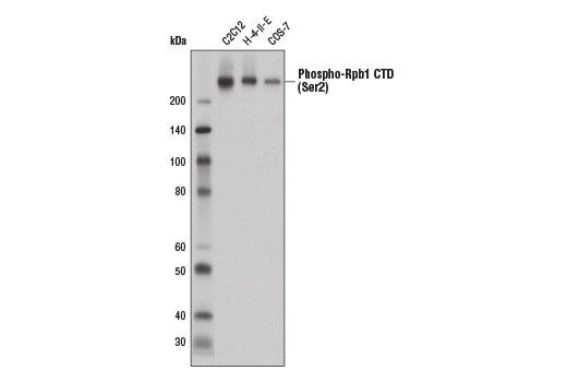
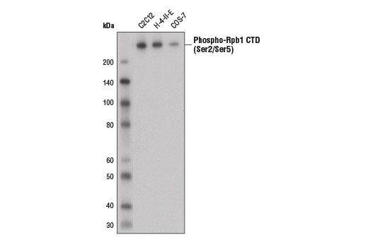
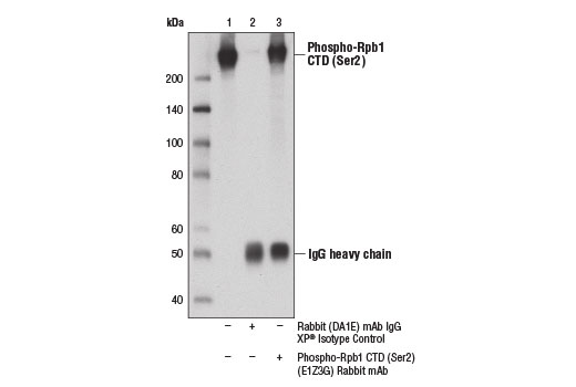
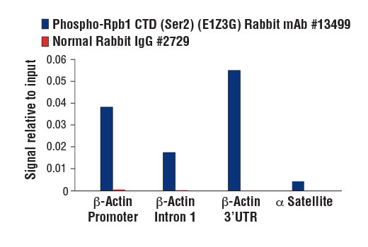
| Phospho-Rpb1 CTD (Ser2) (E1Z3G) Rabbit Monoclonal Antibody | 13499 | 20 µl | 250 kDa | Rabbit IgG |
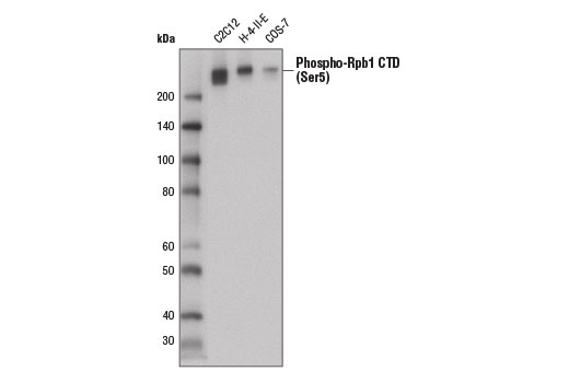
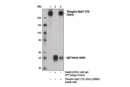
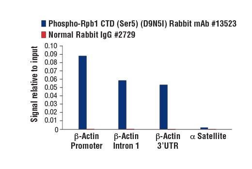
| Phospho-Rpb1 CTD (Ser5) (D9N5I) Rabbit Monoclonal Antibody | 13523 | 20 µl | 250 kDa | Rabbit IgG |
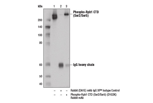
| Phospho-Rpb1 CTD (Ser2/Ser5) (D1G3K) Rabbit Monoclonal Antibody | 13546 | 20 µl | 250 kDa | Rabbit IgG |
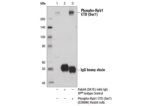
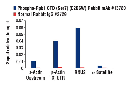
| Phospho-Rpb1 CTD (Ser7) (E2B6W) Rabbit Monoclonal Antibody | 13780 | 20 µl | 250 kDa | Rabbit IgG |
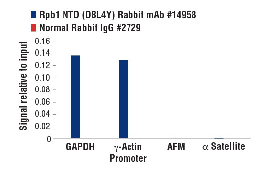
| Rpb1 NTD (D8L4Y) Rabbit Monoclonal Antibody | 14958 | 20 µl | 250 kDa | Rabbit IgG |
| Anti-rabbit IgG, HRP-linked Antibody | 7074 | 100 µl | Goat |
Please visit cellsignal.com for individual component applications, species cross-reactivity, dilutions, protocols, and additional product information.
Description
Storage
Background
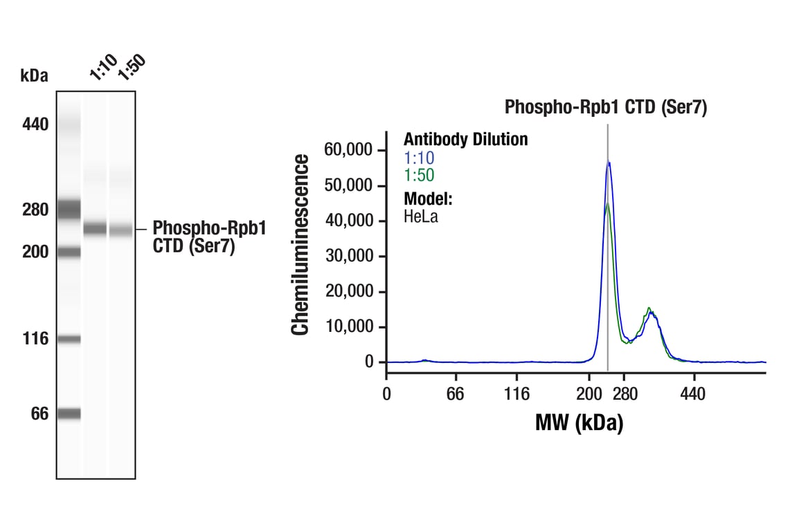
In addition to Ser2/Ser5 phosphorylation, Ser7 of the CTD heptapeptide repeat is also phosphorylated during the active transcription cycle. Phosphorylation at Ser7 is required for efficient transcription of small nuclear (sn) RNA genes (9,10). snRNA genes, which are neither spliced nor poly-adenylated, are structurally different from protein-coding genes. Instead of a poly(A) signal found in protein-coding RNAs, snRNAs contain a conserved 3'-box RNA processing element, which is recognized by the Integrator snRNA 3' end processing complex (11,12). Phosphorylation at Ser7 by CDK7 during the early stages of transcription facilitates recruitment of RPAP2, which dephosphorylates Ser5, creating a dual Ser2/Ser7 phosphorylation mark that facilitates recruitment of the Integrator complex and efficient processing of nascent snRNA transcripts (13-15).
Background References
- Brookes, E. and Pombo, A. (2009) EMBO Rep 10, 1213-9.
- Komarnitsky, P. et al. (2000) Genes Dev 14, 2452-60.
- Ho, C.K. and Shuman, S. (1999) Mol Cell 3, 405-11.
- Ng, H.H. et al. (2003) Mol Cell 11, 709-19.
- Cheng, B. and Price, D.H. (2007) J Biol Chem 282, 21901-12.
- Marshall, N.F. et al. (1996) J Biol Chem 271, 27176-83.
- Krogan, N.J. et al. (2003) Mol Cell Biol 23, 4207-18.
- Proudfoot, N.J. et al. (2002) Cell 108, 501-12.
- Chapman, R.D. et al. (2007) Science 318, 1780-2.
- Egloff, S. et al. (2007) Science 318, 1777-9.
- Egloff, S. et al. (2008) Biochem Soc Trans 36, 590-4.
- Baillat, D. et al. (2005) Cell 123, 265-76.
- Akhtar, M.S. et al. (2009) Mol Cell 34, 387-93.
- Egloff, S. et al. (2010) J Biol Chem 285, 20564-9.
- Egloff, S. et al. (2012) Mol Cell 45, 111-22.
Trademarks and Patents
Cell Signaling Technology is a trademark of Cell Signaling Technology, Inc.
All other trademarks are the property of their respective owners. Visit cellsignal.com/trademarks for more information.
Limited Uses
Except as otherwise expressly agreed in a writing signed by a legally authorized representative of CST, the following terms apply to Products provided by CST, its affiliates or its distributors. Any Customer's terms and conditions that are in addition to, or different from, those contained herein, unless separately accepted in writing by a legally authorized representative of CST, are rejected and are of no force or effect.
Products are labeled with For Research Use Only or a similar labeling statement and have not been approved, cleared, or licensed by the FDA or other regulatory foreign or domestic entity, for any purpose. Customer shall not use any Product for any diagnostic or therapeutic purpose, or otherwise in any manner that conflicts with its labeling statement. Products sold or licensed by CST are provided for Customer as the end-user and solely for research and development uses. Any use of Product for diagnostic, prophylactic or therapeutic purposes, or any purchase of Product for resale (alone or as a component) or other commercial purpose, requires a separate license from CST. Customer shall (a) not sell, license, loan, donate or otherwise transfer or make available any Product to any third party, whether alone or in combination with other materials, or use the Products to manufacture any commercial products, (b) not copy, modify, reverse engineer, decompile, disassemble or otherwise attempt to discover the underlying structure or technology of the Products, or use the Products for the purpose of developing any products or services that would compete with CST products or services, (c) not alter or remove from the Products any trademarks, trade names, logos, patent or copyright notices or markings, (d) use the Products solely in accordance with CST Product Terms of Sale and any applicable documentation, and (e) comply with any license, terms of service or similar agreement with respect to any third party products or services used by Customer in connection with the Products.
Revision 1
Revision 1
Revision 1
Revision 1
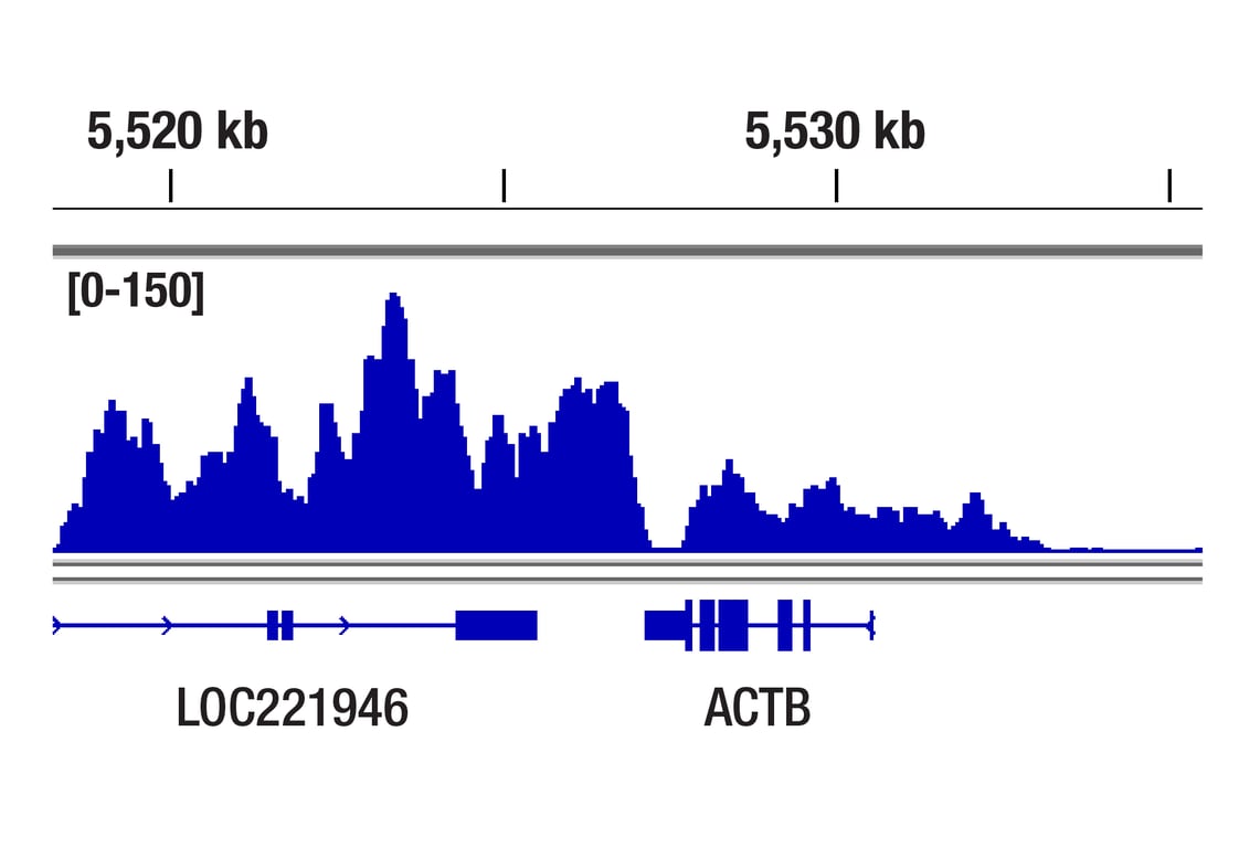
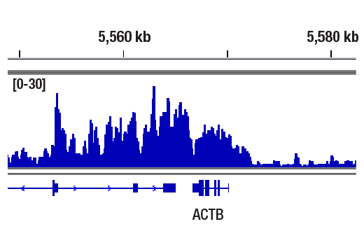
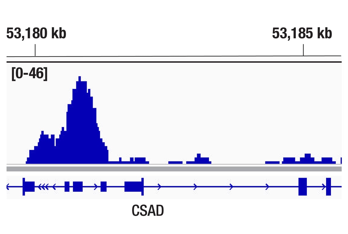
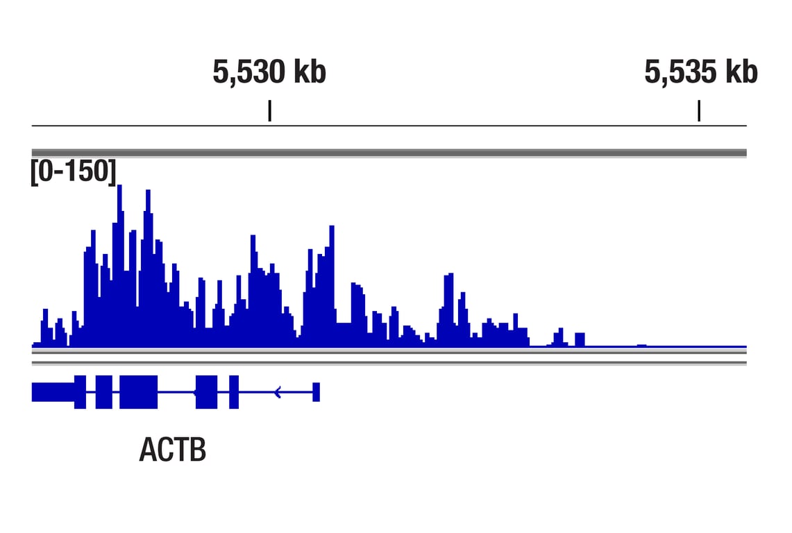
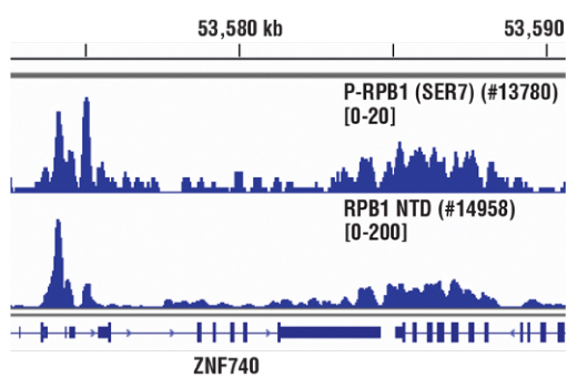
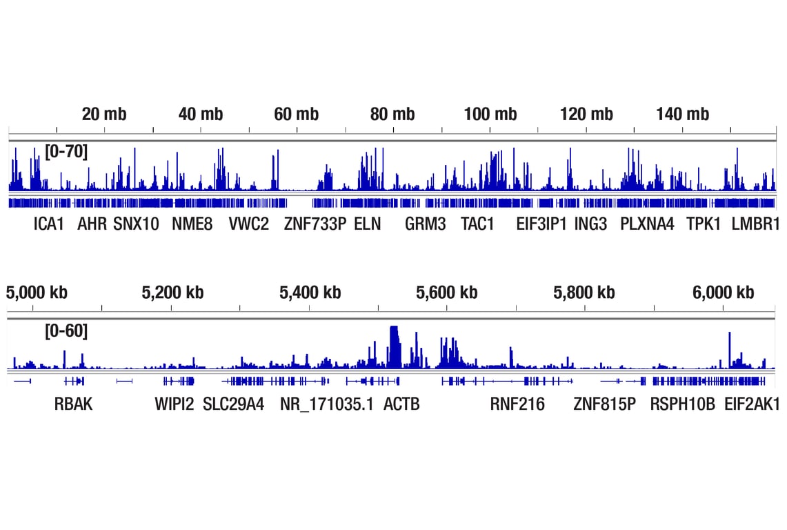
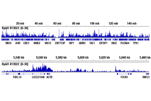
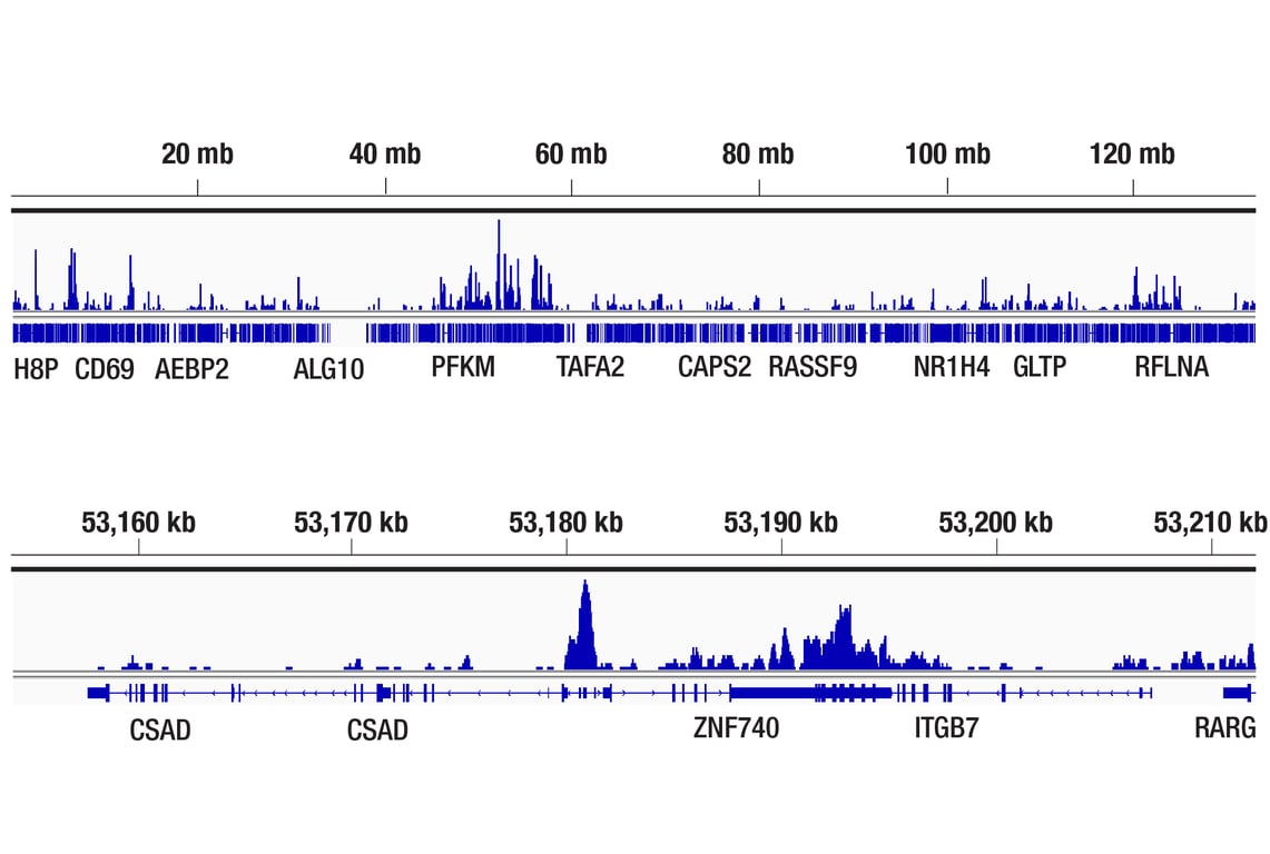
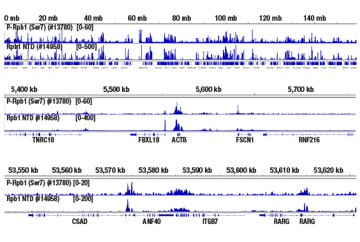
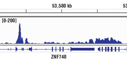
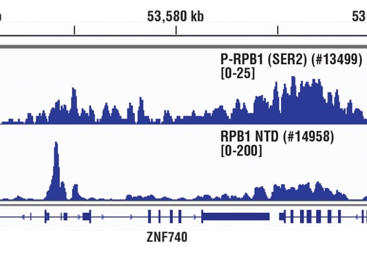
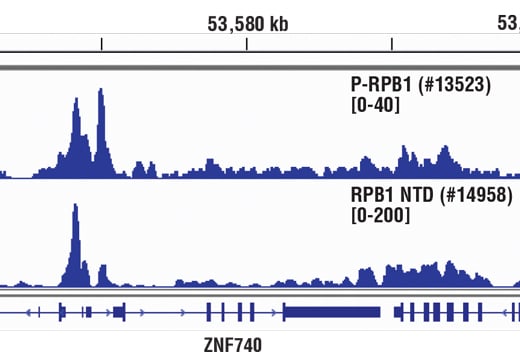
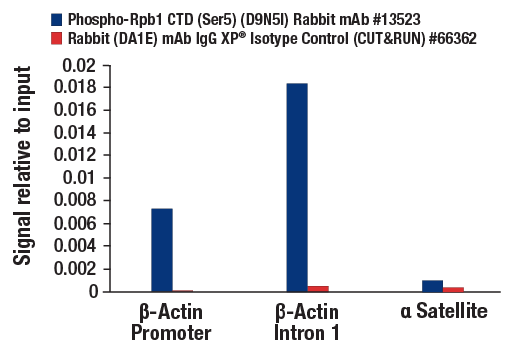
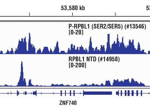
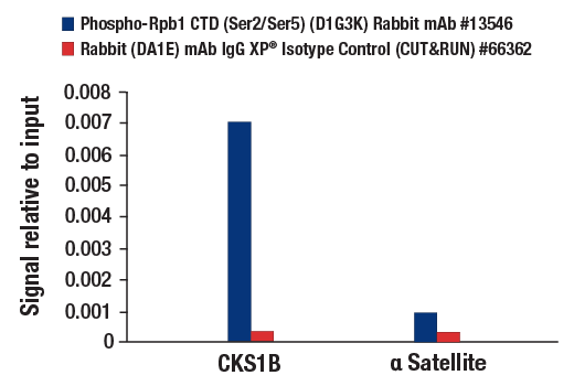
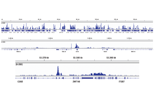
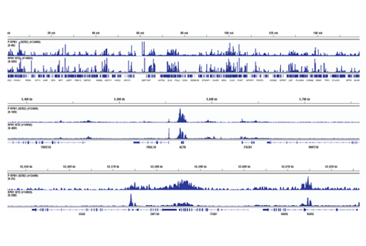
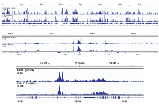
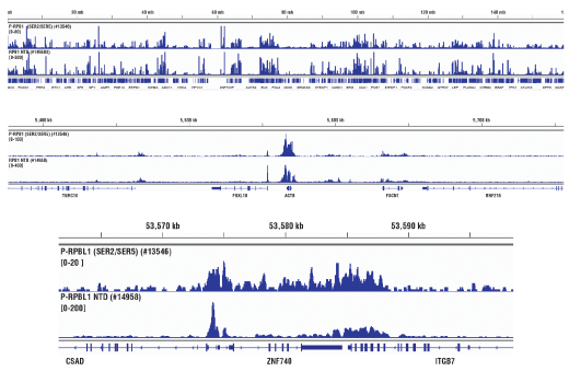
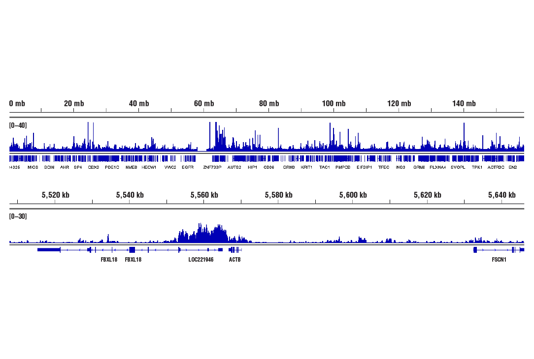
Chromatin immunoprecipitations were performed with cross-linked chromatin from Hela cells and either Phospho-Rpb1 CTD (Ser7) (E2B6W) Rabbit mAb or Rpb1 NTD (D8L4Y) Rabbit mAb #14958, using SimpleChIP® Enzymatic Chromatin IP Kit (Magnetic Beads) #9003. DNA Libraries were prepared using SimpleChIP® ChIP-seq DNA Library Prep Kit for Illumina® #56795. The figure shows binding across the ZNF740 gene on chromosome 12. For additional ChIP-seq tracks, please download the product datasheet.

Revision 1
Revision 1
Revision 1
Revision 1
Revision 1
Revision 1
Revision 1
Revision 1
Revision 1
Revision 1
Revision 1
Revision 1