Revision 5
#5528
Store at -20C
877-616-CELL (2355)
877-678-TECH (8324)
3 Trask Lane | Danvers | Massachusetts | 01923 | USA
For Research Use Only. Not for Use in Diagnostic Procedures.
Applications:
W, W-S, IHC-P, IF-IC, FC-FP
Reactivity:
H M R Mk B Pg
Sensitivity:
Endogenous
MW (kDa):
90
Source/Isotype:
Rabbit IgG
UniProt ID:
#P51812
Entrez-Gene Id:
6197
Product Usage Information
| Application | Dilution |
|---|---|
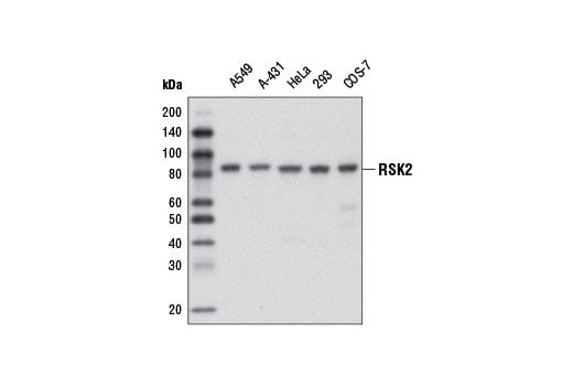
| Western Blotting | 1:1000 |
| Simple Western™ | 1:10 - 1:50 |
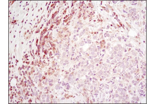
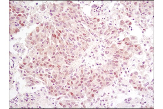
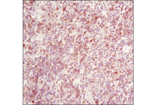
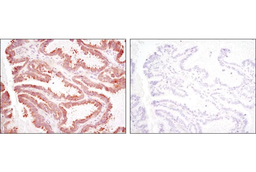
| Immunohistochemistry (Paraffin) | 1:800 - 1:3200 |
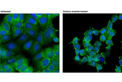
| Immunofluorescence (Immunocytochemistry) | 1:200 - 1:400 |
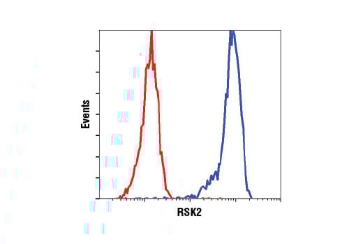
| Flow Cytometry (Fixed/Permeabilized) | 1:50 - 1:200 |
Storage
For a carrier free (BSA and azide free) version of this product see product #99402.
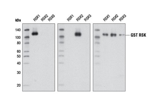
Specificity/Sensitivity
Species predicted to react based on 100% sequence homology
Source / Purification
Background
Stimulation by various growth factors leads to activation of RSK2, which is a critical downstream effector kinase in several pathways. EGF stimulation leads to phosphorylation of CREB at Ser133 and phosphorylation of histone H3 in vivo by RSK2 (4,5). RSK2 phosphorylation of p53 may help regulate chromatin structure and cell cycle (6). RSK2 is prominently expressed in the brain and is essential for cognitive function and learning. During development, RSK2 regulates the differentiation of osteoblasts and skeletal muscle cells (7,8). Mutations in the corresponding gene are associated with Coffin-Lowry syndrome (CLS), an X-linked disorder characterized by mental retardation and the presence of characteristic facial anomalies (9).
Background References
- Fisher, T.L. and Blenis, J. (1996) Mol Cell Biol 16, 1212-9.
- Smith, J.A. et al. (1999) J Biol Chem 274, 2893-8.
- Dalby, K.N. et al. (1998) J Biol Chem 273, 1496-505.
- De Cesare, D. et al. (1998) Proc Natl Acad Sci U S A 95, 12202-7.
- Sassone-Corsi, P. et al. (1999) Science 285, 886-91.
- Cho, Y.Y. et al. (2005) Cancer Res 65, 3596-603.
- Yang, X. et al. (2004) Cell 117, 387-98.
- Cho, Y.Y. et al. (2007) J Biol Chem 282, 8380-92.
- Delaunoy, J.P. et al. (2006) Clin Genet 70, 161-6.
Species Reactivity
Species reactivity is determined by testing in at least one approved application (e.g., western blot).
Western Blot Buffer
IMPORTANT: For western blots, incubate membrane with diluted primary antibody in 5% w/v BSA, 1X TBS, 0.1% Tween® 20 at 4°C with gentle shaking, overnight.
Applications Key
W: Western Blotting W-S: Simple Western™ IHC-P: Immunohistochemistry (Paraffin) IF-IC: Immunofluorescence (Immunocytochemistry) FC-FP: Flow Cytometry (Fixed/Permeabilized)
Cross-Reactivity Key
H: Human M: Mouse R: Rat Mk: Monkey B: Bovine Pg: Pig
Trademarks and Patents
Cell Signaling Technology is a trademark of Cell Signaling Technology, Inc.
All other trademarks are the property of their respective owners. Visit cellsignal.com/trademarks for more information.
Limited Uses
Except as otherwise expressly agreed in a writing signed by a legally authorized representative of CST, the following terms apply to Products provided by CST, its affiliates or its distributors. Any Customer's terms and conditions that are in addition to, or different from, those contained herein, unless separately accepted in writing by a legally authorized representative of CST, are rejected and are of no force or effect.
Products are labeled with For Research Use Only or a similar labeling statement and have not been approved, cleared, or licensed by the FDA or other regulatory foreign or domestic entity, for any purpose. Customer shall not use any Product for any diagnostic or therapeutic purpose, or otherwise in any manner that conflicts with its labeling statement. Products sold or licensed by CST are provided for Customer as the end-user and solely for research and development uses. Any use of Product for diagnostic, prophylactic or therapeutic purposes, or any purchase of Product for resale (alone or as a component) or other commercial purpose, requires a separate license from CST. Customer shall (a) not sell, license, loan, donate or otherwise transfer or make available any Product to any third party, whether alone or in combination with other materials, or use the Products to manufacture any commercial products, (b) not copy, modify, reverse engineer, decompile, disassemble or otherwise attempt to discover the underlying structure or technology of the Products, or use the Products for the purpose of developing any products or services that would compete with CST products or services, (c) not alter or remove from the Products any trademarks, trade names, logos, patent or copyright notices or markings, (d) use the Products solely in accordance with CST Product Terms of Sale and any applicable documentation, and (e) comply with any license, terms of service or similar agreement with respect to any third party products or services used by Customer in connection with the Products.
Revision 5
Simple Western™ analysis of lysates (0.1 mg/mL) from COS-7 untreated cells using RSK2 (D21B2) XP® Rabbit mAb #5528. The virtual lane view (left) shows a single target band (as indicated) at 1:10 and 1:50 dilutions of primary antibody. The corresponding electropherogram view (right) plots chemiluminescence by molecular weight along the capillary at 1:10 (blue line) and 1:50 (green line) dilutions of primary antibody. This experiment was performed under reducing conditions on the Jess™ Simple Western instrument from ProteinSimple, a BioTechne brand, using the 12-230 kDa separation module.

Revision 5
Revision 5
Revision 5