WB, IP, ChIP
H M R
Endogenous
53
Rabbit IgG
#P19793
6256
Product Information
Product Usage Information
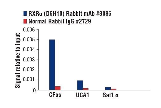
For optimal ChIP results, use 10 μl of antibody and 10 μg of chromatin (approximately 4 x 10⁶ cells) per IP. This antibody has been validated using SimpleChIP® Enzymatic Chromatin IP Kits.
| Application | Dilution |
|---|---|
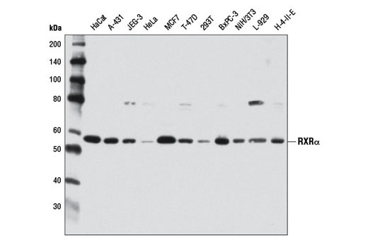
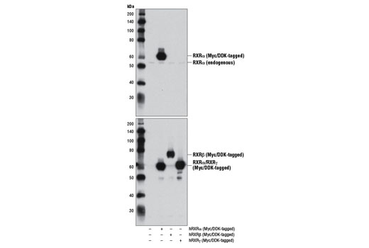
| Western Blotting | 1:1000 |
| Immunoprecipitation | 1:100 |
| Chromatin IP | 1:50 |
Storage
Specificity / Sensitivity
Species Reactivity:
Human, Mouse, Rat
Source / Purification
Monoclonal antibody is produced by immunizing animals with a synthetic peptide corresponding to residues near the amino terminus of human RXRα protein.
Background
The human retinoid X receptors (RXRs) are encoded by three distinct genes (RXRα, RXRβ, and RXRγ) and bind selectively and with high affinity to the vitamin A derivative, 9-cis-retinoic acid. RXRs are type-II nuclear hormone receptors that are largely localized to the nuclear compartment independent of ligand binding. Nuclear RXRs form heterodimers with nuclear hormone receptor subfamily 1 proteins, including thyroid hormone receptor, retinoic acid receptors, vitamin D receptor, peroxisome proliferator-activated receptors, liver X receptors, and farnesoid X receptor (1). Since RXRs heterodimerize with multiple nuclear hormone receptors, they play a central role in transcriptional control of numerous hormonal signaling pathways by binding to cis-acting response elements in the promoter/enhancer region of target genes (2).
Retinoid X receptor α (RXRα) is the founding RXR family member and is predominantly expressed in the liver, kidney, epidermis, intestine, and a variety of tissues (2-4). Knockout of the murine rxrα gene results in embryonic lethality tentatively due to myocardial hypoplasia, which demonstrates the importance of RXRα in retinoid signaling in vivo (5,6). Biochemical evidence suggests that RXRα transcriptional activity is post-translationally regulated through the Ras-Raf-MAPK signaling cascade. MAPK-dependent phosphorylation of RXRα directly abrogates the ability of RXRα to associate with nuclear receptor coactivators (7).
- Gronemeyer, H. et al. (2004) Nat Rev Drug Discov 3, 950-64.
- Mangelsdorf, D.J. et al. (1992) Genes Dev 6, 329-44.
- Mangelsdorf, D.J. et al. (1990) Nature 345, 224-9.
- Dollé, P. et al. (1994) Mech Dev 45, 91-104.
- Kastner, P. et al. (1994) Cell 78, 987-1003.
- Sucov, H.M. et al. (1994) Genes Dev 8, 1007-18.
- Macoritto, M. et al. (2008) J Biol Chem 283, 4943-56.
Species Reactivity
Species reactivity is determined by testing in at least one approved application (e.g., western blot).
Western Blot Buffer
IMPORTANT: For western blots, incubate membrane with diluted primary antibody in 5% w/v nonfat dry milk, 1X TBS, 0.1% Tween® 20 at 4°C with gentle shaking, overnight.
Applications Key
WB: Western Blotting IP: Immunoprecipitation ChIP: Chromatin IP
Cross-Reactivity Key
H: human M: mouse R: rat Hm: hamster Mk: monkey Vir: virus Mi: mink C: chicken Dm: D. melanogaster X: Xenopus Z: zebrafish B: bovine Dg: dog Pg: pig Sc: S. cerevisiae Ce: C. elegans Hr: horse GP: Guinea Pig Rab: rabbit All: all species expected
Trademarks and Patents
Limited Uses
Except as otherwise expressly agreed in a writing signed by a legally authorized representative of CST, the following terms apply to Products provided by CST, its affiliates or its distributors. Any Customer's terms and conditions that are in addition to, or different from, those contained herein, unless separately accepted in writing by a legally authorized representative of CST, are rejected and are of no force or effect.
Products are labeled with For Research Use Only or a similar labeling statement and have not been approved, cleared, or licensed by the FDA or other regulatory foreign or domestic entity, for any purpose. Customer shall not use any Product for any diagnostic or therapeutic purpose, or otherwise in any manner that conflicts with its labeling statement. Products sold or licensed by CST are provided for Customer as the end-user and solely for research and development uses. Any use of Product for diagnostic, prophylactic or therapeutic purposes, or any purchase of Product for resale (alone or as a component) or other commercial purpose, requires a separate license from CST. Customer shall (a) not sell, license, loan, donate or otherwise transfer or make available any Product to any third party, whether alone or in combination with other materials, or use the Products to manufacture any commercial products, (b) not copy, modify, reverse engineer, decompile, disassemble or otherwise attempt to discover the underlying structure or technology of the Products, or use the Products for the purpose of developing any products or services that would compete with CST products or services, (c) not alter or remove from the Products any trademarks, trade names, logos, patent or copyright notices or markings, (d) use the Products solely in accordance with CST Product Terms of Sale and any applicable documentation, and (e) comply with any license, terms of service or similar agreement with respect to any third party products or services used by Customer in connection with the Products.