Revision 1
#45394
Store at -20C
SARS-CoV-2 Virus-Host Interaction Antibody Sampler Kit
1 Kit
(8 x 20 microliters)
877-616-CELL (2355)
877-678-TECH (8324)
3 Trask Lane | Danvers | Massachusetts | 01923 | USA
For Research Use Only. Not for Use in Diagnostic Procedures.
| Product Includes | Product # | Quantity | Mol. Wt | Isotype/Source |
|---|---|---|---|---|
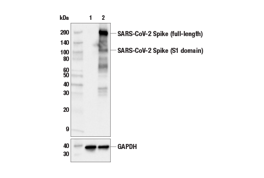
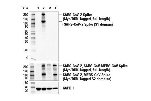
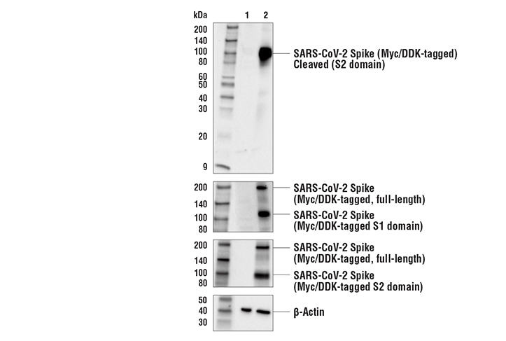
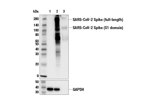
| SARS-CoV-2 Spike Protein (S1) (E5S3V) Rabbit Monoclonal Antibody | 99423 | 20 µl | 110, 220 kDa | Rabbit IgG |
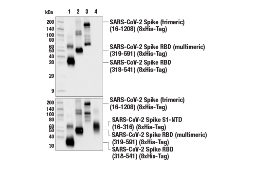
| SARS-CoV-2 Spike Protein (RBD) (E7B3E) Rabbit Monoclonal Antibody | 63847 | 20 µl | 110, 220 kDa | Rabbit IgG |
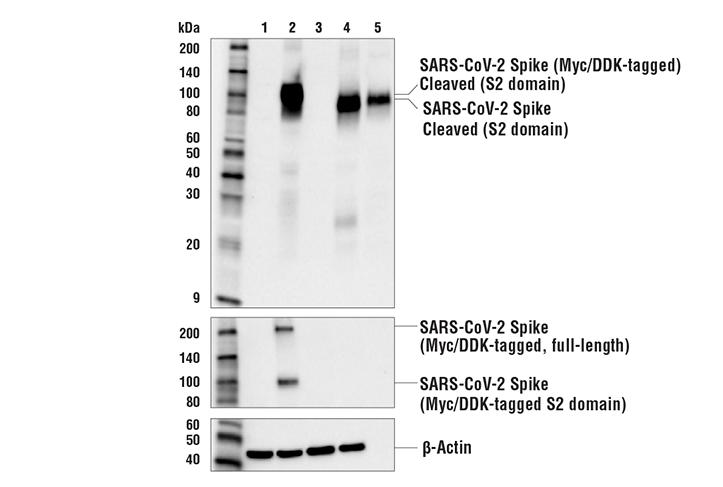
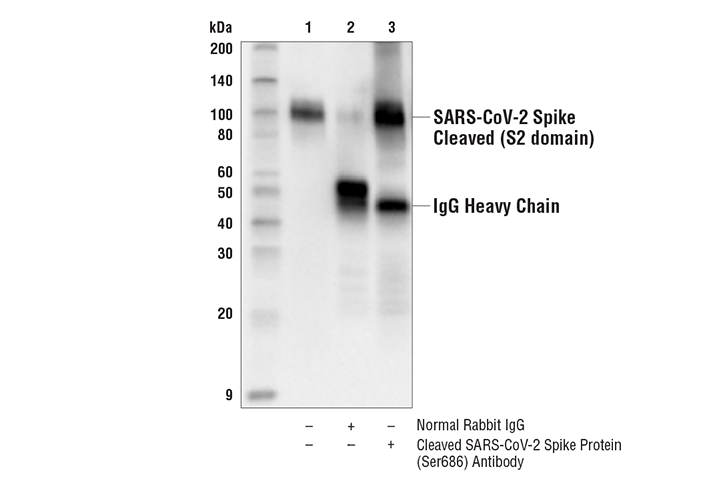
| Cleaved SARS-CoV-2 Spike Protein (Ser686) Antibody | 84534 | 20 µl | 100 kDa | Rabbit |
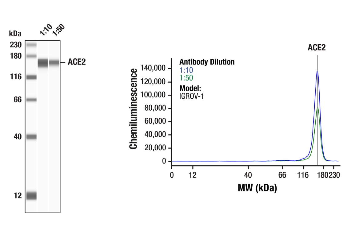
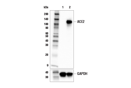
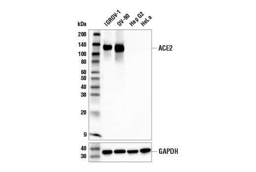
| ACE2 (E5O6J) Rabbit Monoclonal Antibody | 92485 | 20 µl | 120-135 kDa | Rabbit IgG |
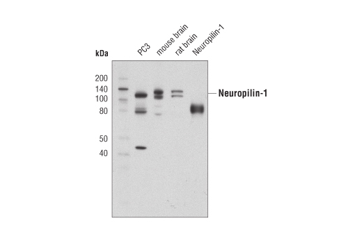
| Neuropilin-1 (D62C6) Rabbit Monoclonal Antibody | 3725 | 20 µl | 120-140 kDa | Rabbit IgG |
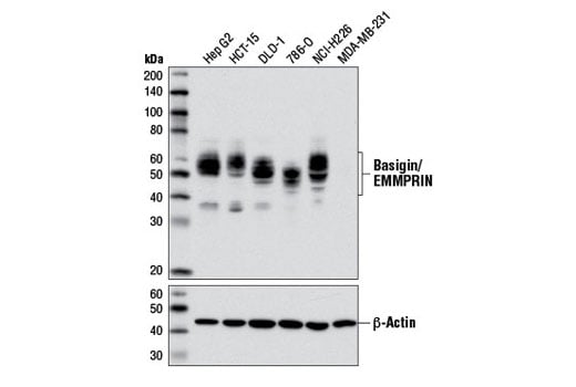
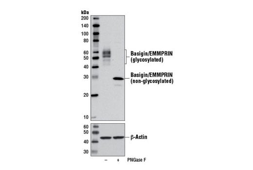
| Basigin/EMMPRIN (E1S1V) Rabbit Monoclonal Antibody | 13287 | 20 µl | 38-58 kDa | Rabbit IgG |
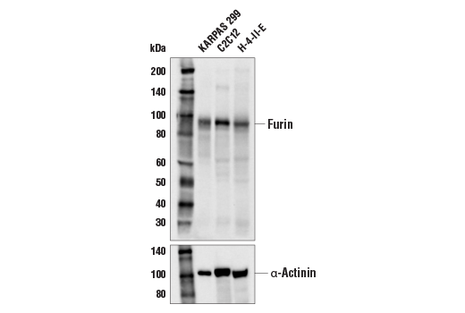
| Furin Antibody | 43996 | 20 µl | 90 kDa | Rabbit |
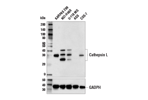
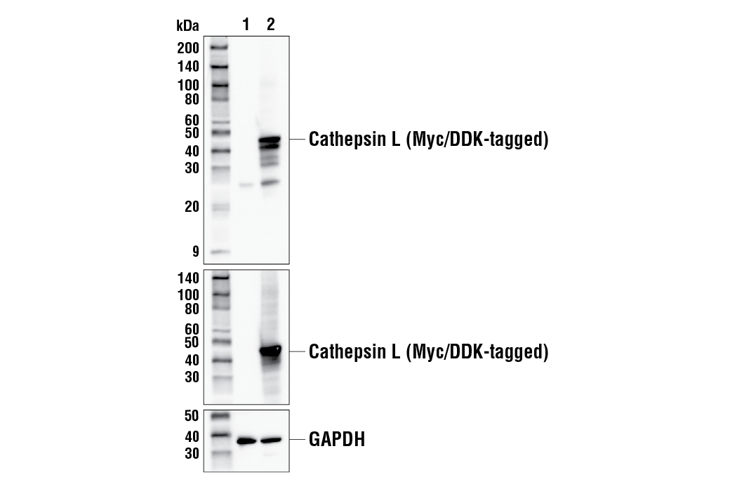
| Cathepsin L Antibody | 71298 | 20 µl | 25-42 kDa | Rabbit |
| Anti-rabbit IgG, HRP-linked Antibody | 7074 | 100 µl | Goat |
Please visit cellsignal.com for individual component applications, species cross-reactivity, dilutions, protocols, and additional product information.
Description
Storage
Background
The SARS-CoV-2 spike protein contains a novel tetrabasic "furin cleavage site" (FCS) at the S1/S2 junction. Research studies suggest this site is cleaved by proprotein convertases (e.g., furin) or lysosomal proteases (e.g., cathepsin L) (5,6). S1/S2 cleavage elicits a confirmational change in the spike protein that positions elements of the trimeric RBD in an exposed "up" position, priming it for interaction with host receptor proteins. Cleavage can occur at multiple steps of the viral lifecycle, including during viral packaging, or upon contact of the intact virion with the host cell surface. This novel cleavage event has been suggested to contribute to the high infectivity rate of the SARS-CoV-2 virus (7).
The SARS-CoV-2 virus has been shown to utilize the angiotensin-converting enzyme 2 (ACE2) protein as its primary receptor for cellular entry (8). However, research studies have suggested that other cell surface proteins may serve as receptors or co-receptors for SARS-CoV-2. These include neuropilin-1 (NPN1), a single-pass transmembrane receptor that can function as part of a semaphorin receptor complex, and as a vascular endothelial growth factor (VEGF) receptor (9), and Basigin/EMMPRIN (CD147), a type I integral membrane receptor belonging to the immunoglobulin superfamily (10).
Background References
- Zhou, P. et al. (2020) Nature 579, 270-273.
- Tortorici, M.A. and Veesler, D. (2019) Adv Virus Res 105, 93-116.
- Li, F. et al. (2006) J Virol 80, 6794-800.
- Li, F. (2016) Annu Rev Virol 3, 237-261.
- Coutard, B. et al. (2020) Antiviral Res 176, 104742.
- Jaimes, J.A. et al. (2020) iScience 23, 101212.
- Hasan, A. et al. (2021) J Biomol Struct Dyn 39, 3025-3033.
- Shang, J. et al. (2020) Nature 581, 221-224.
- Cantuti-Castelvetri, L. et al. (2020) Science 370, 856-860.
- Wang, K. et al. (2020) Signal Transduct Target Ther 5, 283.
Trademarks and Patents
Cell Signaling Technology is a trademark of Cell Signaling Technology, Inc.
All other trademarks are the property of their respective owners. Visit cellsignal.com/trademarks for more information.
Limited Uses
Except as otherwise expressly agreed in a writing signed by a legally authorized representative of CST, the following terms apply to Products provided by CST, its affiliates or its distributors. Any Customer's terms and conditions that are in addition to, or different from, those contained herein, unless separately accepted in writing by a legally authorized representative of CST, are rejected and are of no force or effect.
Products are labeled with For Research Use Only or a similar labeling statement and have not been approved, cleared, or licensed by the FDA or other regulatory foreign or domestic entity, for any purpose. Customer shall not use any Product for any diagnostic or therapeutic purpose, or otherwise in any manner that conflicts with its labeling statement. Products sold or licensed by CST are provided for Customer as the end-user and solely for research and development uses. Any use of Product for diagnostic, prophylactic or therapeutic purposes, or any purchase of Product for resale (alone or as a component) or other commercial purpose, requires a separate license from CST. Customer shall (a) not sell, license, loan, donate or otherwise transfer or make available any Product to any third party, whether alone or in combination with other materials, or use the Products to manufacture any commercial products, (b) not copy, modify, reverse engineer, decompile, disassemble or otherwise attempt to discover the underlying structure or technology of the Products, or use the Products for the purpose of developing any products or services that would compete with CST products or services, (c) not alter or remove from the Products any trademarks, trade names, logos, patent or copyright notices or markings, (d) use the Products solely in accordance with CST Product Terms of Sale and any applicable documentation, and (e) comply with any license, terms of service or similar agreement with respect to any third party products or services used by Customer in connection with the Products.
Revision 1
Revision 1

Revision 1

Revision 1
Revision 1
Revision 1
Revision 1
Revision 1
Revision 1
Revision 1
Revision 1
Revision 1
Revision 1
Revision 1
Revision 1