Revision 1
#25501
Store at -20C
SET1/COMPASS Antibody Sampler Kit
1 Kit
(8 x 20 microliters)
877-616-CELL (2355)
877-678-TECH (8324)
3 Trask Lane | Danvers | Massachusetts | 01923 | USA
For Research Use Only. Not for Use in Diagnostic Procedures.
| Product Includes | Product # | Quantity | Mol. Wt | Isotype/Source |
|---|---|---|---|---|
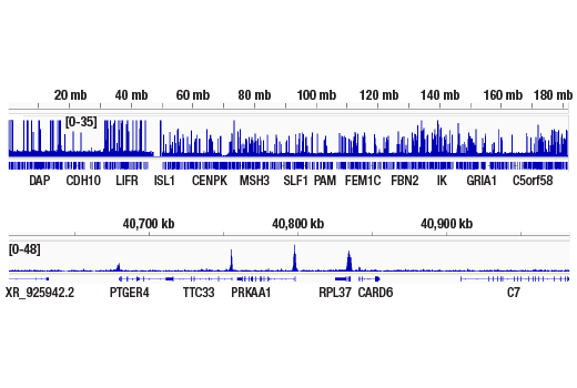
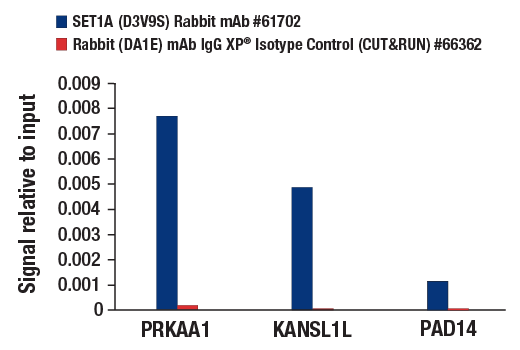
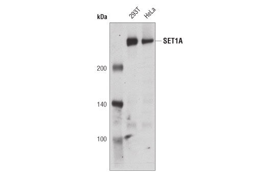
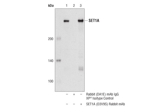
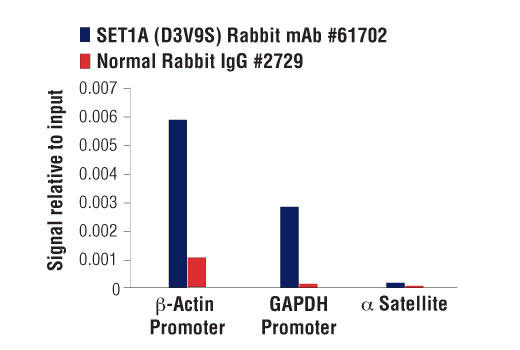
| SET1A (D3V9S) Rabbit Monoclonal Antibody | 61702 | 20 µl | 300 kDa | Rabbit IgG |
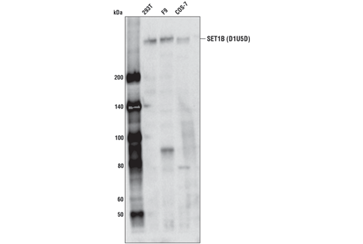
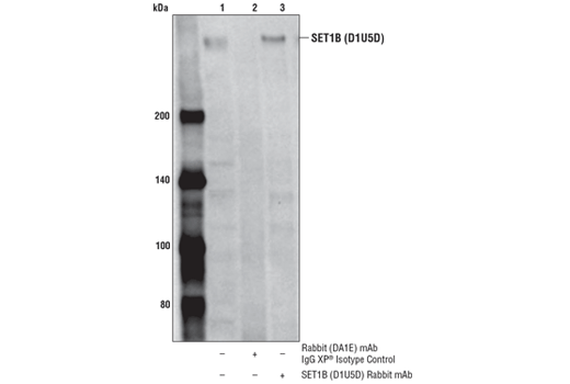
| SET1B (D1U5D) Rabbit Monoclonal Antibody | 44922 | 20 µl | 320 kDa | Rabbit IgG |
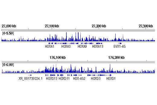
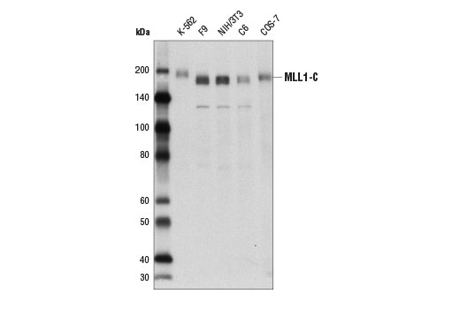
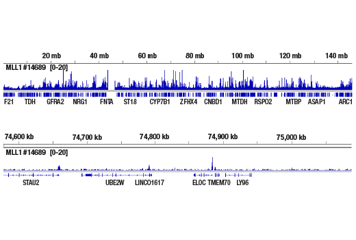
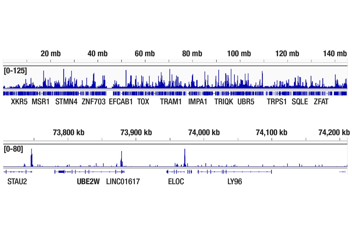
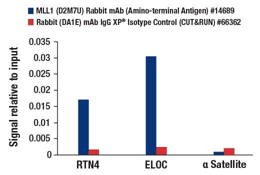
| MLL1 (D2M7U) Rabbit Monoclonal Antibody (Amino-terminal Antigen) | 14689 | 20 µl | 300 kDa | Rabbit IgG |
| MLL1 (D6G8N) Rabbit Monoclonal Antibody (Carboxy-terminal Antigen) | 14197 | 20 µl | 180 kDa | Rabbit IgG |
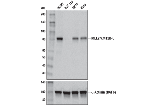
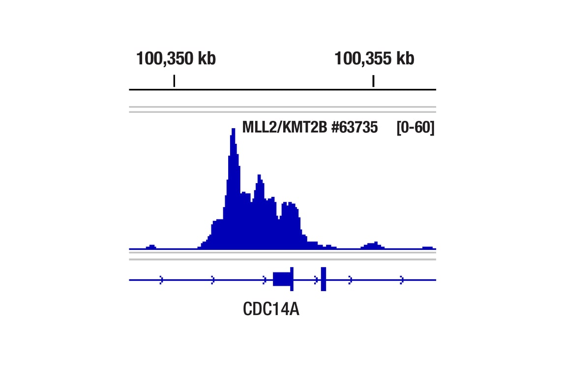
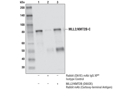
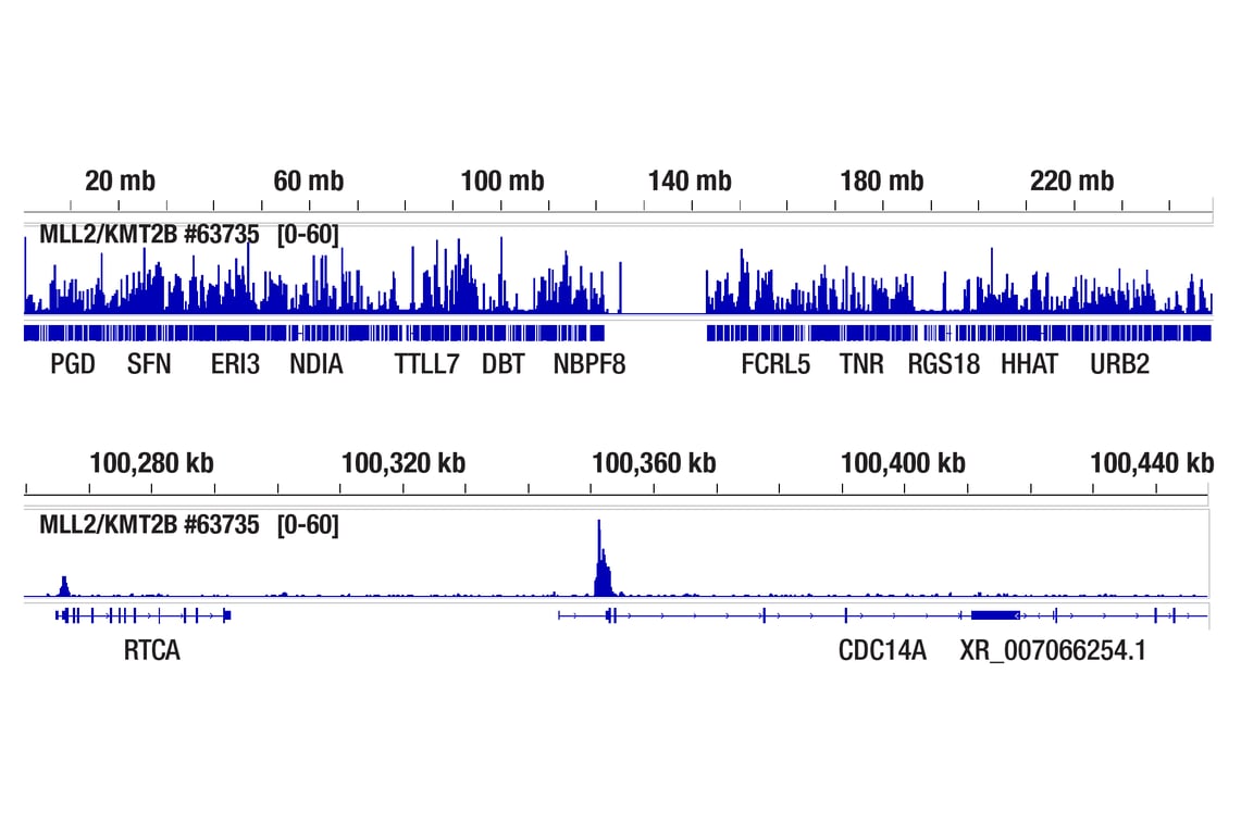
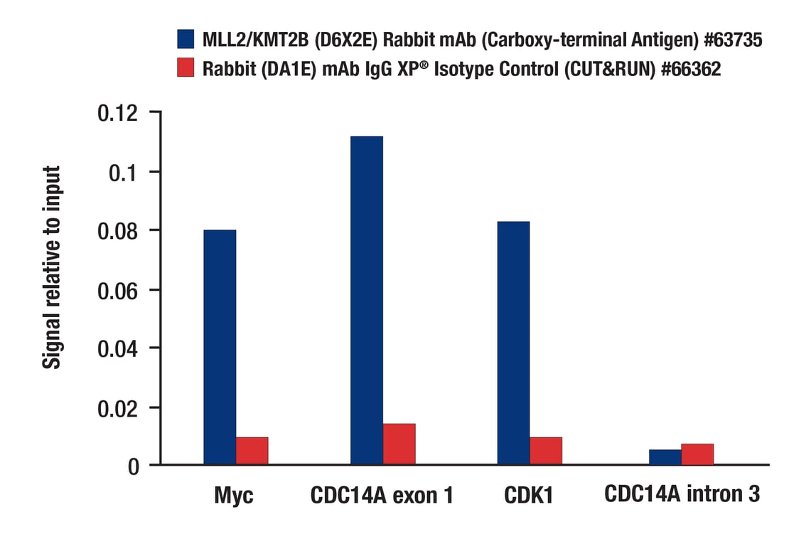
| MLL2/KMT2B (D6X2E) Rabbit Monoclonal Antibody (Carboxy-terminal Antigen) | 63735 | 20 µl | 80 kDa | Rabbit IgG |
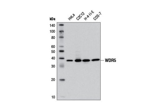
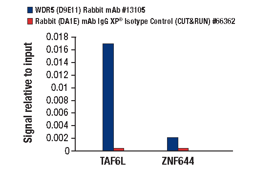
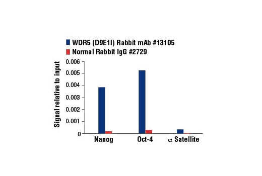
| WDR5 (D9E1I) Rabbit Monoclonal Antibody | 13105 | 20 µl | 37 kDa | Rabbit IgG |
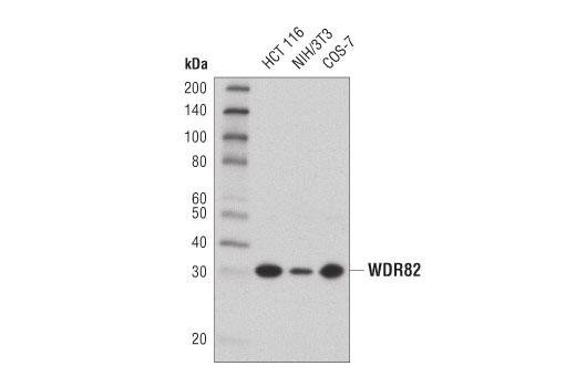
| WDR82 (D2I3B) Rabbit Monoclonal Antibody | 99715 | 20 µl | 30 kDa | Rabbit IgG |
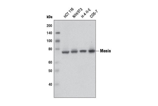
| Menin (D45B1) Rabbit Monoclonal Antibody | 6891 | 20 µl | 76 kDa | Rabbit IgG |
| Anti-rabbit IgG, HRP-linked Antibody | 7074 | 100 µl | Goat |
Please visit cellsignal.com for individual component applications, species cross-reactivity, dilutions, protocols, and additional product information.
Description
Storage
Background
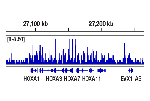
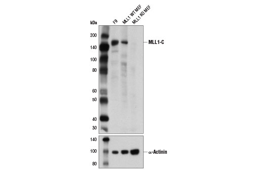
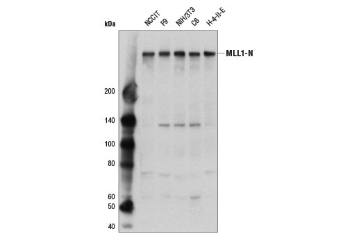
Like yeast Set1, all six Set1-related mammalian proteins methylate histone H3 on lysine 4 (2-6). SET1A, SET1B, MLL1 and MLL2 mediate di- and tri-methylation of histone H3 Lys4 at gene promoters to facilitate transcription activation. MLL3 and MLL4 function primarily to mono-methylate histone H3 Lys4 at gene enhancers. MLL1 and MLL2 function as master regulators of both embryogenesis and hematopoiesis, and are required for proper expression of Hox genes (8-10). MLL1 is a large approximately 4000 amino acid protein that is cleaved by the Taspase 1 threonine endopeptidase to form N-terminal (MLL1-N) and C-terminal MLL1 (MLL1-C) fragments, both of which are subunits of the functional MLL1/COMPASS complex (11,12). MLL1 translocations are found in a large number of hematological malignancies, suggesting that Set1 histone methyltransferase complexes play a critical role in leukemogenesis (6). Like MLL1, MLL2 is also a large, approximately 2700 amino acid protein that is cleaved by the Taspase 1 threonine endopeptidase to form N-terminal (MLL2-N) and C-terminal (MLL2-C) fragments, both of which are subunits of the functional MLL2/COMPASS complex. MLL2 has also been implicated as a modulator of hematological malignancies (13). MLL3 and MLL4 proteins are not cleaved by Taspase 1.
Background References
- Miller, T. et al. (2001) Proc Natl Acad Sci U S A 98, 12902-7.
- Shilatifard, A. (2008) Curr Opin Cell Biol 20, 341-8.
- Tenney, K. and Shilatifard, A. (2005) J Cell Biochem 95, 429-36.
- Lee, J.H. and Skalnik, D.G. (2005) J Biol Chem 280, 41725-31.
- Lee, J.H. et al. (2007) J Biol Chem 282, 13419-28.
- Hughes, C.M. et al. (2004) Mol Cell 13, 587-97.
- Yokoyama, A. et al. (2004) Mol Cell Biol 24, 5639-49.
- Eissenberg, J.C. and Shilatifard, A. (2010) Dev Biol 339, 240-9.
- Smith, E. et al. (2011) Genes Dev 25, 661-72.
- Denissov, S. et al. (2014) Development 141, 526-37.
- Takeda, S. et al. (2006) Genes Dev 20, 2397-409.
- Yokoyama, A. et al. (2002) Blood 100, 3710-8.
- Chen, Y. et al. (2017) Cancer Cell 31, 755-770.e6.
Trademarks and Patents
Cell Signaling Technology is a trademark of Cell Signaling Technology, Inc.
All other trademarks are the property of their respective owners. Visit cellsignal.com/trademarks for more information.
Limited Uses
Except as otherwise expressly agreed in a writing signed by a legally authorized representative of CST, the following terms apply to Products provided by CST, its affiliates or its distributors. Any Customer's terms and conditions that are in addition to, or different from, those contained herein, unless separately accepted in writing by a legally authorized representative of CST, are rejected and are of no force or effect.
Products are labeled with For Research Use Only or a similar labeling statement and have not been approved, cleared, or licensed by the FDA or other regulatory foreign or domestic entity, for any purpose. Customer shall not use any Product for any diagnostic or therapeutic purpose, or otherwise in any manner that conflicts with its labeling statement. Products sold or licensed by CST are provided for Customer as the end-user and solely for research and development uses. Any use of Product for diagnostic, prophylactic or therapeutic purposes, or any purchase of Product for resale (alone or as a component) or other commercial purpose, requires a separate license from CST. Customer shall (a) not sell, license, loan, donate or otherwise transfer or make available any Product to any third party, whether alone or in combination with other materials, or use the Products to manufacture any commercial products, (b) not copy, modify, reverse engineer, decompile, disassemble or otherwise attempt to discover the underlying structure or technology of the Products, or use the Products for the purpose of developing any products or services that would compete with CST products or services, (c) not alter or remove from the Products any trademarks, trade names, logos, patent or copyright notices or markings, (d) use the Products solely in accordance with CST Product Terms of Sale and any applicable documentation, and (e) comply with any license, terms of service or similar agreement with respect to any third party products or services used by Customer in connection with the Products.
Revision 1
Revision 1
Revision 1
Revision 1
Revision 1

Revision 1
Revision 1
Revision 1
Revision 1

Revision 1
Revision 1
Revision 1
Revision 1