View Featured Offers >>
9170
PhosphoPlus® Stat1 (Tyr701) Antibody Kit
Primary Antibodies
PhosphoPlus Antibody Kit

PhosphoPlus® Stat1 (Tyr701) Antibody Kit #9170

Citations (1)
After the primary antibody is bound to the target protein, a complex with HRP-linked secondary antibody is formed. The LumiGLO® is added and emits light during enzyme catalyzed decomposition.
Western blot analysis of extracts from HeLa, A20, and PC-12 cells, untreated or treated with Human Interferon-α1 (hIFN-α1) #8927 (10 ng/ml, 30 min), using Phospho-Stat1 (Tyr701) (D4A7) Rabbit mAb (upper) or Stat1 Antibody #9172 (lower).
Western blot analysis of extracts from A549 cells (lane 1) or STAT1 knock-out cells (lane 2) using Stat1 Antibody #9172 (upper), and β-actin (D6A8) Rabbit mAb #8457 (lower). The absence of signal in the STAT1 knock-out A549 cells confirms specificity of the antibody for STAT1.
Confocal immunofluorescent analysis of HeLa cells, untreated (left) or treated with hIFN-α1 #8927 (100 ng/mL, 30 min; right), using Phospho-Stat1 (Tyr701) (D4A7) Rabbit mAb (green) and β-Tubulin (9F3) Rabbit mAb (Alexa Fluor® 555 Conjugate) #2116 (red).
Western blot analysis of extracts from HeLa cells 48 hours following mock transfection, transfection with non-targeted (control) siRNA or transfection with Stat1 siRNA. Stat1 was detected using Stat1 Antibody #9172, and p42 was detected using p42 MAPK Antibody #9108. The Stat1 Antibody confirms silencing of Stat1 expression, and the p42 MAPK Antibody is used to control for protein loading and siRNA specificity.
Flow cytometric analysis of Jurkat cells, untreated (blue) or treated with hIFN-α1 #8927 (green), using Phospho-Stat1 (Tyr701) (D4A7) Rabbit mAb.
Western blot analysis of extracts from SK-MEL-28 cells, untreated or IFN-alpha-treated (100 ng/ml), using Phospho-Stat1 (Tyr701) Antibody #9171 (upper) or Stat1 Antibody (lower).
Chromatin immunoprecipitations were performed with cross-linked chromatin from HT-1080 cells treated with IFN-γ (50 ng/ml) for 30 minutes and Phospho-Stat1 (Tyr701) (D4A7) Rabbit mAb, using SimpleChIP® Enzymatic Chromatin IP Kit (Magnetic Beads) #9003. DNA Libraries were prepared using DNA Library Prep Kit for Illumina® (ChIP-seq, CUT&RUN) #56795. The figure shows binding across IRF-1, a known target gene of Phospho-Stat1 (see additional figure containing ChIP-qPCR data). For additional ChIP-seq tracks, please download the product datasheet.
Chromatin immunoprecipitations were performed with cross-linked chromatin from HT-1080 cells treated with IFN-γ (50 ng/ml) for 30 minutes and either Stat1 Antibody or Normal Rabbit IgG #2729 using SimpleChIP® Enzymatic Chromatin IP Kit (Magnetic Beads) #9003. The enriched DNA was quantified by real-time PCR using human IRF-1 promoter primers, SimpleChIP® Human TAP1 Promoter Primers #5148, and SimpleChIP® Human α Satellite Repeat Primers #4486. The amount of immunoprecipitated DNA in each sample is represented as signal relative to the total amount of input chromatin, which is equivalent to one.
Chromatin immunoprecipitations were performed with cross-linked chromatin from 4 x 10⁶ HT-1080 cells treated with IFN-γ (50 ng/ml) for 30 minutes and 5 μl of Phospho-Stat1 (Tyr701) (D4A7) Rabbit mAb, using SimpleChIP® Enzymatic Chromatin IP Kit (Magnetic Beads) #9003. DNA Libraries were prepared using DNA Library Prep Kit for Illumina® (ChIP-seq, CUT&RUN) #56795. The figure shows binding across chromosome 5 (upper), including IRF1 (lower), a known target gene of Phospho-Stat1 (see additional figure containing ChIP-qPCR data).
Chromatin immunoprecipitations were performed with cross-linked chromatin from HT-1080 cells treated with IFN-γ (50 ng/ml) for 30 minutes and either Phospho-Stat1 (Tyr701) (D4A7) Rabbit mAb or Normal Rabbit IgG #2729 using SimpleChIP® Enzymatic Chromatin IP Kit (Magnetic Beads) #9003. The enriched DNA was quantified by real-time PCR using human IRF-1 promoter primers, SimpleChIP® Human TAP1 Promoter Primers #5148, and SimpleChIP® Human α Satellite Repeat Primers #4486. The amount of immunoprecipitated DNA in each sample is represented as signal relative to the total amount of input chromatin, which is equivalent to one.
Inquiry Info.# 9170

Specificity / Sensitivity

Phospho-Stat1 (Tyr701) (D4A7) Rabbit mAb recognizes endogenous levels of Stat1 protein only when phosphorylated at Tyr701. Stat1 antibody detects endogenous levels of total Stat1 α (91kDa) and Stat1 β (84kDa) protein.

Source / Purification

Phospho-Stat1 (Tyr701) (D4A7) monoclonal antibody is produced by immunizing animals with a synthetic peptide corresponding to residues surrounding Tyr701 of human Stat1 protein. Stat1 polyclonal antibody is produced using a synthetic peptide corresponding to a sequence of human Stat1 (Stat1 antibody). Polyclonal antibodies are purified by protein A and peptide affinity chromatography.

Background

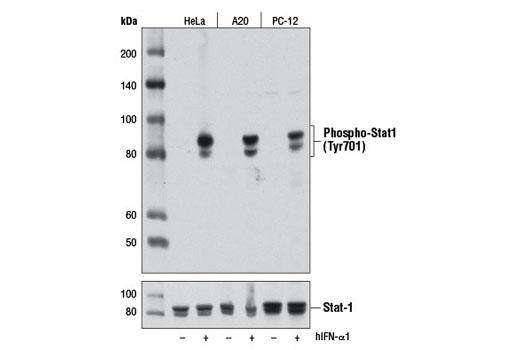
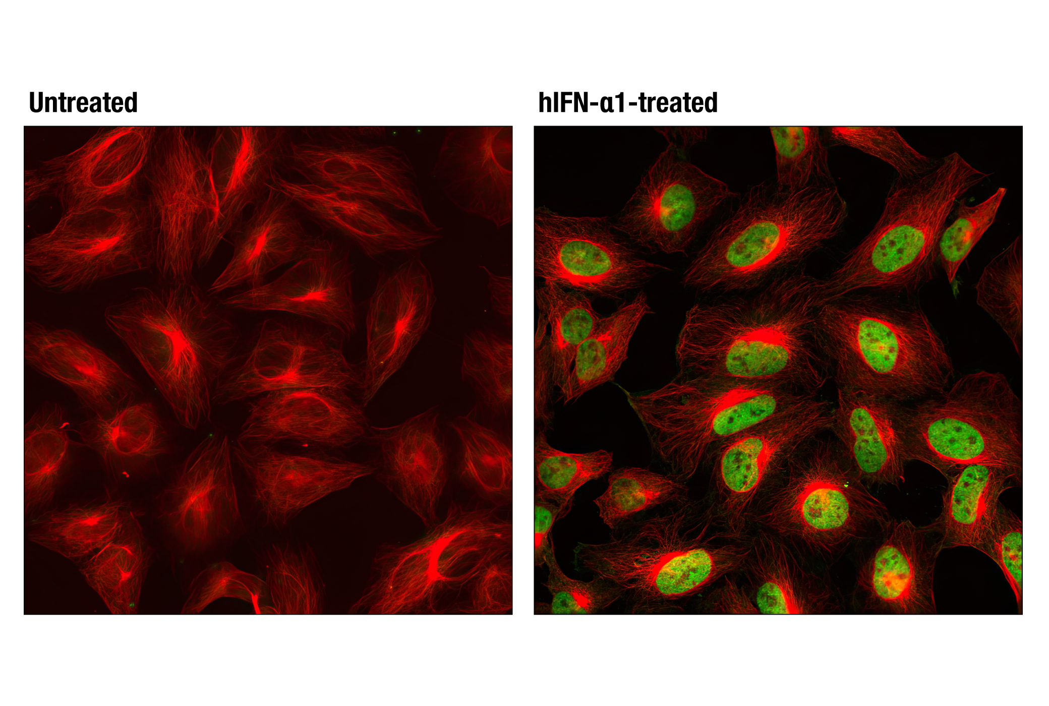
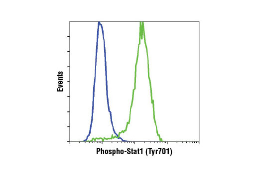
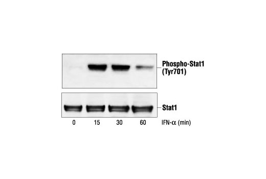
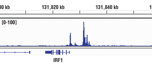
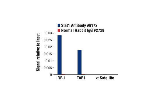
The Stat1 transcription factor is activated in response to a large number of ligands (1) and is essential for responsiveness to IFN-α and IFN-γ (2,3). Phosphorylation of Stat1 at Tyr701 induces Stat1 dimerization, nuclear translocation, and DNA binding (4). Stat1 protein exists as a pair of isoforms, Stat1α (91 kDa) and the splice variant Stat1β (84 kDa). In most cells, both isoforms are activated by IFN-α, but only Stat1α is activated by IFN-γ. The inappropriate activation of Stat1 occurs in many tumors (5). In addition to tyrosine phosphorylation, Stat1 is also phosphorylated at Ser727 through a p38 mitogen-activated protein kinase (MAPK)-dependent pathway in response to IFN-α and other cellular stresses (6). Serine phosphorylation may be required for the maximal induction of Stat1-mediated gene activation.

Pathways

Explore pathways related to this product.

Limited Uses

Except as otherwise expressly agreed in a writing signed by a legally authorized representative of CST, the following terms apply to Products provided by CST, its affiliates or its distributors. Any Customer's terms and conditions that are in addition to, or different from, those contained herein, unless separately accepted in writing by a legally authorized representative of CST, are rejected and are of no force or effect.

Products are labeled with For Research Use Only or a similar labeling statement and have not been approved, cleared, or licensed by the FDA or other regulatory foreign or domestic entity, for any purpose. Customer shall not use any Product for any diagnostic or therapeutic purpose, or otherwise in any manner that conflicts with its labeling statement. Products sold or licensed by CST are provided for Customer as the end-user and solely for research and development uses. Any use of Product for diagnostic, prophylactic or therapeutic purposes, or any purchase of Product for resale (alone or as a component) or other commercial purpose, requires a separate license from CST. Customer shall (a) not sell, license, loan, donate or otherwise transfer or make available any Product to any third party, whether alone or in combination with other materials, or use the Products to manufacture any commercial products, (b) not copy, modify, reverse engineer, decompile, disassemble or otherwise attempt to discover the underlying structure or technology of the Products, or use the Products for the purpose of developing any products or services that would compete with CST products or services, (c) not alter or remove from the Products any trademarks, trade names, logos, patent or copyright notices or markings, (d) use the Products solely in accordance with CST Product Terms of Sale and any applicable documentation, and (e) comply with any license, terms of service or similar agreement with respect to any third party products or services used by Customer in connection with the Products.

For Research Use Only. Not for Use in Diagnostic Procedures.
Cell Signaling Technology is a trademark of Cell Signaling Technology, Inc.
PhosphoPlus is a registered trademark of Cell Signaling Technology, Inc.
All other trademarks are the property of their respective owners. Visit our Trademark Information page.