Revision 7
#12800
Store at -20C
877-616-CELL (2355)
877-678-TECH (8324)
3 Trask Lane | Danvers | Massachusetts | 01923 | USA
For Research Use Only. Not for Use in Diagnostic Procedures.
Applications:
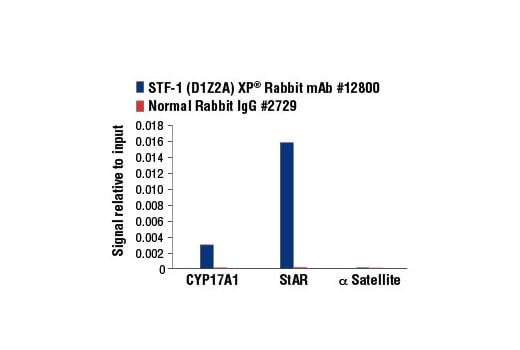
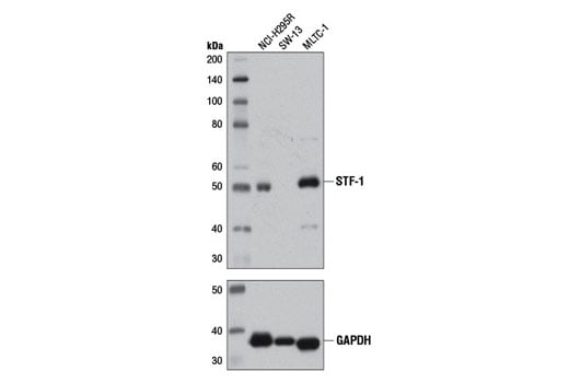
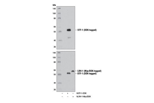
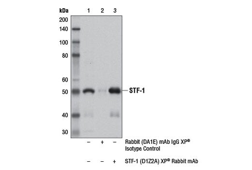
W, IP, IF-IC, ChIP, ChIP-seq
Reactivity:
H M
Sensitivity:
Endogenous
MW (kDa):
50
Source/Isotype:
Rabbit IgG
UniProt ID:
#Q13285
Entrez-Gene Id:
2516
Product Usage Information
| Application | Dilution |
|---|---|
| Western Blotting | 1:1000 |
| Immunoprecipitation | 1:100 |
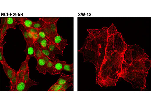
| Immunofluorescence (Immunocytochemistry) | 1:100 |
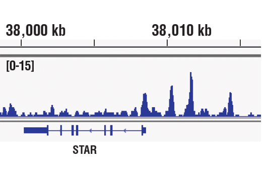
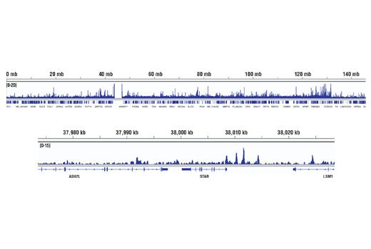
| Chromatin IP | 1:50 |
| Chromatin IP-seq | 1:50 |
Storage
Specificity/Sensitivity
Species predicted to react based on 100% sequence homology
Source / Purification
Background
Like other nuclear hormone receptors, STF-1 has a modular domain structure composed of an amino-terminal zinc finger DNA-binding domain, a ligand-binding domain, a carboxy-terminal AF-2 activation domain, and a hinge region with AF-1-like activation activity. STF-1 also contains a fushi tarazu factor 1 box, which functions as an accessory DNA binding domain (11). STF-1 is primarily phosphorylated at Ser203, which is thought to enhance its transcriptional activity by promoting complex formation with transcriptional cofactors (12). In addition to phosphorylation at Ser203, STF-1 is subject to SUMO conjugation and acetylation at ε-amino groups of target lysine residues. Whereas SUMOylation represses STF-1 function (13,14), acetylation enhances its transcriptional activity (15).
Background References
- Parker, K.L. and Schimmer, B.P. (1997) Endocr Rev 18, 361-77.
- Luo, X. et al. (1994) Cell 77, 481-90.
- Zhao, L. et al. (2001) Development 128, 147-54.
- Jeyasuria, P. et al. (2004) Mol Endocrinol 18, 1610-9.
- Pelusi, C. et al. (2008) Biol Reprod 79, 1074-83.
- Zhao, L. et al. (2008) Mol Endocrinol 22, 1403-15.
- Achermann, J.C. et al. (1999) Nat Genet 22, 125-6.
- Lourenço, D. et al. (2009) N Engl J Med 360, 1200-10.
- Bulun, S.E. et al. (2009) Mol Cell Endocrinol 300, 104-8.
- Figueiredo, B.C. et al. (2005) J Clin Endocrinol Metab 90, 615-9.
- Little, T.H. et al. (2006) Mol Endocrinol 20, 831-43.
- Hammer, G.D. et al. (1999) Mol Cell 3, 521-6.
- Chen, W.Y. et al. (2004) J Biol Chem 279, 38730-5.
- Lee, F.Y. et al. (2011) Dev Cell 21, 315-27.
- Chen, W.Y. et al. (2005) Mol Cell Biol 25, 10442-53.
Species Reactivity
Species reactivity is determined by testing in at least one approved application (e.g., western blot).
Western Blot Buffer
IMPORTANT: For western blots, incubate membrane with diluted primary antibody in 5% w/v nonfat dry milk, 1X TBS, 0.1% Tween® 20 at 4°C with gentle shaking, overnight.
Applications Key
W: Western Blotting IP: Immunoprecipitation IF-IC: Immunofluorescence (Immunocytochemistry) ChIP: Chromatin IP ChIP-seq: Chromatin IP-seq
Cross-Reactivity Key
H: Human M: Mouse
Trademarks and Patents
Cell Signaling Technology is a trademark of Cell Signaling Technology, Inc.
SimpleChIP is a registered trademark of Cell Signaling Technology, Inc.
All other trademarks are the property of their respective owners. Visit cellsignal.com/trademarks for more information.
Limited Uses
Except as otherwise expressly agreed in a writing signed by a legally authorized representative of CST, the following terms apply to Products provided by CST, its affiliates or its distributors. Any Customer's terms and conditions that are in addition to, or different from, those contained herein, unless separately accepted in writing by a legally authorized representative of CST, are rejected and are of no force or effect.
Products are labeled with For Research Use Only or a similar labeling statement and have not been approved, cleared, or licensed by the FDA or other regulatory foreign or domestic entity, for any purpose. Customer shall not use any Product for any diagnostic or therapeutic purpose, or otherwise in any manner that conflicts with its labeling statement. Products sold or licensed by CST are provided for Customer as the end-user and solely for research and development uses. Any use of Product for diagnostic, prophylactic or therapeutic purposes, or any purchase of Product for resale (alone or as a component) or other commercial purpose, requires a separate license from CST. Customer shall (a) not sell, license, loan, donate or otherwise transfer or make available any Product to any third party, whether alone or in combination with other materials, or use the Products to manufacture any commercial products, (b) not copy, modify, reverse engineer, decompile, disassemble or otherwise attempt to discover the underlying structure or technology of the Products, or use the Products for the purpose of developing any products or services that would compete with CST products or services, (c) not alter or remove from the Products any trademarks, trade names, logos, patent or copyright notices or markings, (d) use the Products solely in accordance with CST Product Terms of Sale and any applicable documentation, and (e) comply with any license, terms of service or similar agreement with respect to any third party products or services used by Customer in connection with the Products.
Revision 7
Revision 7
Revision 7