Revision 1
#8357
Store at -20C
Stress and Apoptosis Antibody Sampler Kit
1 Kit
(8 x 20 microliters)
877-616-CELL (2355)
877-678-TECH (8324)
3 Trask Lane | Danvers | Massachusetts | 01923 | USA
For Research Use Only. Not for Use in Diagnostic Procedures.
| Product Includes | Product # | Quantity | Mol. Wt | Isotype/Source |
|---|---|---|---|---|
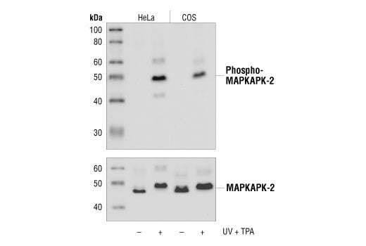
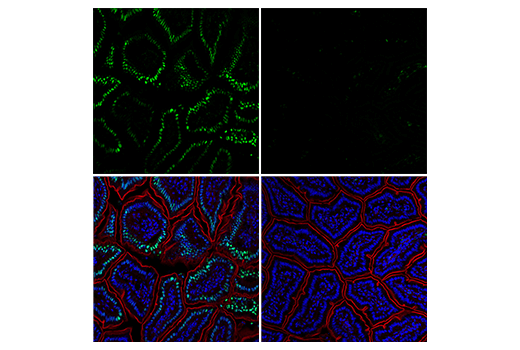
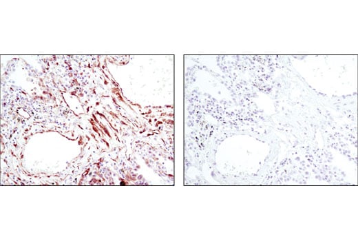
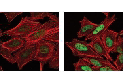
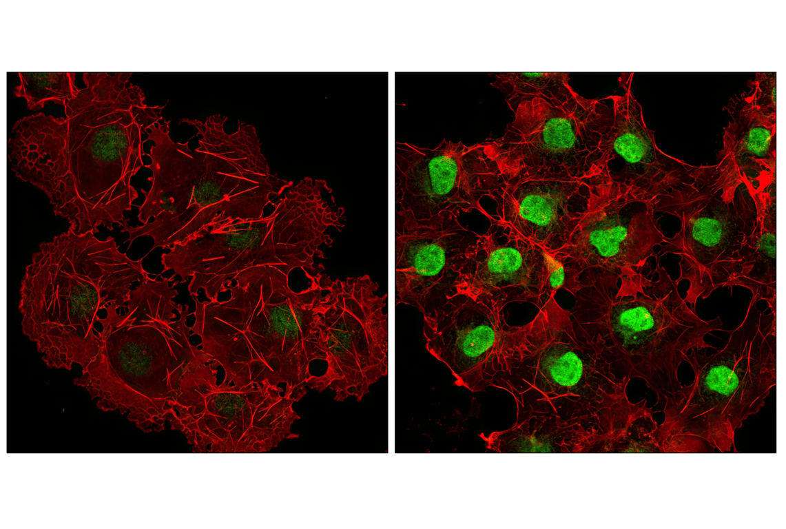
| Phospho-MAPKAPK-2 (Thr334) (27B7) Rabbit Monoclonal Antibody | 3007 | 20 µl | 49 kDa | Rabbit IgG |
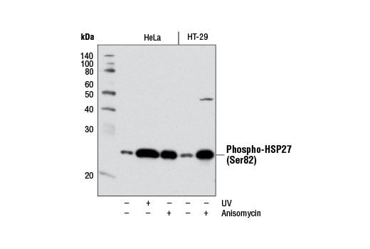
| Phospho-HSP27 (Ser82) (D1H2F6) Rabbit Monoclonal Antibody | 9709 | 20 µl | 27 kDa | Rabbit IgG |
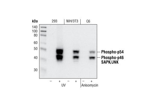
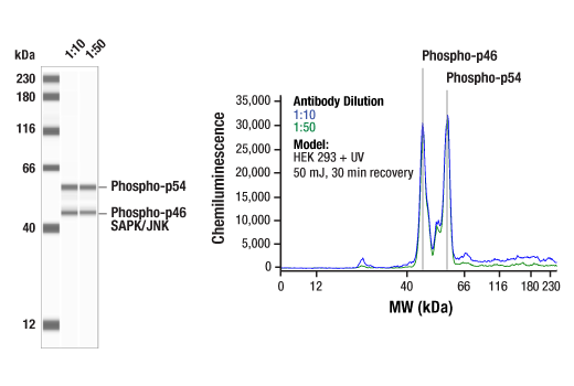
| Phospho-SAPK/JNK (Thr183/Tyr185) (81E11) Rabbit Monoclonal Antibody | 4668 | 20 µl | 46, 54 kDa | Rabbit IgG |
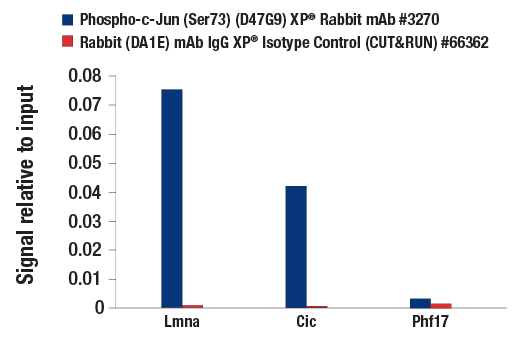
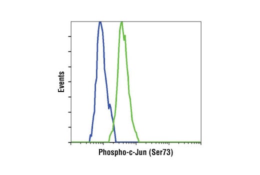
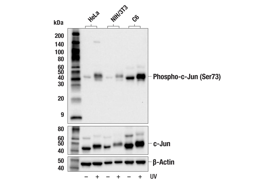
| Phospho-c-Jun (Ser73) (D47G9) Rabbit Monoclonal Antibody | 3270 | 20 µl | 48 kDa | Rabbit IgG |
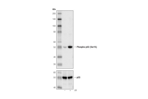
| Phospho-p53 (Ser15) (16G8) Mouse Monoclonal Antibody | 9286 | 20 µl | 53 kDa | Mouse IgG1 |
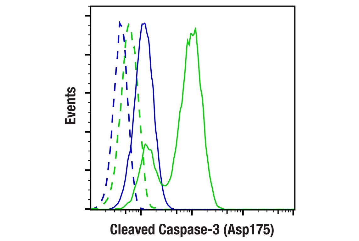
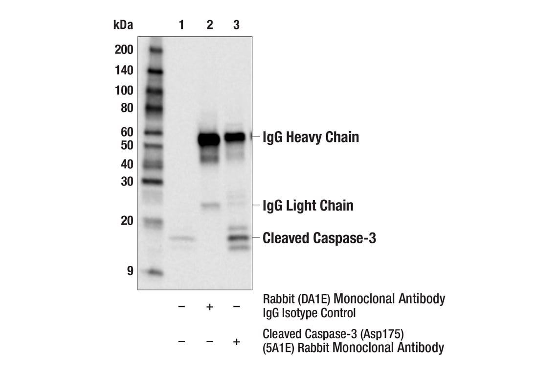
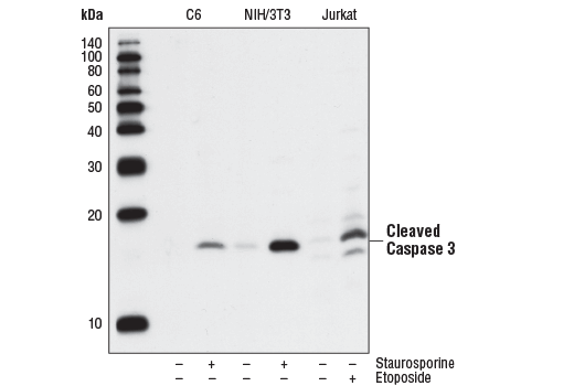
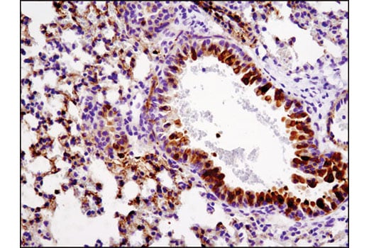
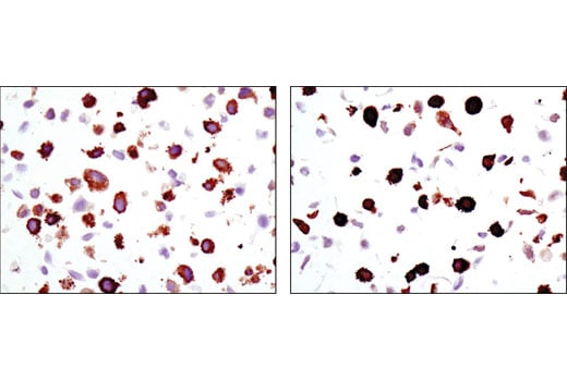
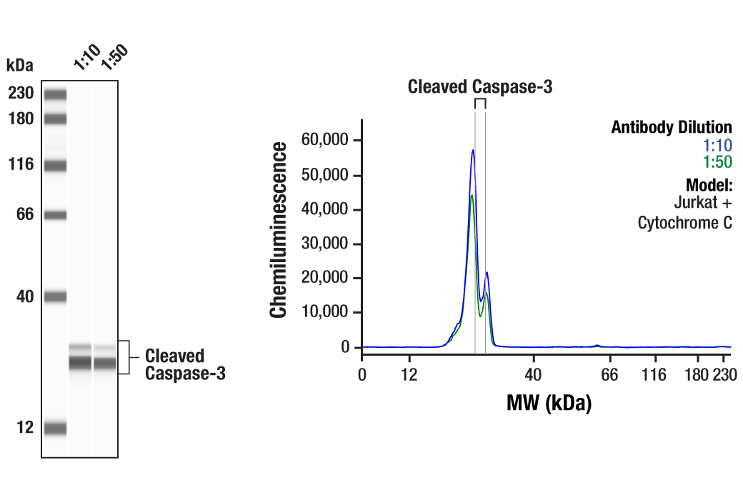
| Cleaved Caspase-3 (Asp175) (5A1E) Rabbit Monoclonal Antibody | 9664 | 20 µl | 17, 19 kDa | Rabbit IgG |
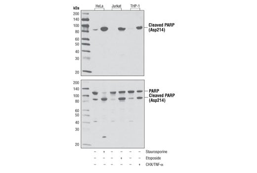
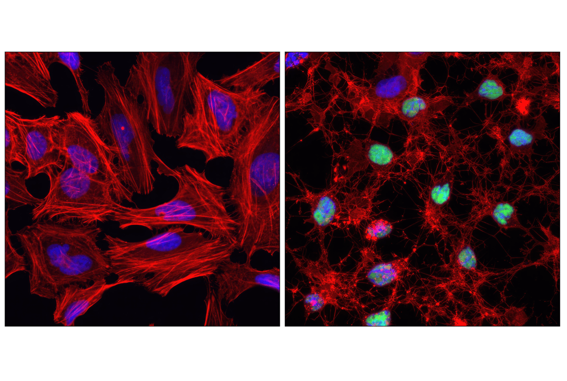
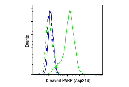
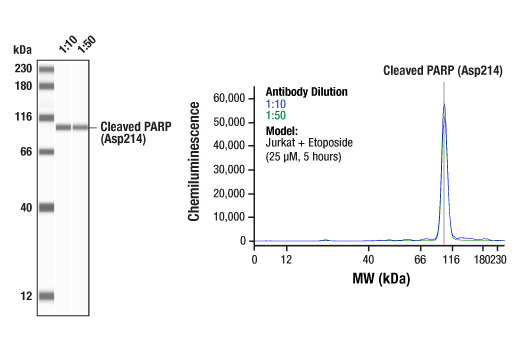
| Cleaved PARP (Asp214) (D64E10) Rabbit Monoclonal Antibody | 5625 | 20 µl | 89 kDa | Rabbit IgG |
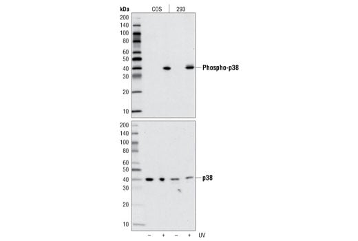
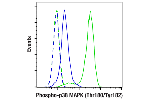
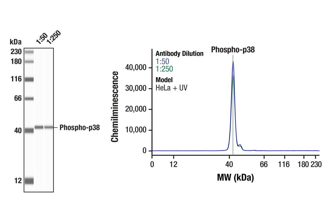
| Phospho-p38 MAPK (Thr180/Tyr182) (D3F9) Rabbit Monoclonal Antibody | 4511 | 20 µl | 43 kDa | Rabbit IgG |
| Anti-rabbit IgG, HRP-linked Antibody | 7074 | 100 µl | Goat | |
| Anti-mouse IgG, HRP-linked Antibody | 7076 | 100 µl | Horse |
Please visit cellsignal.com for individual component applications, species cross-reactivity, dilutions, protocols, and additional product information.
Description
Storage
Background
Background References
- Fernandes-Alnemri, T. et al. (1994) J Biol Chem 269, 30761-4.
- Satoh, M.S. and Lindahl, T. (1992) Nature 356, 356-8.
- Lazebnik, Y.A. et al. (1994) Nature 371, 346-7.
- Cohen, G.M. (1997) Biochem J 326 ( Pt 1), 1-16.
- Nicholson, D.W. et al. (1995) Nature 376, 37-43.
- Tewari, M. et al. (1995) Cell 81, 801-9.
- Oliver, F.J. et al. (1998) J Biol Chem 273, 33533-9.
- Levine, A.J. (1997) Cell 88, 323-31.
- Shieh, S.Y. et al. (1997) Cell 91, 325-34.
- Chehab, N.H. et al. (1999) Proc Natl Acad Sci U S A 96, 13777-82.
- Honda, R. et al. (1997) FEBS Lett 420, 25-7.
- Kyriakis, J.M. and Avruch, J. (2001) Physiol Rev 81, 807-69.
- Leppä, S. and Bohmann, D. (1999) Oncogene 18, 6158-62.
- Davis, R.J. (2000) Cell 103, 239-52.
- Shaulian, E. and Karin, M. (2002) Nat Cell Biol 4, E131-6.
- Weiss, C. and Bohmann, D. (2004) Cell Cycle 3, 111-3.
- Rouse, J. et al. (1994) Cell 78, 1027-37.
- Han, J. et al. (1994) Science 265, 808-11.
- Lee, J.C. et al. Nature 372, 739-46.
- Freshney, N.W. et al. (1994) Cell 78, 1039-49.
- Ben-Levy, R. et al. (1995) EMBO J 14, 5920-30.
- Landry, J. et al. (1992) J Biol Chem 267, 794-803.
Trademarks and Patents
Cell Signaling Technology is a trademark of Cell Signaling Technology, Inc.
All other trademarks are the property of their respective owners. Visit cellsignal.com/trademarks for more information.
Limited Uses
Except as otherwise expressly agreed in a writing signed by a legally authorized representative of CST, the following terms apply to Products provided by CST, its affiliates or its distributors. Any Customer's terms and conditions that are in addition to, or different from, those contained herein, unless separately accepted in writing by a legally authorized representative of CST, are rejected and are of no force or effect.
Products are labeled with For Research Use Only or a similar labeling statement and have not been approved, cleared, or licensed by the FDA or other regulatory foreign or domestic entity, for any purpose. Customer shall not use any Product for any diagnostic or therapeutic purpose, or otherwise in any manner that conflicts with its labeling statement. Products sold or licensed by CST are provided for Customer as the end-user and solely for research and development uses. Any use of Product for diagnostic, prophylactic or therapeutic purposes, or any purchase of Product for resale (alone or as a component) or other commercial purpose, requires a separate license from CST. Customer shall (a) not sell, license, loan, donate or otherwise transfer or make available any Product to any third party, whether alone or in combination with other materials, or use the Products to manufacture any commercial products, (b) not copy, modify, reverse engineer, decompile, disassemble or otherwise attempt to discover the underlying structure or technology of the Products, or use the Products for the purpose of developing any products or services that would compete with CST products or services, (c) not alter or remove from the Products any trademarks, trade names, logos, patent or copyright notices or markings, (d) use the Products solely in accordance with CST Product Terms of Sale and any applicable documentation, and (e) comply with any license, terms of service or similar agreement with respect to any third party products or services used by Customer in connection with the Products.
Revision 1

Revision 1
Revision 1


Revision 1
Revision 1
Revision 1
Revision 1
Revision 1


Revision 1


Revision 1
Revision 1
Revision 1
Revision 1
Revision 1
Revision 1
Revision 1

Revision 1
Revision 1