Revision 8
#16551
Store at -20C
877-616-CELL (2355)
877-678-TECH (8324)
3 Trask Lane | Danvers | Massachusetts | 01923 | USA
For Research Use Only. Not for Use in Diagnostic Procedures.
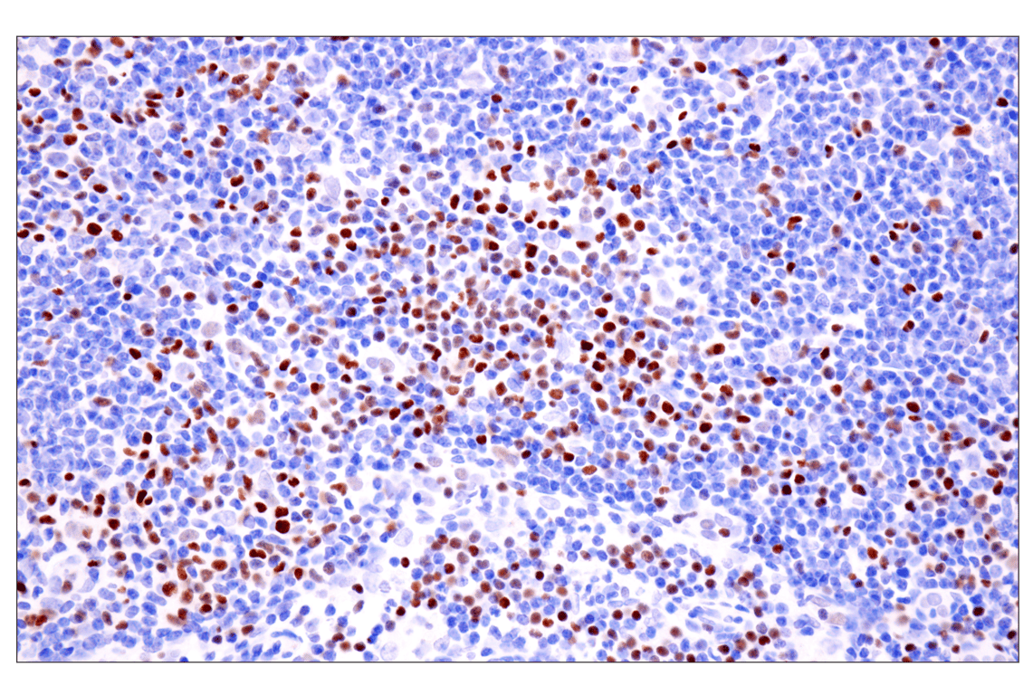
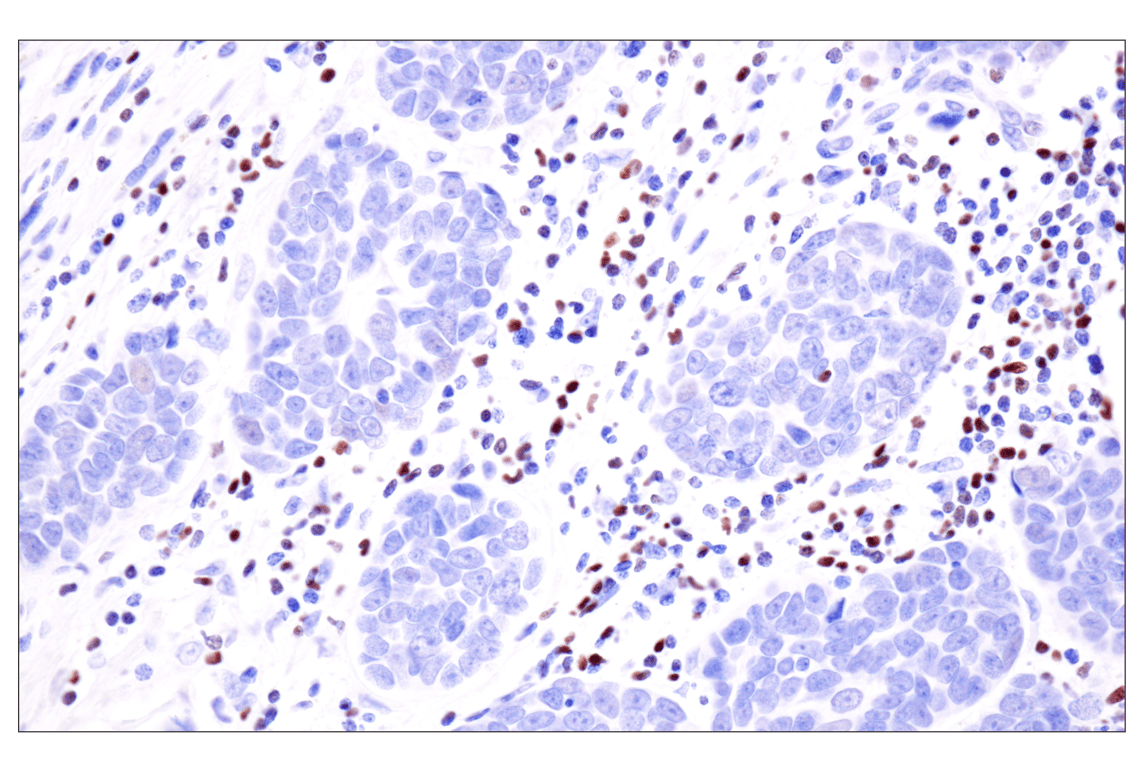
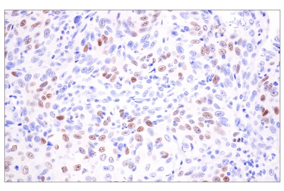
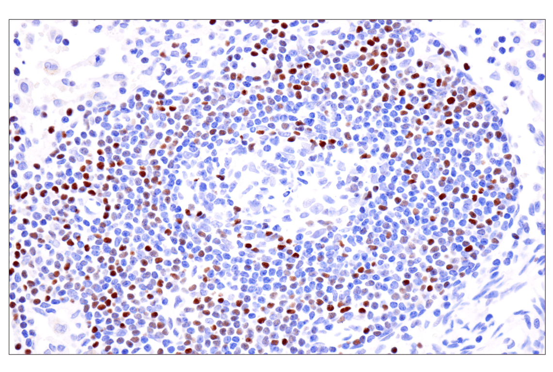
Applications:
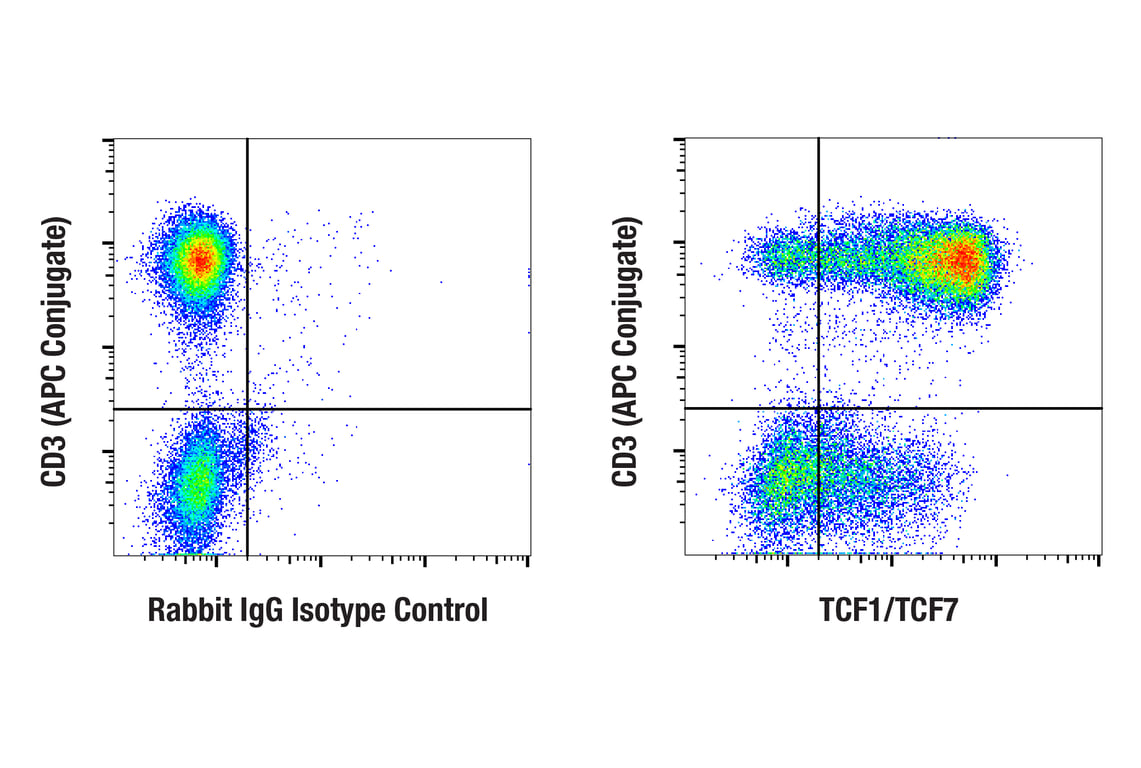
W, IHC-P, IF-F, IF-IC, FC-FP, ChIP, C&R, C&T
Reactivity:
H M R
Sensitivity:
Endogenous
MW (kDa):
28-50
Source/Isotype:
Rabbit IgG
UniProt ID:
#Q00417
Entrez-Gene Id:
21414
Product Usage Information
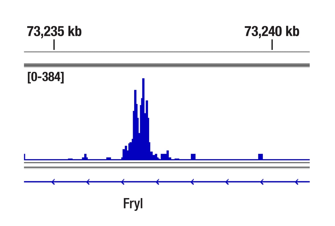
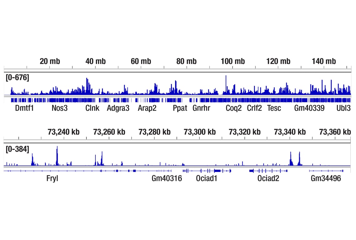
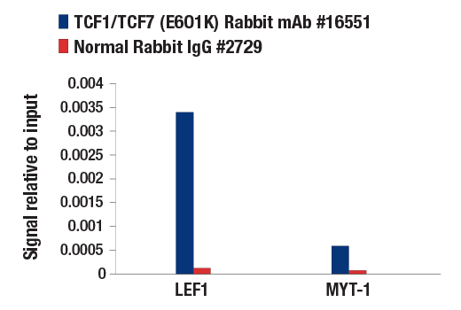
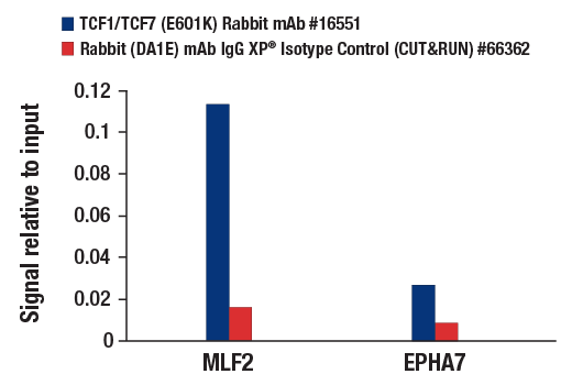
For optimal ChIP results, use 10 μl of antibody and 10 μg of chromatin (approximately 4 x 10⁶ cells) per IP. This antibody has been validated using SimpleChIP® Enzymatic Chromatin IP Kits.
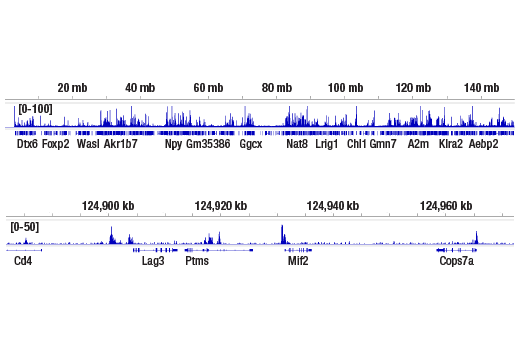
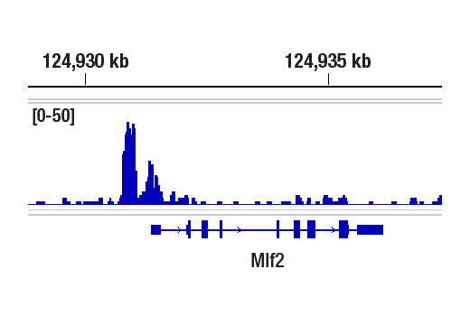
The CUT&RUN dilution was determined using CUT&RUN Assay Kit #86652.
The CUT&Tag dilution was determined using CUT&Tag Assay Kit #77552.
| Application | Dilution |
|---|---|
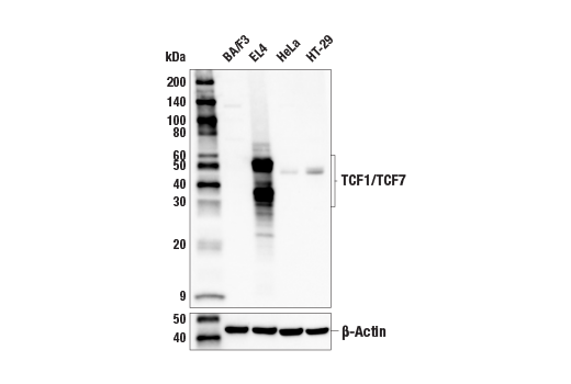
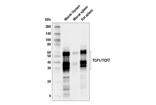
| Western Blotting | 1:1000 |
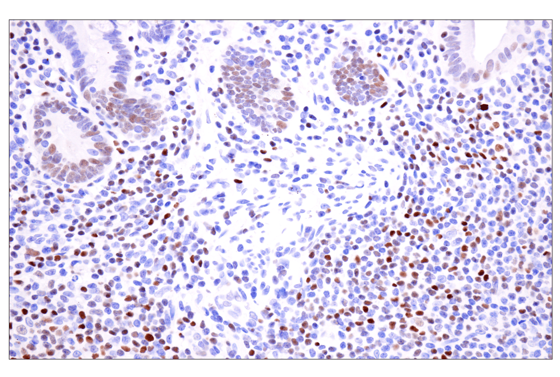
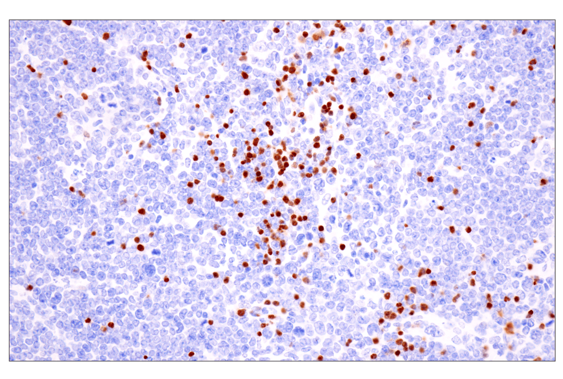
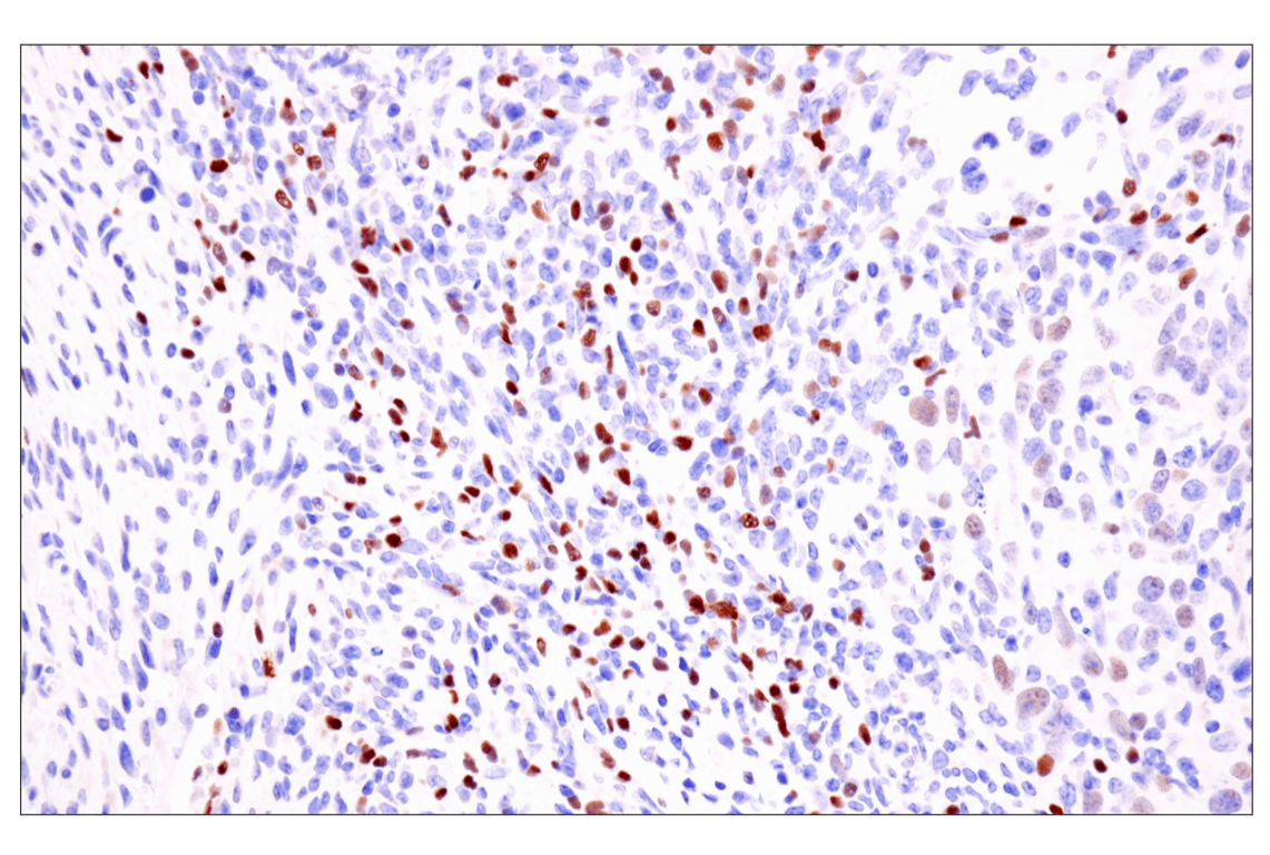
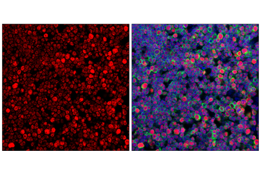
| Immunohistochemistry (Paraffin) | 1:200 - 1:800 |
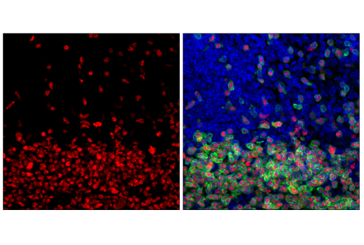
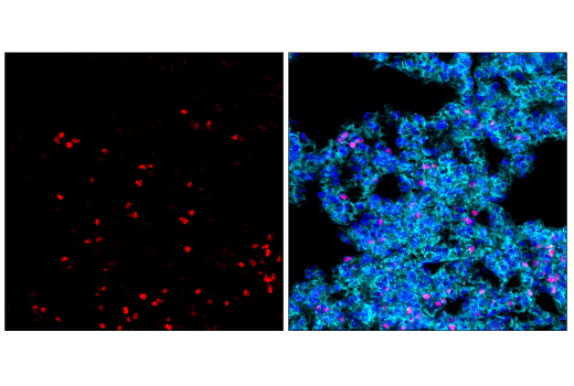
| Immunofluorescence (Frozen) | 1:400 - 1:800 |
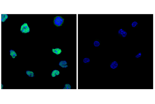
| Immunofluorescence (Immunocytochemistry) | 1:1600 - 1:3200 |
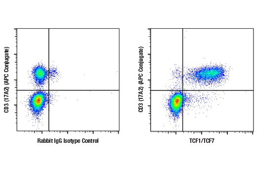
| Flow Cytometry (Fixed/Permeabilized) | 1:100 - 1:400 |
| Chromatin IP | 1:50 |
| CUT&RUN | 1:50 |
| CUT&Tag | 1:50 |
Storage
For a carrier free (BSA and azide free) version of this product see product #56853.
Specificity/Sensitivity
Source / Purification
Background
TCF1/TCF7 has several isoforms due to alternative splicing and transcription from an alternative promoter. The isoforms generated by the alternative promoter do not contain the amino-terminal β-catenin binding domain and therefore may function in a dominant negative manner (6). TCF1/TCF7 displays dynamic expression both in the total amount and the type of isoforms expressed in T cells during development and differentiation (7).
Background References
- Waterman, M.L. (2004) Cancer Metastasis Rev 23, 41-52.
- Schilham, M.W. and Clevers, H. (1998) Semin Immunol 10, 127-32.
- Brantjes, H. et al. (2002) Biol Chem 383, 255-61.
- Reya, T. and Clevers, H. (2005) Nature 434, 843-50.
- Logan, C.Y. and Nusse, R. (2004) Annu Rev Cell Dev Biol 20, 781-810.
- Waterman, M.L. (2004) Cancer Metastasis Rev 23, 41-52.
- Willinger, T. et al. (2006) J Immunol 176, 1439-46.
Species Reactivity
Species reactivity is determined by testing in at least one approved application (e.g., western blot).
Western Blot Buffer
IMPORTANT: For western blots, incubate membrane with diluted primary antibody in 5% w/v nonfat dry milk, 1X TBS, 0.1% Tween® 20 at 4°C with gentle shaking, overnight.
Applications Key
W: Western Blotting IHC-P: Immunohistochemistry (Paraffin) IF-F: Immunofluorescence (Frozen) IF-IC: Immunofluorescence (Immunocytochemistry) FC-FP: Flow Cytometry (Fixed/Permeabilized) ChIP: Chromatin IP C&R: CUT&RUN C&T: CUT&Tag
Cross-Reactivity Key
H: Human M: Mouse R: Rat
Trademarks and Patents
Cell Signaling Technology is a trademark of Cell Signaling Technology, Inc.
SimpleChIP is a registered trademark of Cell Signaling Technology, Inc.
All other trademarks are the property of their respective owners. Visit cellsignal.com/trademarks for more information.
Limited Uses
Except as otherwise expressly agreed in a writing signed by a legally authorized representative of CST, the following terms apply to Products provided by CST, its affiliates or its distributors. Any Customer's terms and conditions that are in addition to, or different from, those contained herein, unless separately accepted in writing by a legally authorized representative of CST, are rejected and are of no force or effect.
Products are labeled with For Research Use Only or a similar labeling statement and have not been approved, cleared, or licensed by the FDA or other regulatory foreign or domestic entity, for any purpose. Customer shall not use any Product for any diagnostic or therapeutic purpose, or otherwise in any manner that conflicts with its labeling statement. Products sold or licensed by CST are provided for Customer as the end-user and solely for research and development uses. Any use of Product for diagnostic, prophylactic or therapeutic purposes, or any purchase of Product for resale (alone or as a component) or other commercial purpose, requires a separate license from CST. Customer shall (a) not sell, license, loan, donate or otherwise transfer or make available any Product to any third party, whether alone or in combination with other materials, or use the Products to manufacture any commercial products, (b) not copy, modify, reverse engineer, decompile, disassemble or otherwise attempt to discover the underlying structure or technology of the Products, or use the Products for the purpose of developing any products or services that would compete with CST products or services, (c) not alter or remove from the Products any trademarks, trade names, logos, patent or copyright notices or markings, (d) use the Products solely in accordance with CST Product Terms of Sale and any applicable documentation, and (e) comply with any license, terms of service or similar agreement with respect to any third party products or services used by Customer in connection with the Products.
Revision 8
Revision 8
Revision 8

Revision 8

Revision 8
Revision 8
Revision 8
Revision 8