Revision 2
#77397
Store at -20C
TGF-beta Fibrosis Pathway Antibody Sampler Kit
1 Kit
(9 x 20 microliters)
877-616-CELL (2355)
877-678-TECH (8324)
3 Trask Lane | Danvers | Massachusetts | 01923 | USA
For Research Use Only. Not for Use in Diagnostic Procedures.
| Product Includes | Product # | Quantity | Mol. Wt | Isotype/Source |
|---|---|---|---|---|
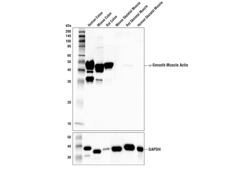
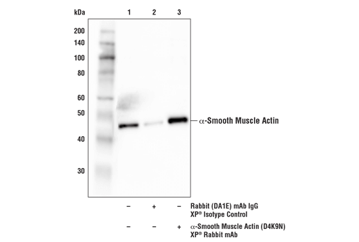
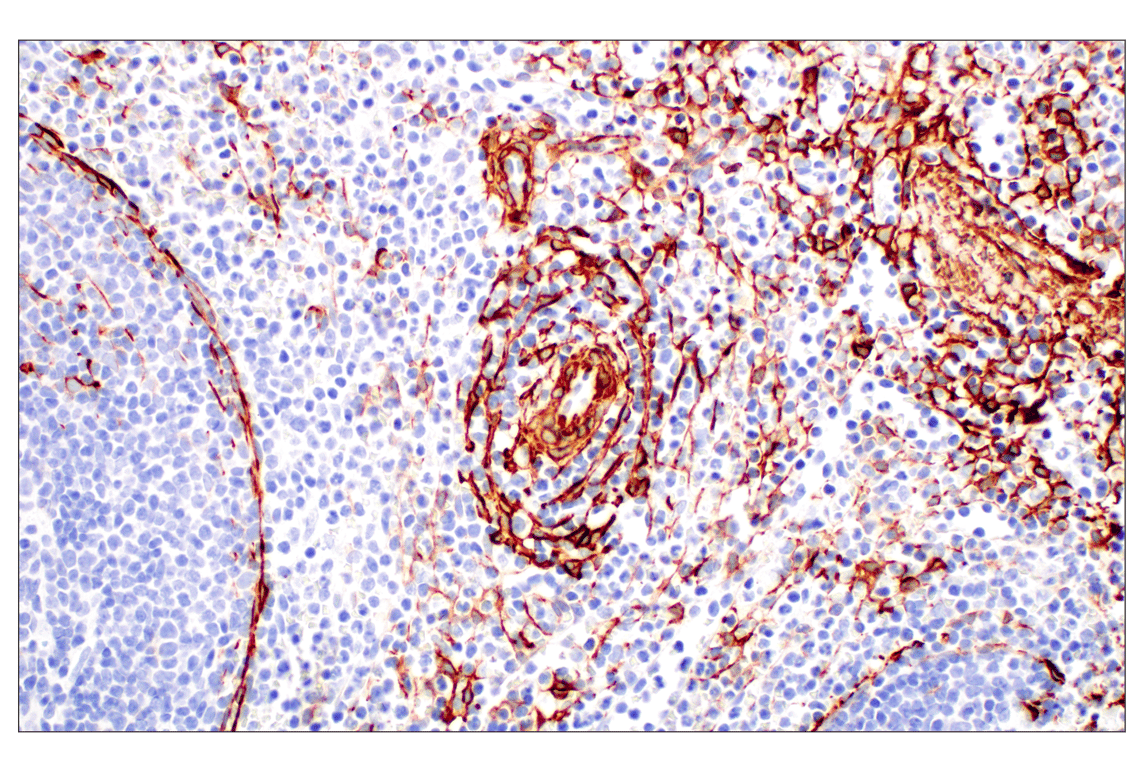
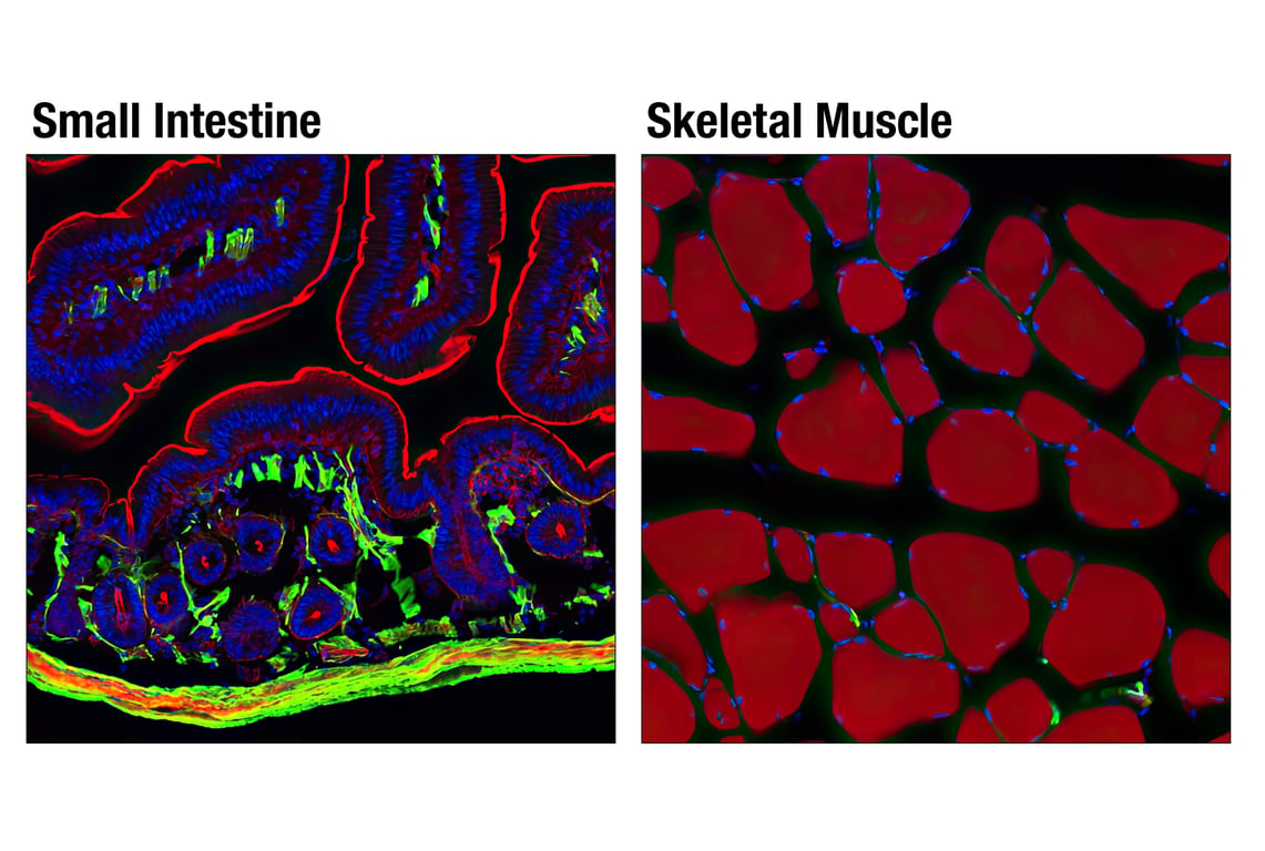
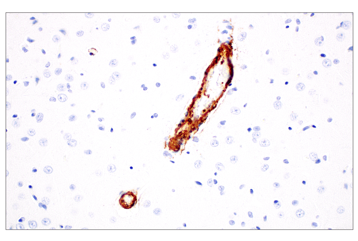
| alpha-Smooth Muscle Actin (D4K9N) Rabbit Monoclonal Antibody | 19245 | 20 µl | 42 kDa | Rabbit IgG |
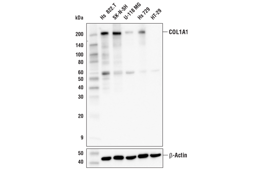
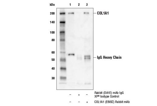
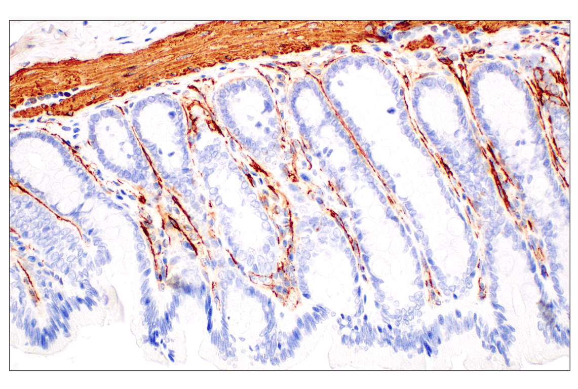
| COL1A1 (E8I9Z) Rabbit Monoclonal Antibody | 91144 | 20 µl | 220 kDa | Rabbit IgG |
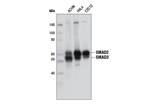
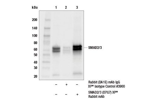
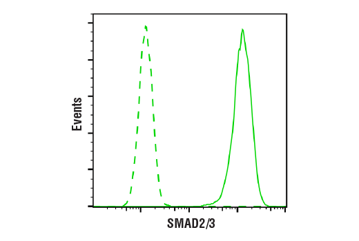
| SMAD2/3 (D7G7) Rabbit Monoclonal Antibody | 8685 | 20 µl | 52, 60 kDa | Rabbit IgG |
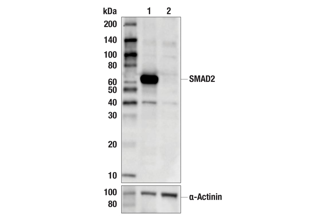
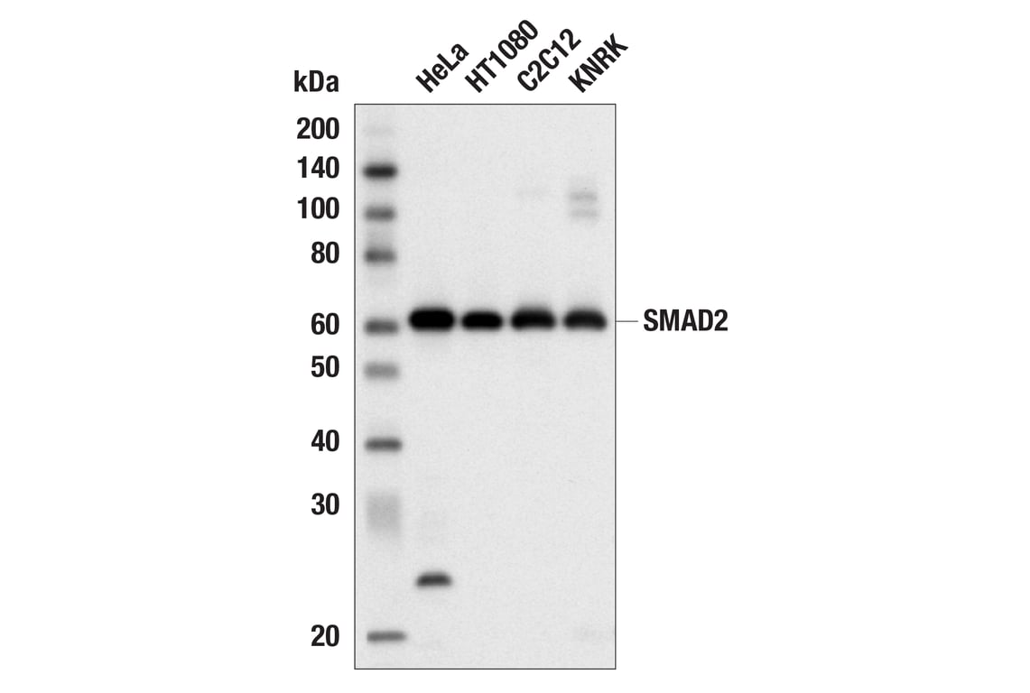
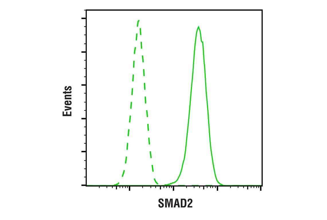
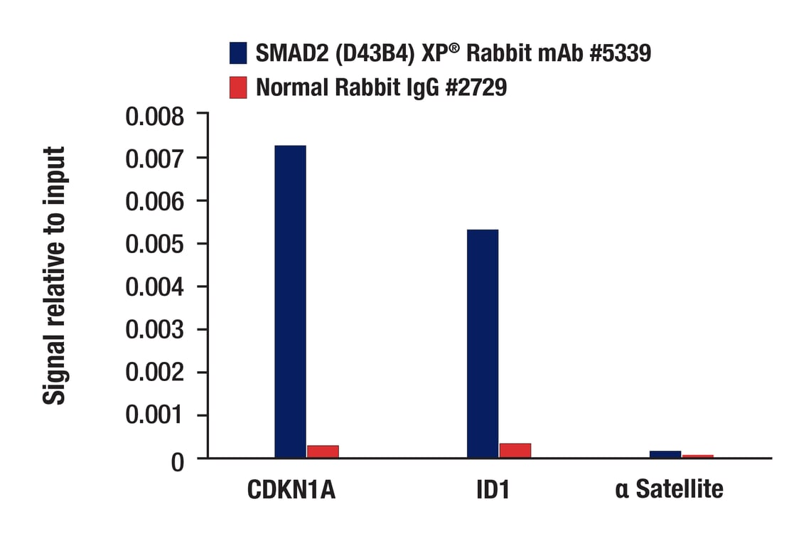
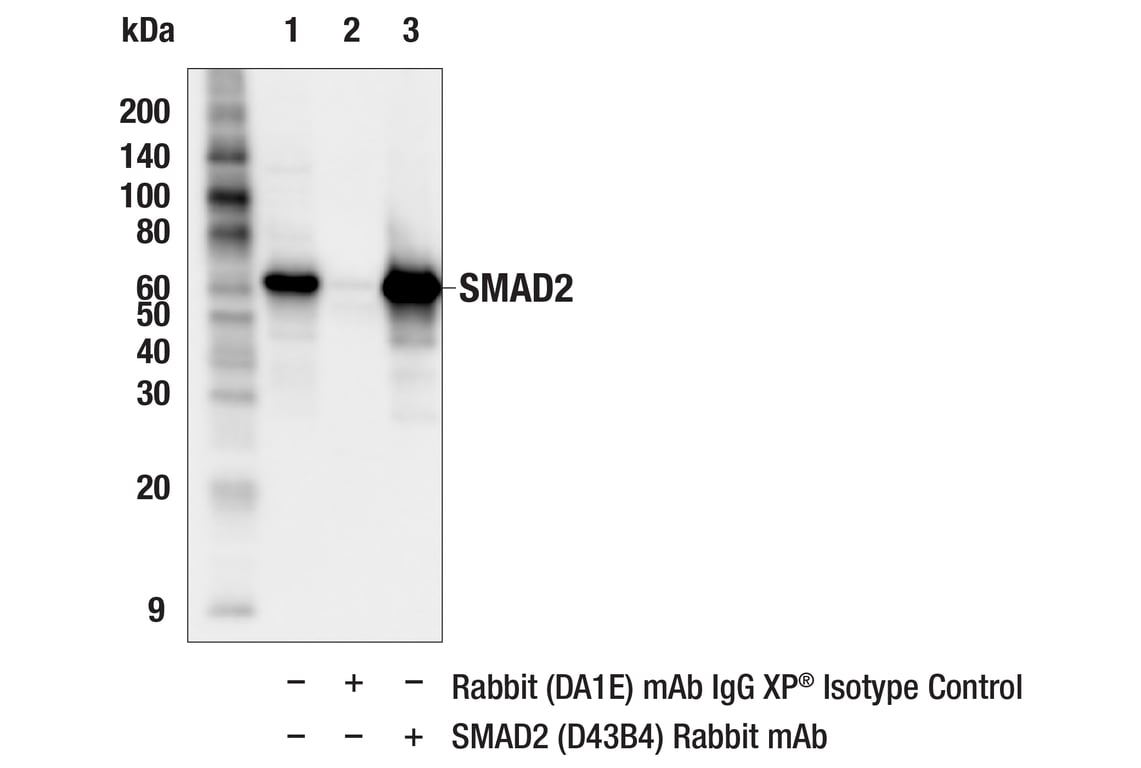
| SMAD2 (D43B4) Rabbit Monoclonal Antibody | 5339 | 20 µl | 60 kDa | Rabbit IgG |
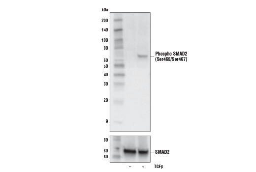
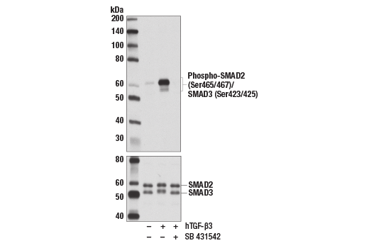
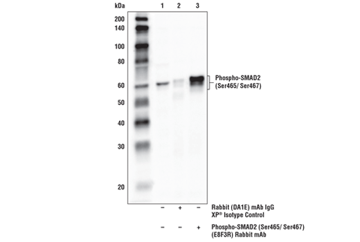
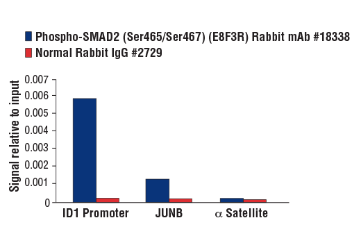
| Phospho-SMAD2 (Ser465/Ser467) (E8F3R) Rabbit Monoclonal Antibody | 18338 | 20 µl | 60 kDa | Rabbit IgG |
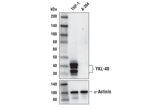
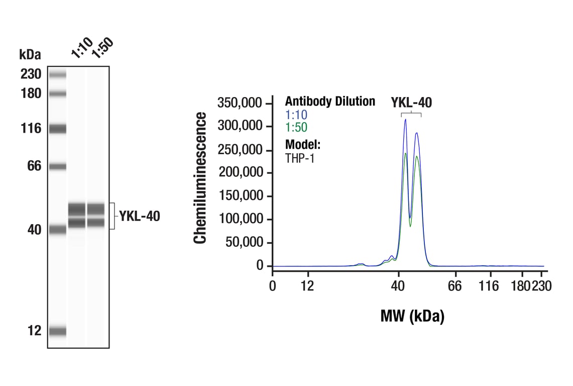
| YKL-40 (E2L1M) Rabbit Monoclonal Antibody | 47066 | 20 µl | 30-40 kDa | Rabbit IgG |
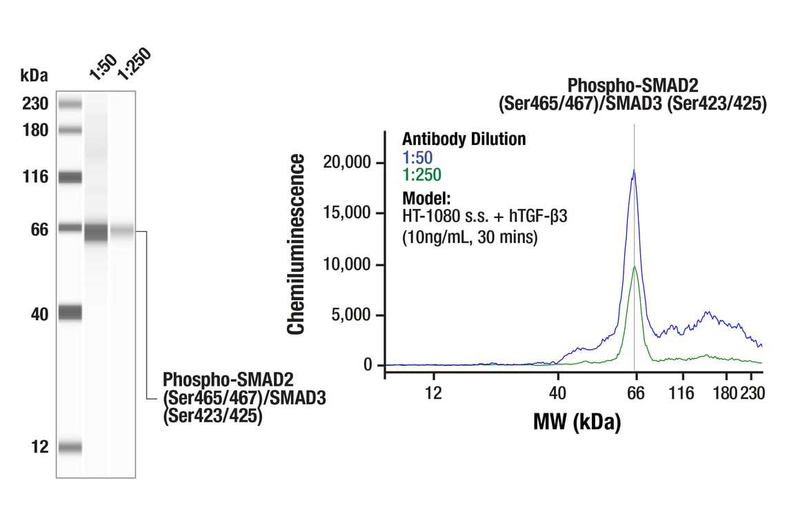
| Phospho-SMAD2 (Ser465/467)/SMAD3 (Ser423/425) (D27F4) Rabbit Monoclonal Antibody | 8828 | 20 µl | 52, 60 kDa | Rabbit IgG |
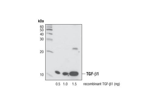
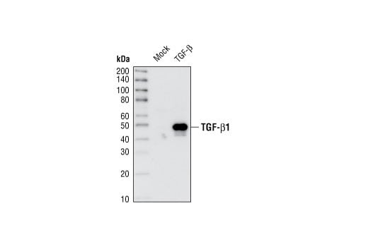
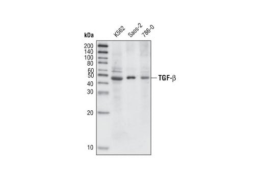
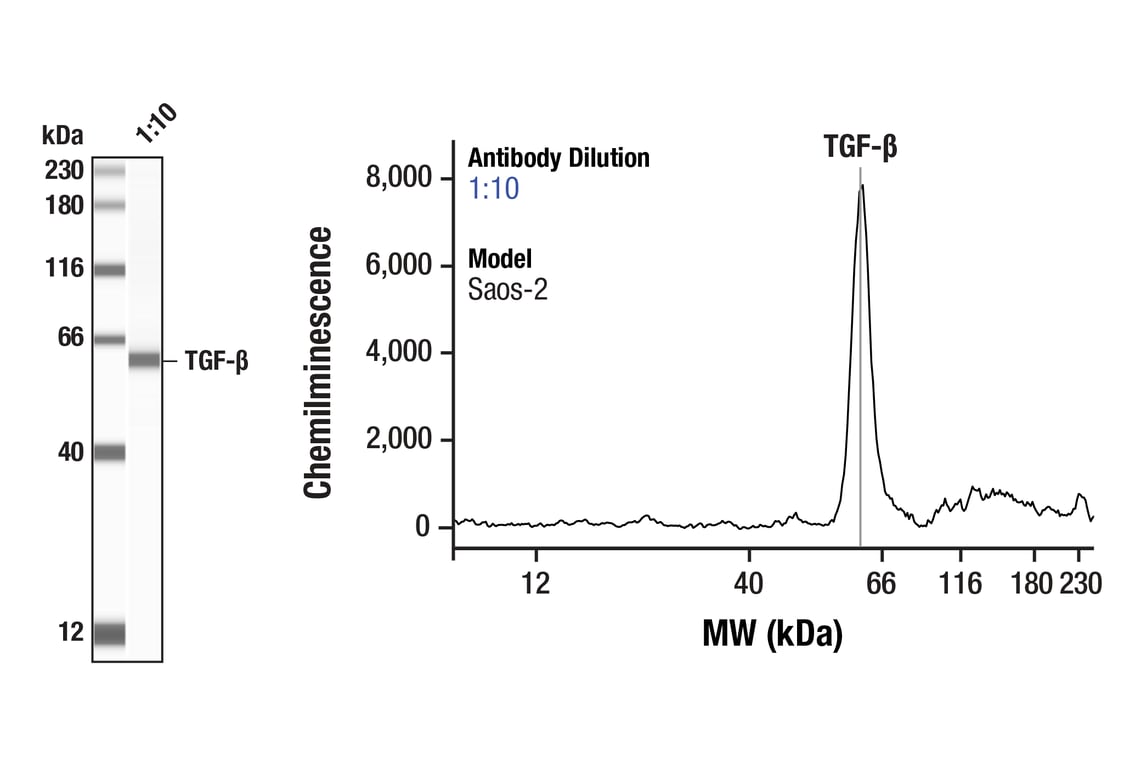
| TGF-beta (56E4) Rabbit Monoclonal Antibody | 3709 | 20 µl | 12, 45-60 kDa | Rabbit IgG |
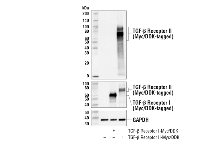
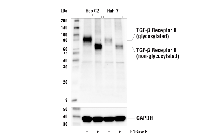
| TGF-beta Receptor II (E5M6F) Rabbit Monoclonal Antibody | 41896 | 20 µl | 85 kDa | Rabbit IgG |
| Anti-rabbit IgG, HRP-linked Antibody | 7074 | 100 µl | Goat |
Please visit cellsignal.com for individual component applications, species cross-reactivity, dilutions, protocols, and additional product information.
Description
Storage
Background
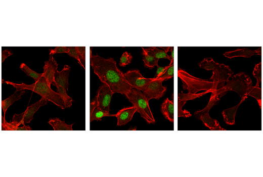
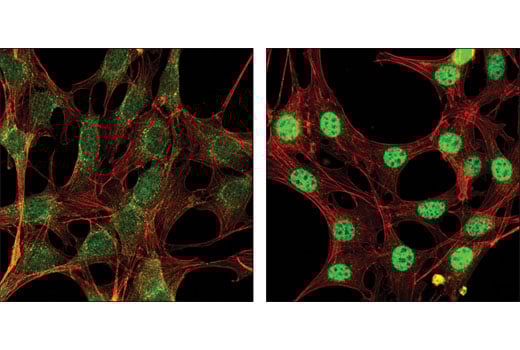
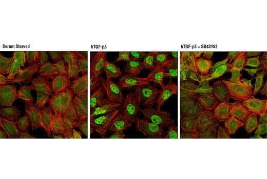
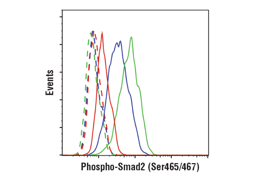
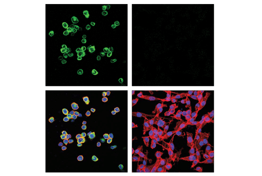
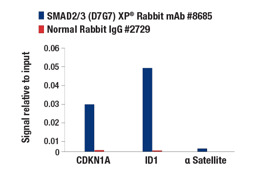
TGF-β elicits signaling through three cell surface receptors: type I (RI), type II (RII), and type III (RIII). In response to ligand binding, the type II receptors form stable heterotrimeric complexes with the type I receptors, allowing phosphorylation and activation of type I receptor kinase. Activated type I receptors associate with SMAD2/3 and phosphorylate them on a conserved carboxy terminal SSXS motif. The phosphorylated SMADs dissociate from the receptor and form a heterotrimeric complex with the co-Smad (Smad4), allowing translocation of the complex to the nucleus. Once in the nucleus, phosphorylated SMAD2/3 targets a subset of DNA binding proteins to regulate the transcriptional program (6-8).
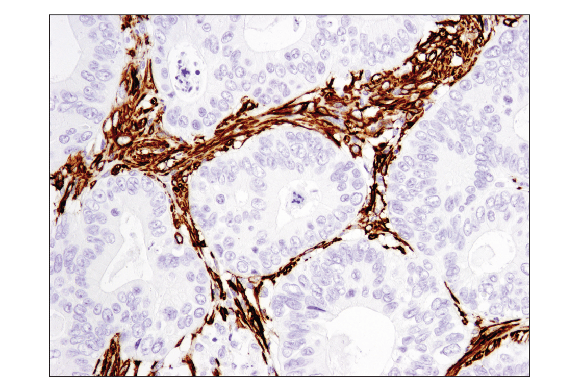
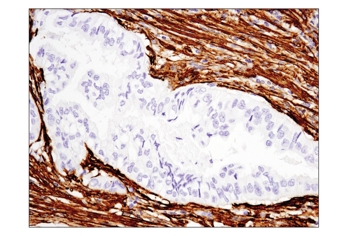
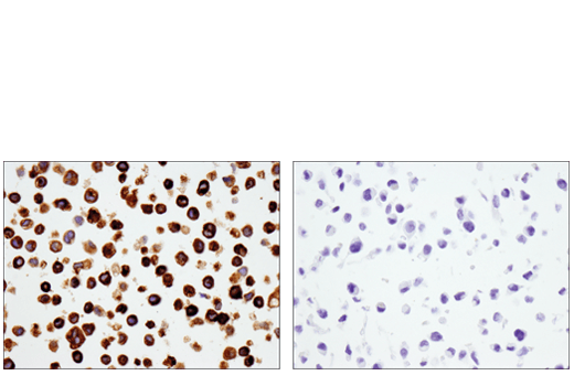
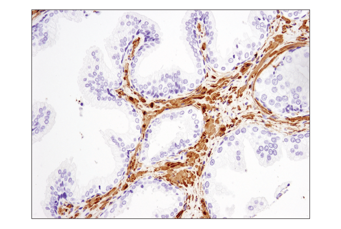
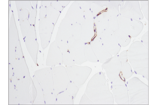
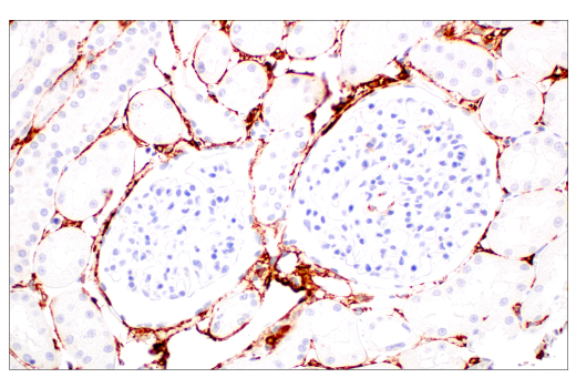
In the context of fibrosis, SMAD2/3 activation upregulates expression of profibrotic genes such as COL1A1 and other ECM modulators that modify the extracellular matrix of the tissue. (9). TGF-β/ SMAD2/3 signaling also induces expression of α-Smooth Muscle Actin in fibroblasts, causing transformation of these cells to myofibroblasts (10). Myofibroblasts further modify the ECM, causing excessive accumulation of collagens and other ECM components. Injury to the tissue attracts macrophages and other immune cells and the fibrotic tissue soon becomes a site of inflammation (11). In this pro-fibrotic, pro-inflammatory environment, YKL-40, also known as Chitinase-3-like protein 1 (CHI3L1), is secreted. YKL-40 is a pro-inflammatory glycoprotein that also contributes to the progression of fibrosis (12). Measurement of collagen content, α-Smooth Muscle Actin, and the release of YKL-40 are predictive of fibrotic activity.
Background References
- Massagué, J. et al. (2000) Cell 103, 295-309.
- de Caestecker, M.P. et al. (2000) J Natl Cancer Inst 92, 1388-402.
- Derynck, R. et al. (2001) Nat Genet 29, 117-29.
- Miyazono, K. et al. (2000) Adv Immunol 75, 115-57.
- Meng, X.M. et al. (2016) Nat Rev Nephrol 12, 325-38.
- Wu, G. et al. (2000) Science 287, 92-7.
- Attisano, L. and Wrana, J.L. (2002) Science 296, 1646-7.
- Moustakas, A. et al. (2001) J Cell Sci 114, 4359-69.
- Bagalad, B.S. et al. J Oral Maxillofac Pathol 21, 462-3.
- Mack, M. (2018) Matrix Biol 68-69, 106-21.
- Johansen, J.S. (2006) Dan Med Bull 53, 172-209.
Trademarks and Patents
Cell Signaling Technology is a trademark of Cell Signaling Technology, Inc.
CST is a registered trademark of Cell Signaling Technology, Inc.
U.S. Patent No. 5,675,063.
All other trademarks are the property of their respective owners. Visit cellsignal.com/trademarks for more information.
Limited Uses
Except as otherwise expressly agreed in a writing signed by a legally authorized representative of CST, the following terms apply to Products provided by CST, its affiliates or its distributors. Any Customer's terms and conditions that are in addition to, or different from, those contained herein, unless separately accepted in writing by a legally authorized representative of CST, are rejected and are of no force or effect.
Products are labeled with For Research Use Only or a similar labeling statement and have not been approved, cleared, or licensed by the FDA or other regulatory foreign or domestic entity, for any purpose. Customer shall not use any Product for any diagnostic or therapeutic purpose, or otherwise in any manner that conflicts with its labeling statement. Products sold or licensed by CST are provided for Customer as the end-user and solely for research and development uses. Any use of Product for diagnostic, prophylactic or therapeutic purposes, or any purchase of Product for resale (alone or as a component) or other commercial purpose, requires a separate license from CST. Customer shall (a) not sell, license, loan, donate or otherwise transfer or make available any Product to any third party, whether alone or in combination with other materials, or use the Products to manufacture any commercial products, (b) not copy, modify, reverse engineer, decompile, disassemble or otherwise attempt to discover the underlying structure or technology of the Products, or use the Products for the purpose of developing any products or services that would compete with CST products or services, (c) not alter or remove from the Products any trademarks, trade names, logos, patent or copyright notices or markings, (d) use the Products solely in accordance with CST Product Terms of Sale and any applicable documentation, and (e) comply with any license, terms of service or similar agreement with respect to any third party products or services used by Customer in connection with the Products.
Revision 2
Revision 2
Revision 2

Revision 2
Revision 2
Revision 2
Revision 2
Revision 2
Revision 2
Revision 2
Revision 2
Revision 2
Revision 2
Revision 2
Revision 2
Revision 2
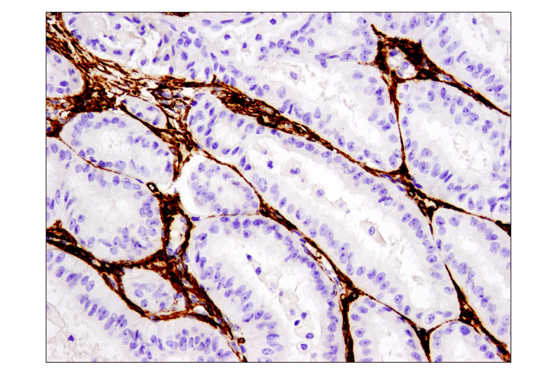
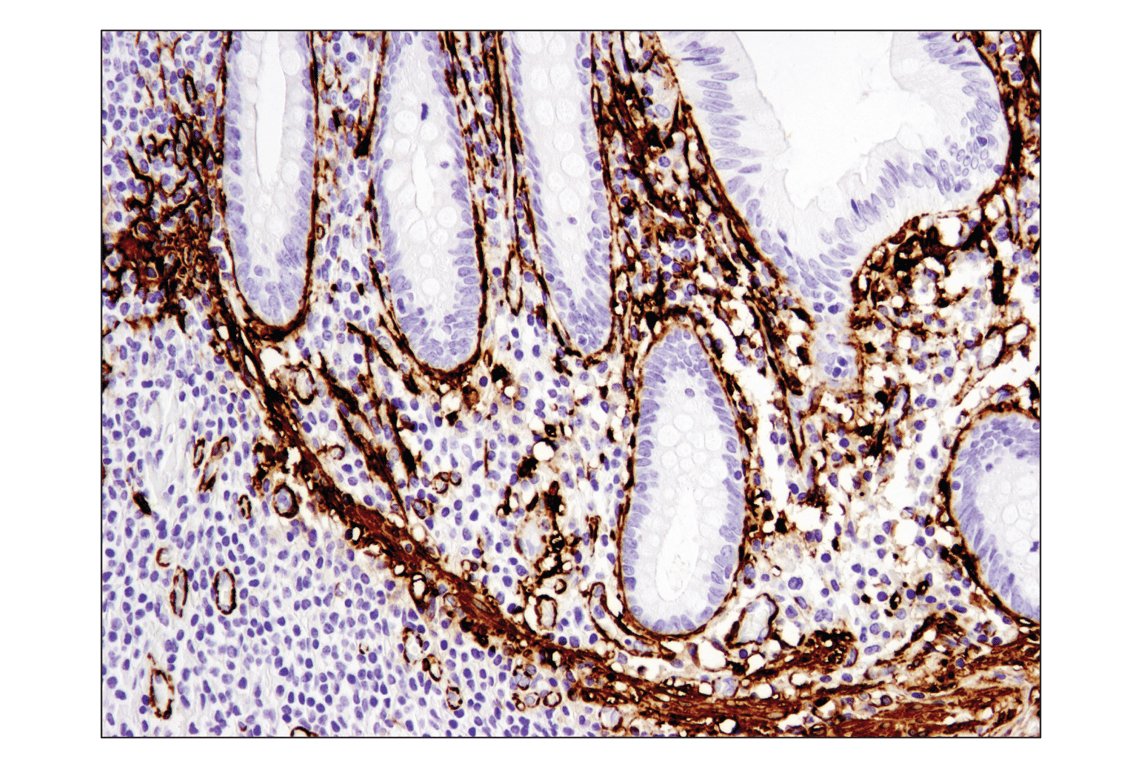
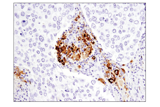
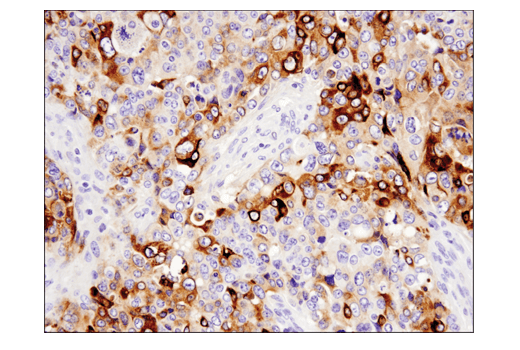
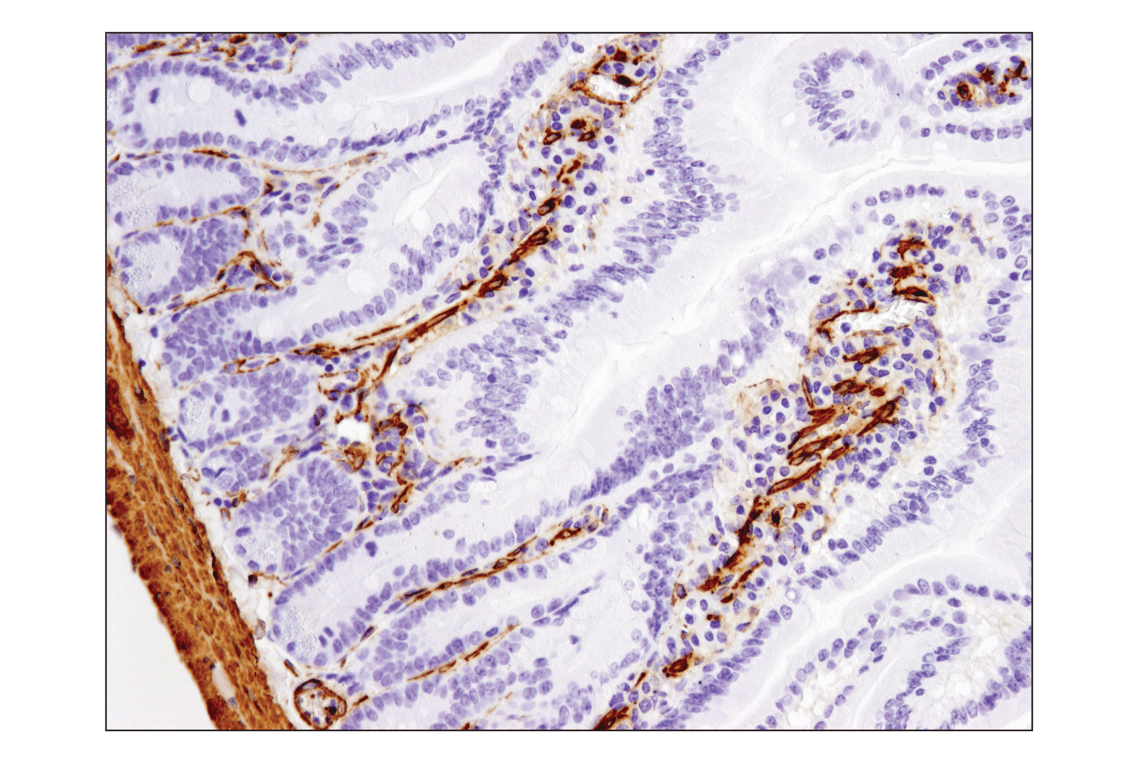
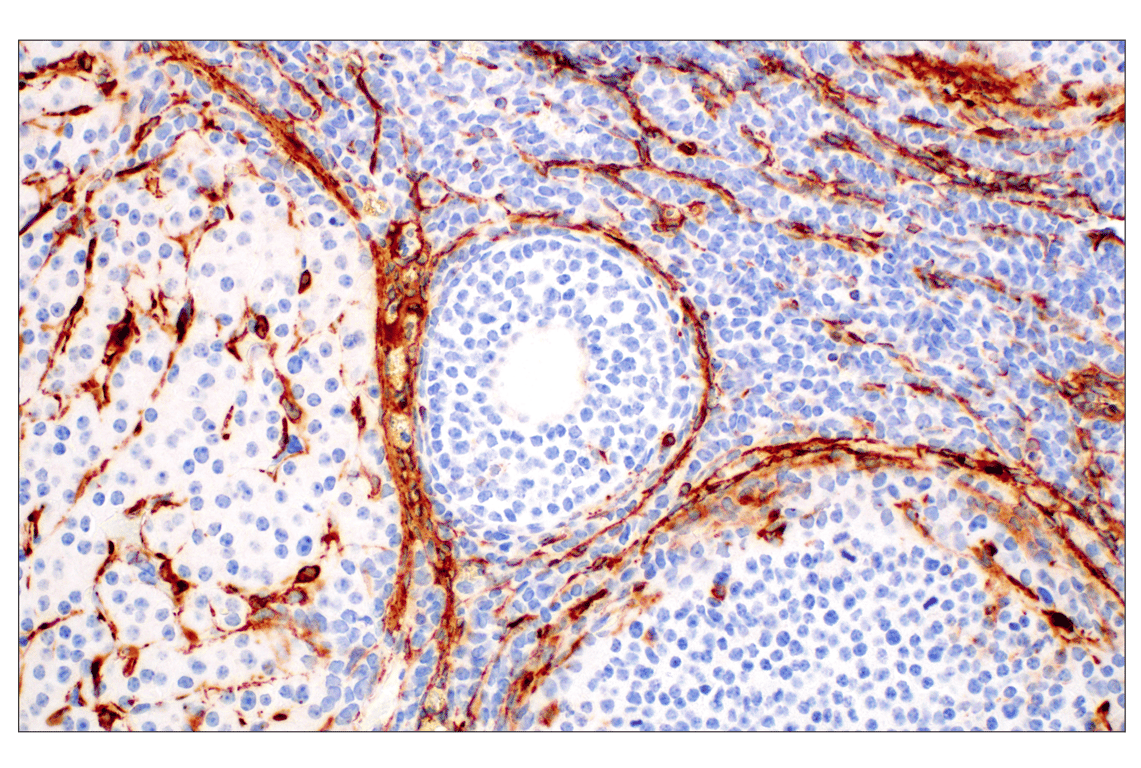
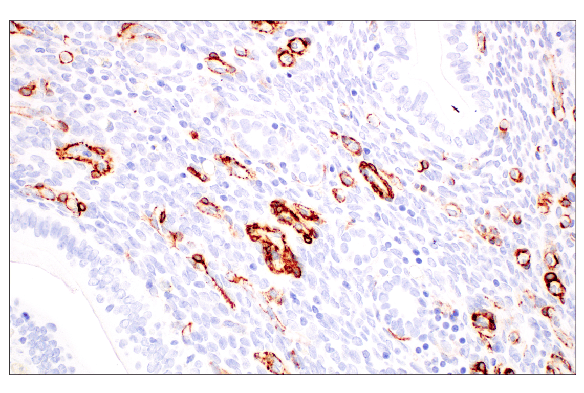
Immunohistochemical analysis of paraffin-embedded rat brain using alpha-Smooth Muscle Actin (D4K9N) XP® Rabbit mAb.
Revision 2
Revision 2
Revision 2