Revision 4
#13674
Store at -20C
877-616-CELL (2355)
877-678-TECH (8324)
3 Trask Lane | Danvers | Massachusetts | 01923 | USA
For Research Use Only. Not for Use in Diagnostic Procedures.
Applications:
W, IP, IF-IC, FC-FP
Reactivity:
H
Sensitivity:
Endogenous
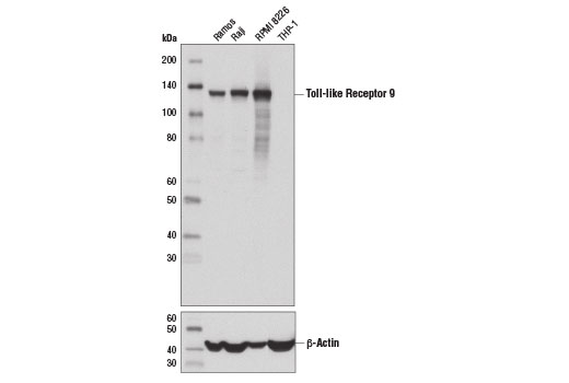
MW (kDa):
130
Source/Isotype:
Rabbit IgG
UniProt ID:
#Q9NR96
Entrez-Gene Id:
54106
Product Usage Information
| Application | Dilution |
|---|---|
| Western Blotting | 1:1000 |
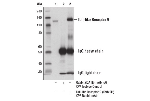
| Immunoprecipitation | 1:50 |
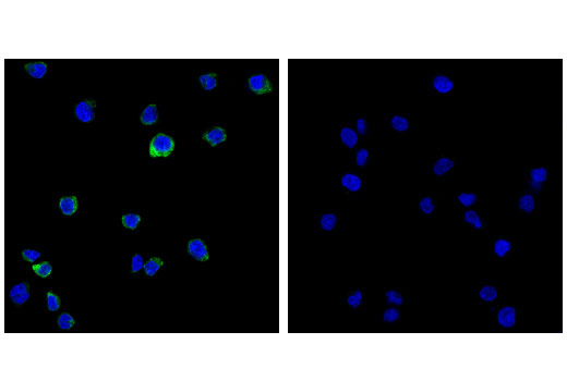
| Immunofluorescence (Immunocytochemistry) | 1:400 - 1:1600 |
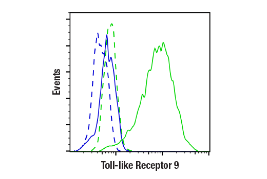
| Flow Cytometry (Fixed/Permeabilized) | 1:100 - 1:400 |
Storage
Specificity/Sensitivity
Source / Purification
Background
TLR9 is highly expressed in macrophages, dendritic cells, and B lymphocytes, and in humans has five isoforms generated by alternative splicing (15,16). TLR9 binds to unmethylated CpG motifs present on bacterial DNA and stimulates NF-κB via the MyD88 adaptor protein (17-19). In contrast to most TLR family members that are localized to the plasma membrane, TLR9 is an intracellular receptor localized to the ER in resting cells (20). Upon binding to CpG DNA, TLR9 is proteolytically processed and translocates to endo-lysosomal compartments where it binds MyD88, initiating downstream signaling (21-23).
Background References
- Akira, S. (2003) J Biol Chem 278, 38105-8.
- Beutler, B. (2004) Nature 430, 257-63.
- Dunne, A. and O'Neill, L.A. (2003) Sci STKE 2003, re3.
- Medzhitov, R. et al. (1997) Nature 388, 394-7.
- Schwandner, R. et al. (1999) J Biol Chem 274, 17406-9.
- Takeuchi, O. et al. (1999) Immunity 11, 443-51.
- Alexopoulou, L. et al. (2001) Nature 413, 732-8.
- Zhang, F.X. et al. (1999) J Biol Chem 274, 7611-4.
- Horng, T. et al. (2001) Nat Immunol 2, 835-41.
- Oshiumi, H. et al. (2003) Nat Immunol 4, 161-7.
- Muzio, M. et al. (1997) Science 278, 1612-5.
- Wesche, H. et al. (1997) Immunity 7, 837-47.
- Suzuki, N. et al. (2002) Nature 416, 750-6.
- Irie, T. et al. (2000) FEBS Lett 467, 160-4.
- Du, X. et al. (2000) Eur Cytokine Netw 11, 362-71.
- Chuang, T.H. and Ulevitch, R.J. (2000) Eur Cytokine Netw 11, 372-8.
- Hemmi, H. et al. (2000) Nature 408, 740-5.
- Bauer, S. et al. (2001) Proc Natl Acad Sci U S A 98, 9237-42.
- Takeshita, F. et al. (2001) J Immunol 167, 3555-8.
- Latz, E. et al. (2004) Nat Immunol 5, 190-8.
- Park, B. et al. (2008) Nat Immunol 9, 1407-14.
- Ewald, S.E. et al. (2008) Nature 456, 658-62.
- Sepulveda, F.E. et al. (2009) Immunity 31, 737-48.
Species Reactivity
Species reactivity is determined by testing in at least one approved application (e.g., western blot).
Western Blot Buffer
IMPORTANT: For western blots, incubate membrane with diluted primary antibody in 5% w/v BSA, 1X TBS, 0.1% Tween® 20 at 4°C with gentle shaking, overnight.
Applications Key
W: Western Blotting IP: Immunoprecipitation IF-IC: Immunofluorescence (Immunocytochemistry) FC-FP: Flow Cytometry (Fixed/Permeabilized)
Cross-Reactivity Key
H: Human
Trademarks and Patents
Cell Signaling Technology is a trademark of Cell Signaling Technology, Inc.
Alexa Fluor is a registered trademark of Life Technologies Corporation.
All other trademarks are the property of their respective owners. Visit cellsignal.com/trademarks for more information.
Limited Uses
Except as otherwise expressly agreed in a writing signed by a legally authorized representative of CST, the following terms apply to Products provided by CST, its affiliates or its distributors. Any Customer's terms and conditions that are in addition to, or different from, those contained herein, unless separately accepted in writing by a legally authorized representative of CST, are rejected and are of no force or effect.
Products are labeled with For Research Use Only or a similar labeling statement and have not been approved, cleared, or licensed by the FDA or other regulatory foreign or domestic entity, for any purpose. Customer shall not use any Product for any diagnostic or therapeutic purpose, or otherwise in any manner that conflicts with its labeling statement. Products sold or licensed by CST are provided for Customer as the end-user and solely for research and development uses. Any use of Product for diagnostic, prophylactic or therapeutic purposes, or any purchase of Product for resale (alone or as a component) or other commercial purpose, requires a separate license from CST. Customer shall (a) not sell, license, loan, donate or otherwise transfer or make available any Product to any third party, whether alone or in combination with other materials, or use the Products to manufacture any commercial products, (b) not copy, modify, reverse engineer, decompile, disassemble or otherwise attempt to discover the underlying structure or technology of the Products, or use the Products for the purpose of developing any products or services that would compete with CST products or services, (c) not alter or remove from the Products any trademarks, trade names, logos, patent or copyright notices or markings, (d) use the Products solely in accordance with CST Product Terms of Sale and any applicable documentation, and (e) comply with any license, terms of service or similar agreement with respect to any third party products or services used by Customer in connection with the Products.
Revision 4
Revision 4