View Featured Offers >>
5565
PTMScan® Phospho-PKA Substrate Motif (RRXS*/T*) Kit
Proteomic Analysis Products
PTMScan

PTMScan® Phospho-PKA Substrate Motif (RRXS*/T*) Kit #5565

Citations (0)
The Motif Logo shows the amino acid distributions around the sites recognized by the antibody.
Chart showing the proportions of underlying sequence motifs found in a PTMScan study using PKA substrate antibody and Orbitrap LC-MS analysis. Combined analysis of two experiments (#10619, #10620) on peptides from PC-12 cells gave 187 non-redundant sites containing PKA substrate and related motifs. The primary motifs are highlighted in white. The proportion containing phosphoserine was 82%, and the proportion with arginine residues at both the -3 and -2 positions (PKA substrate motif) was 64%. Although this antibody has a strong preference for the PKA substrate motif, it also recognizes peptides with just an arginine residue at the -3 position.
Product Includes Volume (with Count)
PTMScan® Phospho-PKA Substrate Motif (K/RK/RXS*/T*) Immunoaffinity Beads 10 x 80 µl
PTMScan® IAP Buffer (10X) 9993 10 x 600 µl

Product Usage Information

Cells are lysed in a urea-containing buffer, cellular proteins are digested by proteases, and the resulting peptides are purified by reversed-phase, solid-phase extraction. Peptides are then subjected to immunoaffinity purification using a PTMScan® Motif antibody conjugated to protein A agarose beads. Unbound peptides are removed through washing, and the captured PTM-containing peptides are eluted with dilute acid. Reversed-phase purification is performed on microtips to desalt and separate peptides from antibody prior to concentrating the enriched peptides for LC-MS/MS analysis. CST recommends the use of PTMScan® IAP Buffer #9993 included in the kit.

Storage

Antibody beads supplied in IAP buffer containing 50% glycerol. Store at -20°C. Do not aliquot the antibody.

Protocol

PRINT

View >Collapse >

A. Solutions and Reagents

Reagents Not Included:

  1. 200 mM HEPES, pH 8.0 (Cell Signaling Technology, #44686)
  2. Sodium pyrophosphate (Sigma, S-6422)
  3. β-glycerophosphate (Sigma, G-9891)
  4. Urea, Ultrapure, PTMScan® Qualified (Cell Signaling Technology, #60055)
  5. Sodium orthovanadate (Sigma, S-6508)
  6. Iodoacetamide, PTMScan® Qualified (Cell Signaling Technology, #88931)
  7. DTT (Dithiothreitol) (Cell Signaling Technology, #7016)
  8. PTMScan® Trypsin, Mass Spectrometry Grade (Cell Signaling Technology, #72969)
  9. PTMScan® Lys-C Protease (Cell Signaling Technology, #39003)
  10. Trifluoroacetic acid (TFA), Sequanal grade (Thermo Scientific, 28903)
  11. Trifluoroacetic Acid (TFA), Reagent grade (American Bioanalytical, AB02010
  12. Acetonitrile (Thermo Scientific, 51101)
  13. PTMScan® Peptide Purification Kit (Cell Signaling Technology, #35741)
  14. Water, LC-MS Grade (Burdick and Jackson) (Cell Signaling Technology, #27732)
  15. Phosphate Buffered Saline (PBS-20X) (Cell Signaling Technology, #9808)
  16. 1 mM Hydrochloric acid (HCL)
  17. Ammonium Bicarbonate, 1M in HPLC-Grade Water (Cell Signaling Technology, #78450)

NOTE: Prepare solutions for cell lysis (Section I), C18 column purification (Section II), and IAP enrichment (Section III) with reverse osmosis deionized (RODI) or equivalent grade water. Prepare solutions using HPLC grade water (Burdick and Jackson water) for the peptide concentration steps (Section IV and V).

Stock Solutions:

  1. HEPES, pH 8.0 (200mM): Dissolve 23.8 g HEPES in approximately 450 ml water, adjust to pH 8.0 with 5 M NaOH, and bring to a final volume of 500 ml. Filter through a 0.22 µM filter. Store at 4°C for use up to six months.
  2. Sodium pyrophosphate: Make 50X stock (125 mM): 1.1 g/20 ml. Store at 4°C for up to six months.
  3. β-glycerophosphate: Make 1000X stock: 2.2 g/10 ml. Divide into 100 µl aliquots and store at -20°C.
  4. Sodium orthovanadate: Make 100X stock: 1.84 g/100 ml. Sodium orthovanadate must be depolymerized (activated) according to the following protocol:
    1. For a 100 ml solution, fill up with water to approximately 90 ml. Adjust the pH to 10.0 using 1 M NaOH with stirring. At this pH, the solution will be yellow.
    2. Boil the solution until it turns colorless and cool to room temperature (put on ice for cooling).
    3. Readjust the pH to 10.0 and repeat step 2 until the solution remains colorless and the pH stabilizes at 10.0 (usually it takes two rounds). Adjust the final volume to 100 ml with water.
    4. Store the activated sodium orthovanadate in 1 ml aliquots at -20°C for up to six months. Thaw one aliquot for each experiment; do not refreeze thawed vial.
  5. Dithiothreitol (DTT): Make 1.25 M stock: 19.25 g/100 ml. Divide into 200 µl aliquots. Store at -20°C for up to one year. Thaw one aliquot for each experiment.
  6. Lysyl Endopeptidase (LysC): Store dry powder up to 2 years at -80°C. Seal the cap of the LysC container with parafilm to avoid collecting moisture, which can lead to degradation of the reagent. Prepare 5 mg/ml stock in 20 mM HEPES pH 8.0. Divide into single use aliquots, store at -80°C for up to one year.

I. Cell Lysis and Protein Digestion

A. Solutions and Reagents

NOTE: Prepare solutions with RODI or equivalent grade water.

  1. Urea Lysis Buffer: 20 mM HEPES pH 8.0, 9 M urea, 1 mM sodium orthovanadate, 2.5 mM sodium pyrophosphate, 1 mM β-glycerophosphate.
  2. NOTE: The Urea Lysis Buffer should be prepared fresh prior to each experiment. Do not include protease inhibitors.

    NOTE: Dissolving urea is an endothermic reaction. Urea Lysis Buffer preparation can be facilitated by placing a stir bar in the beaker and by using a warm (not hot) water bath on a stir plate. 9 M urea is used so that upon lysis, the final concentration is approximately 8 M. The urea lysis buffer should be used at room temperature. Placing the urea lysis buffer on ice will cause the urea to precipitate out of solution.

  3. DTT solution (1.25 M) (see stock solutions for preparation)
  4. Iodoacetamide solution: Dissolve 95 mg of iodoacetamide in water to a final volume of 5 ml. After weighing the powder, store in the dark and add water only immediately before use. The iodoacetamide solution should be prepared fresh prior to each experiment.

B. Preparation of Cell Lysate, Suspension Cells

  1. Grow approximately 1-2 x 10⁸ cells for each experimental condition (enough cells to produce approximately 10-20 mg of soluble protein).
  2. Harvest cells by centrifugation at 130 x g, for 5 min at room temperature. Carefully remove supernatant, wash cells with 20 ml of cold 1x PBS, centrifuge, remove PBS wash, and add 10 ml Urea Lysis Buffer (room temperature) to the cell pellet. Pipet the slurry up and down a few times (do not cool lysate on ice as this may cause precipitation of the urea).
  3. NOTE: If desired, the PTMScan® protocol may be interrupted at this stage. The harvested cells can be frozen and stored at -80°C for several weeks.

  4. Using a microtip, sonicate lysate at 15 W output with 3 bursts of 15 sec each. Cool on ice for 1 min between each burst. Clear the lysate by centrifugation at 20,000 x g for 15 min at room temperature and transfer the protein extract (supernatant) into a new tube.
  5. NOTE: Centrifugation is performed at room temperature to prevent urea from precipitating out of solution.

    NOTE: Lysate sonication fragments DNA and reduces sample viscosity. Ensure that the sonicator tip is submerged in the lysate. If the sonicator tip is not submerged properly, it may induce foaming and degradation of your sample.

C. Preparation of Cell Lysate, Adherent Cells

  1. Grow 1-2 x 10⁸ cells for each experimental condition (enough cells to produce approximately 10-20 mg of soluble protein). The cell number corresponds to approximately 10 x 150 mm culture dishes (depending on the cell type), grown to 70-80% confluence.
  2. Harvest all 10 x 150 mm culture dishes for one sample, remove media from the first dish by decanting, and let stand in a tilted position for 30 seconds so the remaining medium flows to the bottom edge. Remove the remainder of the medium at the bottom edge with a P-1000 micropipettor. Rinse each dish with 5 ml of cold PBS. Remove PBS as described above.
  3. Add 10 ml of Urea Lysis Buffer (at room temperature) to the first dish, scrape the cells into the buffer, and let the dish stand in tilted position after scraping the buffer to the bottom edge of the tilted dish. Remove the medium from the second dish as above. Transfer the lysis buffer from the first dish to the second dish using a 10 ml pipette, then tilt the first dish with the lid on for 30 sec and remove remaining buffer from the dish and collect. Scrape cells from the second dish and repeat the process until the cells from all the dishes have been scraped into the lysis buffer. Collect all lysate in a 50 ml conical tube.
  4. NOTE: DO NOT place Urea Lysis Buffer or culture dishes on ice during harvesting. Harvest cells using Urea Lysis Buffer at room temperature. During lysis, the buffer becomes viscous due to DNA released from the cells.

  5. The yield will be approximately 9-12 ml lysate after harvesting all the culture plates.
  6. NOTE: If desired, the PTMScan® protocol may be interrupted at this stage. The cell lysate can be frozen and stored at -80°C for several weeks.

  7. Using a microtip, sonicate lysate at 15 W output with 3 bursts of 15 sec each. Cool on ice for 1 min between each burst. Clear the lysate by centrifugation at 20,000 x g for 15 min at room temperature and transfer the protein extract (supernatant) into a new tube.
  8. NOTE: Lysate sonication fragments DNA and reduces sample viscosity. Ensure that the sonicator tip is submerged in the lysate. If the sonicator tip is not submerged properly, it may induce foaming and degradation of your sample.

D. Reduction and Alkylation of Proteins

  1. Add 1/278 volume of 1.25 M DTT to the cleared cell supernatant (e.g. 36 µl of 1.25 M DTT for 10 ml of protein extract), mix well and incubate at room temperature for 60 min.
  2. Add 1/10 volume of iodoacetamide solution to the cleared cell supernatant, mix well, and incubate for 15 min at room temperature in the dark.

E. Protease Digestion

  1. Dilute 4-fold with 20 mM HEPES pH 8.0 to a final concentration of approximately 2 M urea, 20 mM HEPES, pH 8.0. For example, add 30 ml 20 mM HEPES pH 8.0 for 10 ml of lysate.
  2. * To view an updated protease digestion reference table, please visit http://www.cellsignal.com/services/ptmscan_kits.html

    NOTE: For LysC-digested material, a second protease digestion is required after the C18 tip purification of enriched peptides (see the protocol after C18 tip Purification).

    NOTE: Alternative proteases such as GluC, chymotrypsin, and others can be used in addition to the protease digests outlined in the reference table to expand the coverage of modified peptides from each Motif Antibody. When considering the use of additional protease digests it should be compatible with the respective Motif Antibody by not cleaving residues within the designated sequence motif. Alternate protease digests that generate larger proteolytic peptides may not be ideal if the resulting peptides do not ionize well in the mass spectrometer.

F. LysC Digestion

  1. Prepare 5 mg/ml stock solution of LysC in 20 mM HEPES pH 8.0. Aliquot for single use and store at -80°C.
  2. Add LysC solution to peptides at 1:250 (w:w). For 20 mg sample, use 20 mg ÷ 250 = 80 µg x 1 µl/5 µg = 16 µl LysC and digest overnight at room temperature.
  3. Analyze the lysate before and after digest by SDS-PAGE to check for complete digestion.
  4. Continue through the C18 column purification, IAP, and C18 tip protocols before conducting the SECONDARY DIGESTION with trypsin (see end of protocol, Trypsin Digestion of Enriched LysC peptides)

II. C18 Purification of Lysate Peptides

NOTE: Purification of peptides is performed at room temperature on C18 reversed-phase columns from PTMScan® Peptide Purification Kit (Cell Signaling Technology, #35741).

NOTE: C18 purification uses reversed-phase (hydrophobic) solid-phase extraction. Peptides and lipids bind to the chromatographic material. Large molecules such as DNA, RNA, and most protein, as well as hydrophilic molecules such as many small metabolites are separated from peptides using this technique. Peptides are eluted from the column with 40% acetonitrile (ACN) and separated from lipids and proteins, which elute at approximately 60% ACN and above.

NOTE: About 20 mg of protease-digested peptides can be purified from one C18 column. Purify peptides immediately after proteolytic digestion.

A. Solutions and Reagents

NOTE: Prepare solutions with RODI or equivalent grade water. Use Trifluoroacetic acid (TFA), Reagent grade (American Bioanalytical, AB02010) and Pierce Acetonitrile (ACN), LC-MS Grade (Thermo Scientific, 51101) when preparing solutions. All percentage specifications for solutions are vol/vol.

  1. 20% trifluoroacetic acid (TFA): add 10 ml TFA to water to a total volume of 50 ml.
  2. Solvent A (0.1% TFA): add 5 ml of 20% TFA to 995 ml water.
  3. Solvent B (0.1% TFA, 40% acetonitrile): add 400 ml of acetonitrile (ACN) and 5 ml of 20% TFA to 500 ml of water, adjust final volume to 1 l with water.
  4. Wash buffer (0.1% TFA, 5% acetonitrile): For 100 ml of wash buffer, add 0.5 ml of 20% TFA to 50 ml of water, then add 5 ml of acetonitrile, adjust final volume to 100 ml with water.
  5. NOTE: Organic solvents are volatile. Tubes containing small volumes of these solutions should be prepared immediately before use and should be kept capped as much as possible because the organic components evaporate quickly.

B. Acidification of Digested Cell Lysate

NOTE: Before loading the peptides from the digested sample on the column, they must be acidified with TFA for efficient peptide binding. The acidification step helps remove fatty acids from the digested peptide mixture.

  1. Add 1/20 volume of 20% TFA to the digest for a final concentration of 1% TFA. Check the pH by spotting a small amount of peptide sample on a pH strip (the pH should be under 3). After acidification, allow precipitate to form by letting sample stand for 15 min on ice.
  2. Centrifuge the acidified peptide solution for 15 min at 1,780 x g at room temperature to remove the precipitate. Transfer peptide-containing supernatant into a new 50 ml conical tube without dislodging the precipitated material.

C. Peptide Purification

NOTE: Application of all solutions should be performed by gravity flow.

  1. Connect a 10 cc syringe (remove plunger) to the SHORT END of the C18 column.
  2. Pre-wet the column with 5 ml 100% ACN.
  3. NOTE: Each time solution is applied to the column, air bubbles form in the junction where the 10 cc syringe meets the narrow inlet of the column. These must be removed with a gel-loading tip placed on a P-200 micropipettor, otherwise the solution will not flow through the column efficiently. Always check for appropriate flow.

  4. Wash sequentially with 1 ml, 3 ml, and 6 ml of Solvent A (0.1% TFA).
  5. Load acidified and cleared digest (from Section B).
  6. NOTE: In rare cases, if the flow rates decrease dramatically upon (or after) loading of sample, the purification procedure can be accelerated by gently applying pressure to the column using the 10 cc plunger after cleaning it with organic solvent. Again make sure to remove air bubbles from the narrow inlet of the column before doing so. Do not apply vacuum.

  7. Wash sequentially with 1 ml, 5 ml, and 6 ml of Solvent A (0.1% TFA).
  8. Wash with 2 ml of wash buffer.
  9. Place columns above new 15 or 50 ml polypropylene tubes to collect eluate. Elute peptides with a sequential wash of 3 x 2 ml of Solvent B (0.1% TFA, 40% acetonitrile).
  10. Freeze the eluate on dry ice (or -80°C freezer) for 2 hr to overnight and lyophilize frozen peptide solution for a minimum of 2 days to assure TFA has been removed from the peptide sample.
  11. NOTE: The lyophilization should be performed in a standard lyophilization apparatus. DO NOT USE a SPEED-VAC apparatus at this stage of the protocol.

    NOTE: The lysate digest may have a much higher volume than the 10 cc reservoir will hold (up to 50-60 ml from adherent cells) and therefore the peptides must be applied in several fractions. If available, a 60 cc syringe may be used in place of a 10 cc syringe to allow all sample to be loaded into the syringe at once.

    NOTE: Lyophilized, digested peptides are stable at -80°C for several months (seal the closed tube with parafilm for storage). The PTMScan® procedure can be interrupted before or after lyophilization. Once the lyophilized peptide is dissolved in IAP buffer (see next step), continue to the end of the procedure.

III. Immunoaffinity Purification (IAP)

A. Solutions and Reagents

NOTE: Prepare solutions with RODI or equivalent grade water. Trifluoroacetic acid should be of the highest grade. All percentage specifications for solutions are vol/vol.

  1. Materials Provided in the PTMScan Kit: 10X IAP buffer; dilute with RODI or equivalent water to 1X concentration before use. Store 1X buffer up to one month at 4°C.

B. Procedure

  1. Centrifuge the tube containing lyophilized peptide for 5 minutes at 2,000 x g at room temperature to collect all material for dilution in IAP buffer. Add 1.4 ml IAP buffer. Resuspend pellets mechanically by pipetting repeatedly with a P-1000 micropipettor taking care not to introduce excessive bubbles into the solution. Transfer solution to a 1.7 ml Eppendorf tube.
  2. NOTE: After dissolving the peptide, check the pH of the peptide solution by spotting a small volume on pH indicator paper (The pH should be close to neutral, or no lower than 6.0. In the rare case that the pH is more acidic (due to insufficient removal of TFA from the peptide under sub-optimal conditions of lyophilization), titrate the peptide solution with 1 M Tris base solution that has not been adjusted for pH. 5-10 µl is usually sufficient to neutralize the solution.

  3. Clear solution by centrifugation for 5 min at 10,000 x g at 4°C in a microcentrifuge. The insoluble pellet may appear considerable. This will not pose a problem since most of the peptide will be soluble. Cool on ice.
  4. Centrifuge the vial of antibody-bead slurry at 2,000 x g for 30 sec and remove all buffer from the beads. Wash antibody beads four times with 1 ml of 1X PBS. Centrifuge at 2,000 x g after each wash. Resuspend beads in 40 µl PBS in the provided vial.
  5. Transfer the peptide solution into the vial containing motif antibody beads. Pipet sample directly on top of the beads at the bottom of the tube to ensure immediate mixing. Avoid creating bubbles upon pipetting.
  6. Tighten the cap on the vial. Seal the top of the vial with parafilm to avoid leakage. Incubate on a rotator for 2 hr at 4°C.
  7. Centrifuge at 2,000 x g for 30 sec and transfer the supernatant with a P-1000 micropipettor to a labeled Eppendorf tube to save at -80°C for future use. Flow-through material can be used for subsequent IAPs.
  8. NOTE: Some Phenol Red pH indicator may remain (it co-elutes during the C18 purification of peptides) and color the peptide solution yellow. This coloration has no effect on the immunoaffinity purification step.

    NOTE: Perform all subsequent wash steps at 2-4°C. For all the washes except the final wash, avoid removing the last few microliters, since this may cause inadvertent removal of the beads.

  9. Add 1 ml of IAP buffer to the beads, mix by inverting tube 5 times, centrifuge for 30 sec at 2,000 x g, and remove supernatant with a P-1000 micropipettor.
  10. Repeat step 7 once for a total of TWO IAP buffer washes.
  11. NOTE: All steps from this point forward should be performed with solutions prepared with Burdick and Jackson or other HPLC grade water.

  12. Add 1 ml chilled HPLC water to the beads, mix by inverting tube 5 times, centrifuge for 30 sec at 2,000 x g, and remove supernatant with a P-1000 micropipettor.
  13. Repeat step 9 two times for a total of THREE water washes. During the last water wash, the tube may need to be shaken while inverting in order to ensure efficient mixing.
  14. NOTE: After the last wash step, remove supernatant with a P-1000 micropipettor as before, then centrifuge for 5 sec at 2,000 x g to remove fluid from the tube walls, and carefully remove all remaining supernatant with a gel loading tip attached to a P-200 micropipettor.

  15. Add 55 µl of 0.15% TFA to the beads, tap the bottom of the tube several times (do not vortex), and let stand at room temperature for 10 min, mixing gently every 2-3 min.
  16. NOTE: In this step, the post-translationally modified peptides of interest will be in the eluent.

  17. Centrifuge 30 sec at 2,000 x g in a microcentrifuge and transfer supernatant to a new 1.7 ml Eppendorf tube.
  18. Add 50 µl of 0.15% TFA to the beads, and repeat the centrifugation/elution step. Combine both eluents in the same 1.7 ml tube. Briefly centrifuge the eluent to pellet any remaining beads and carefully transfer eluent to a new 1.7 ml tube taking care not to transfer any beads.

IV. Concentration and Purification of Peptides for LC-MS Analysis

NOTE: We recognize there are many other routine methods for concentrating peptides using commercial products such as C18 tips (see below) that have been optimized for peptide desalting/ concentration. Regardless of the particular method, we recommend that the method of choice be optimized for recovery and be amenable for peptide loading capacities of at least 10 µg.

C18 tips: Thermo Scientific, part number 84850

V. Concentration and Purification of Peptides for LC-MS on StageTip

A. Solutions and Reagents

NOTE: Prepare solutions with Burdick and Jackson water or other HPLC grade water. Organic solvents (trifluoroacetic acid, acetonitrile) should be of the highest grade. Pierce Trifluoroacetic Acid (TFA), Sequencing grade (Thermo Scientific, 28903) and Pierce Acetonitrile (ACN), LC-MS Grade (Thermo Scientific, 51101) are recommended.

  1. Solvent C (0.1% trifluoroacetic acid, 50% acetonitrile): add 0.1 ml trifluoroacetic acid to 40 ml HPLC water, then add 50 ml acetonitrile, adjust the final volume to 100 ml with HPLC water.
  2. Solvent D (0.1% trifluoroacetic acid): add 0.1 ml trifluoroacetic acid to 50 ml HPLC water, adjust the final volume to 100 ml with HPLC water.
  3. Solvent E (0.1% trifluoroacetic acid, 40% acetonitrile): add 0.1 ml trifluoroacetic acid to 30 ml HPLC water, then add 40 ml acetonitrile, adjust the final volume to 100 ml with HPLC water.
  4. NOTE: Organic solvents are volatile. Tubes containing small volumes of these solutions should be prepared immediately before use and should be kept capped as much as possible, because the organic components evaporate quickly.

B. Procedure

  1. Equilibrate the C18 tip by passing 50 µl of Solvent C through (once) followed by 50 µl of Solvent D two times.
  2. Load sample by passing IP eluent through the C18 tip. Load IAP eluent in two steps using 50 µl in each step.
  3. Wash the C18 tip by passing 55 µl of Solvent D through two times.
  4. Elute peptides off the C18 tip by passing 10 µl of Solvent E through two times. Pool the resulting eluent.
  5. Dry down the C18 tip eluent in a vacuum concentrator (Speed-Vac). Proceed with the secondary trypsin digestion protocol below.

VI. Trypsin Digestion of Enriched LysC Peptides

NOTE: Trypsin digestion of enriched LysC peptides is recommended for all basophilic motif antibodies.

  1. Prepare fresh 1 M ammonium bicarbonate stock solution.
  2. Prepare digestion buffer, 50 mM ammonium bicarbonate containing 5% acetonitrile.
  3. Dilute a stock solution of mass-spectrometry grade trypsin with digestion buffer from 0.4 µg/µl to a final concentration of 25 ng/µl.
  4. Resuspend the dried, LysC digested peptides generated from the C18 tip concentration protocol above with 10 µl of trypsin solution (25 ng/µl, 250 ng total). Vortex 3 times to redissolve the peptides and microfuge the sample to collect peptides/trypsin solution at the bottom of the microfuge tube as the final step.
  5. Incubate the solution at 37°C for 2 hr.
  6. After trypsin digestion, add 1 µl of 5% TFA to the digest solution. Vortex to mix and briefly centrifuge to collect peptide solution at the bottom of the microfuge tube.
  7. Transfer the acidified peptide solution to a newly conditioned C18 tip, rinse the 0.5 ml Eppendorf tube once with 40 µl of 0.1% TFA, and apply the rinse solution to the C18 tip.
  8. Perform the C18 tip desalting of the peptide digest and elute the peptides into an Eppendorf tube, HPLC autosampler vial insert, or 96-well plate. Dry purified peptides under vacuum prior to LC-MS analysis (as described above).

Protocol Id: 2727

Product Description

PTMScan® Technology employs a proprietary methodology from Cell Signaling Technology (CST) for peptide enrichment by immunoprecipitation using a specific bead-conjugated antibody in conjunction with liquid chromatography (LC) tandem mass spectrometry (MS/MS) for quantitative profiling of post-translational modification (PTM) sites in cellular proteins. These include phosphorylation (PhosphoScan®), ubiquitination (UbiScan®), acetylation (AcetylScan®), and methylation (MethylScan®), among others. PTMScan® Technology enables researchers to isolate, identify, and quantitate large numbers of post-translationally modified cellular peptides with a high degree of specificity and sensitivity, providing a global overview of PTMs in cell and tissue samples without preconceived biases about where these modified sites occur (1). For more information on PTMScan® Proteomics Services, please visit www.cellsignal.com/common/content/content.jsp?id=ptmscan-services.

Background

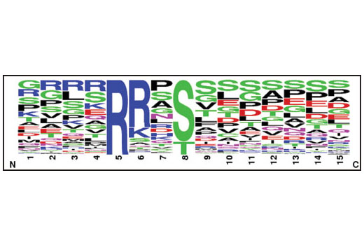
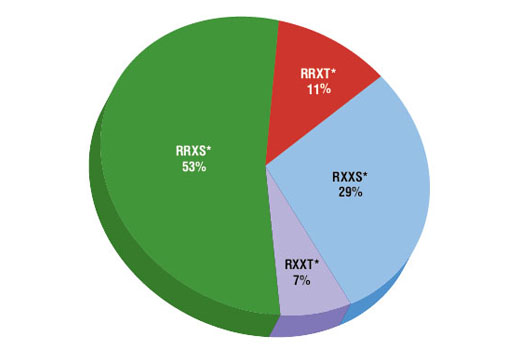
An important class of kinases, refered to as Arg-directed kinases or AGC-family kinases, includes cAMP-dependent protein kinase (PKA), cGMP-dependent protein kinase (PKG), protein kinase C, Akt, and RSK. These kinases share a substrate specificity characterized by Arg at position -3 relative to the phosphorylated Ser or Thr (2,3). Phospho-PKA substrate-specific antibodies from CST are powerful tools for investigating the regulation of phosphorylation by PKA and other Arg-directed kinases, as well as for high throughput kinase drug discovery.In this assay, PTMScan® (RRXS

Limited Uses

Except as otherwise expressly agreed in a writing signed by a legally authorized representative of CST, the following terms apply to Products provided by CST, its affiliates or its distributors. Any Customer's terms and conditions that are in addition to, or different from, those contained herein, unless separately accepted in writing by a legally authorized representative of CST, are rejected and are of no force or effect.

Products are labeled with For Research Use Only or a similar labeling statement and have not been approved, cleared, or licensed by the FDA or other regulatory foreign or domestic entity, for any purpose. Customer shall not use any Product for any diagnostic or therapeutic purpose, or otherwise in any manner that conflicts with its labeling statement. Products sold or licensed by CST are provided for Customer as the end-user and solely for research and development uses. Any use of Product for diagnostic, prophylactic or therapeutic purposes, or any purchase of Product for resale (alone or as a component) or other commercial purpose, requires a separate license from CST. Customer shall (a) not sell, license, loan, donate or otherwise transfer or make available any Product to any third party, whether alone or in combination with other materials, or use the Products to manufacture any commercial products, (b) not copy, modify, reverse engineer, decompile, disassemble or otherwise attempt to discover the underlying structure or technology of the Products, or use the Products for the purpose of developing any products or services that would compete with CST products or services, (c) not alter or remove from the Products any trademarks, trade names, logos, patent or copyright notices or markings, (d) use the Products solely in accordance with CST Product Terms of Sale and any applicable documentation, and (e) comply with any license, terms of service or similar agreement with respect to any third party products or services used by Customer in connection with the Products.

For Research Use Only. Not for Use in Diagnostic Procedures.
Cell Signaling Technology is a trademark of Cell Signaling Technology, Inc.
AcetylScan is a registered trademark of Cell Signaling Technology, Inc.
MethylScan is a registered trademark of Cell Signaling Technology, Inc.
PTMScan is a registered trademark of Cell Signaling Technology, Inc.
UbiScan is a registered trademark of Cell Signaling Technology, Inc.
All other trademarks are the property of their respective owners. Visit our Trademark Information page.