Cell proliferation is an increase in cell number due to cell division, or cytokinesis, the final step of the cell cycle. Cell proliferation is necessary for normal tissue development and maintenance over the lifespan.
Cell proliferation is a tightly regulated process, with many different proteins controlling cell cycle checkpoints. Genetic mutations found in cancer cells cause uncontrolled cellular proliferation.
The cell cycle is composed of multiple phases, including the G1 phase where cells increase in size, the S phase where DNA is newly synthesized, the G2 phase where further cell growth occurs, and finally the M, or mitosis, phase where the cell ultimately divides. Some cells exit the cell cycle and stop dividing altogether; this is known as quiescence or resting state (G0)
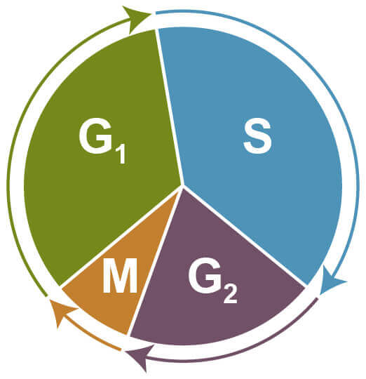
Phases of the Cell Cycle: G1 - increasing size; S - DNA synthesis and replication; G2 - growth; M - mitosis and cell division; G0 - quiescent.
Two of the most common ways of measuring cell proliferation include cell cycle assays and proliferation assays, which rely on the incorporation of propidium propidium iodide or BrdU into DNA:
| Assay | What is Measured | |
|---|---|---|
| Measures the level of DNA synthesis occuring during cellular proliferation | ||
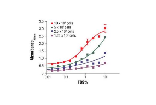
BrdU Cell Proliferation Assay Kit #6813: C2C12 cells were seeded at varying density in serum free medium in a 96-well plate and incubated overnight. Serum was added to the plate at various concentrations and cells were incubated for 24 hr. Finally, 10 μM BrdU was added to the plate and cells were incubated for 4 hr. |
||
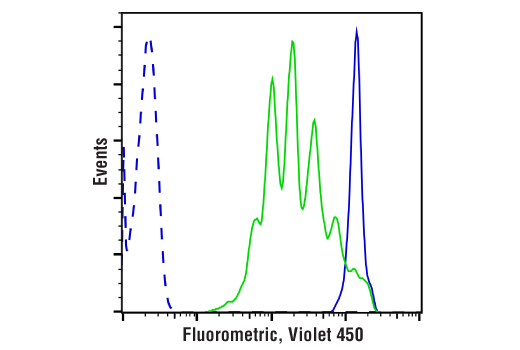
| Measures cell division using amine-reactive dyes and the principle of dye dilution to trace multiple rounds of cell proliferation, via flow cytometry | ||
|
||
| Quantifies the number of cells at different stages of the cell cycle based on the profile of their DNA. | ||
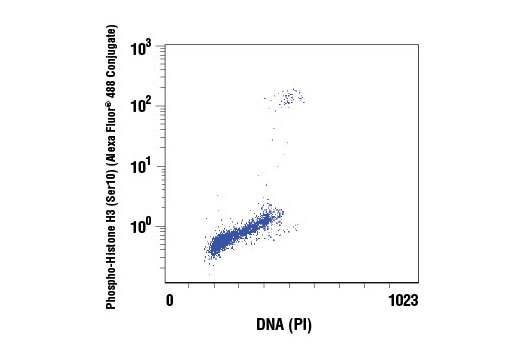
Propidium Iodide (PI)/RNase Staining Solution #4087: Flow cytometric analysis of untreated Jurkat cells, using Phospho-Histone H3 (Ser10) Antibody (Alexa Fluor® 488 Conjugate) #9708 and Propidium Iodide (PI)/RNase Staining Solution (DNA content). |
||
Another common method for measuring cell proliferation is by examining the expression levels of cell cycle proteins and other proteins required for proliferation. Some common protein markers for proliferating cells include:
| Proliferation Marker | Proliferation Marker Description | |
|---|---|---|
| Proliferating cell nuclear antigen (PNCA) | DNA sliding clamp protein that plays an important role in DNA replication | |
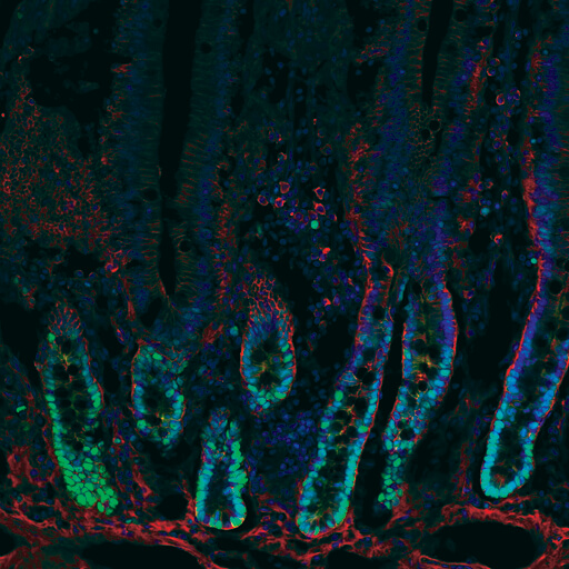
PCNA (D3H8P) XP® Rabbit mAb #1311: Confocal immunofluorescent analysis of rat small intestine using PCNA (D3H8P) XP® Rabbit mAb (green) showing staining of proliferative cells in intestinal crypts and β-Catenin (L54E2) Mouse mAb (IF Preferred) #2677 (red). Blue pseudocolor = DRAQ5 #4084 (fluorescent DNA dye). |
||
| Ki-67 | Non-nuclear histone protein only expressed at high levels in proliferating cells | |
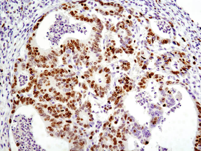
Ki-67 (D2H10) Rabbit mAb (IHC Specific) #9027: Immunohistochemical analysis of paraffin-embedded human colon carcinoma using Ki-67 (D2H10) Rabbit mAb (IHC Specific). |
||
| Phospho-histone H3 | Nuclear histone protein that is phosphorylated in proliferating cells | |
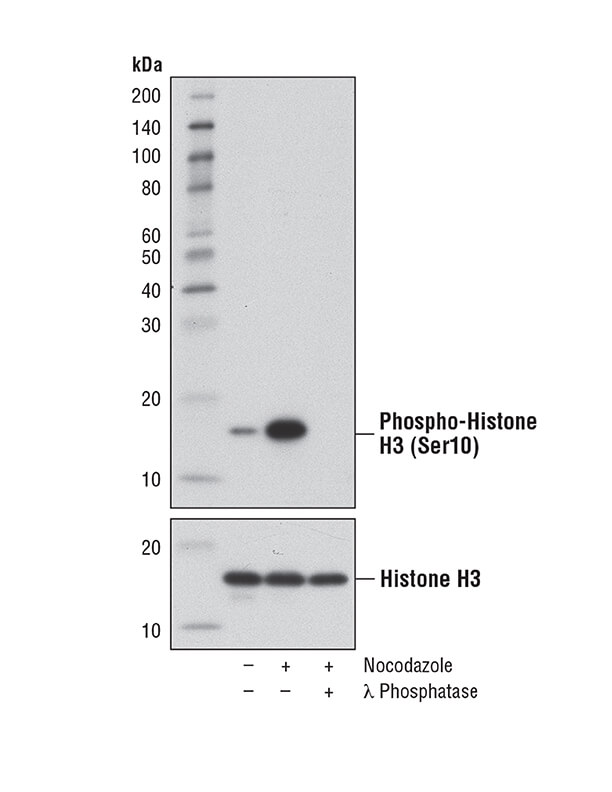
Phospho-Histone H3 (Ser10) (D7N8E) XP® Rabbit mAb #53348: Western blot analysis of extracts from HeLa cells, either untreated or treated with Nocodazole #2190 (100 ng/ml, 16 hr), using Phospho-Histone H3 (Ser10) (D7N8E) XP® Rabbit mAb (upper) or Histone H3 (D1H2) Rabbit mAb #4499 (lower). Phospho-specificity of the antibody is shown by further treatment of the lysate with λ phosphatase. |
||
| β-galactosidase activity | β-galactosidase activity is associated with senescent cells that are not proliferating | |
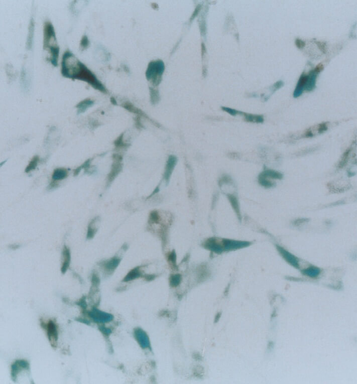
Senescence β-Galactosidase Staining Kit #9860: β-Galactosidase staining at pH 6 on senescent WI38 cells at population doubling 36 (right). |
||