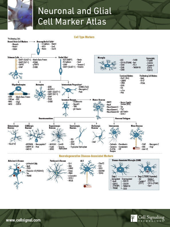
Neurons are specialized CNS cells that are able to receive and transmit electrical impulses to process electrochemical signals. The anatomy of a neuron is conducive to sending information to neurons close by or at a distance. Identifying mature neurons is accomplished by observing expression of MAP2, NeuN, Neurofilament-H, Neurofilament-L and Neurofilament-M.
Several lines of evidence suggest that aggregated proteins or long-term stimuli can result in chronic inflammation that is harmful to the cells and can contribute to the pathogenesis of neurodegenerative diseases, including Alzheimer’s disease (AD), Parkinson’s disease (PD), and amyotrophic lateral sclerosis (ALS).
In order to maintain proper brain function, the nervous system is comprised of not only neurons, but also other cells including microglia, oligodendrocytes, and astrocytes.
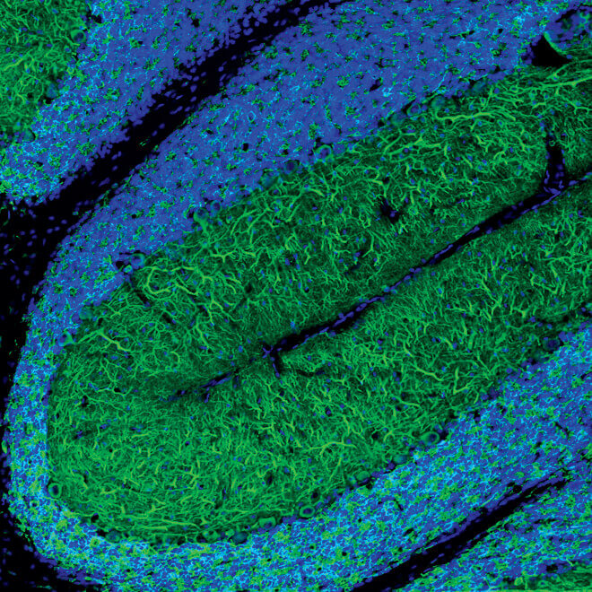
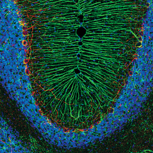
Confocal immunofluorescent analysis of frozen rat cerebellum using MAP2 (D5G1) XP® Rabbit mAb (green). Blue pseudocolor = DRAQ5 #4084 (fluorescent DNA dye).
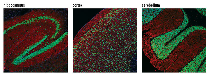
Confocal immunofluorescent analysis of mouse hippocampus (left), cortex (middle), and cerebellum (right) using NeuN (D4G4O) XP® Rabbit mAb (green). Actin filaments were labeled with DyLight 554 Phalloidin #13054 (red). Blue pseudocolor = DRAQ5 #4084 (fluorescent DNA dye).
Confocal immunofluorescent image of mouse cerebellum labeled with Neurofilament-H (RMdO 20) Mouse mAb (green) and Calbindin Antibody #2136 (red). Blue pseudocolor = DRAQ5 (fluorescent DNA dye).
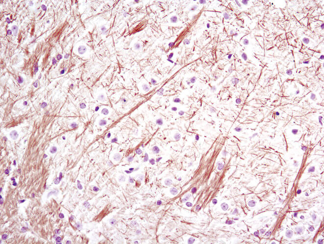
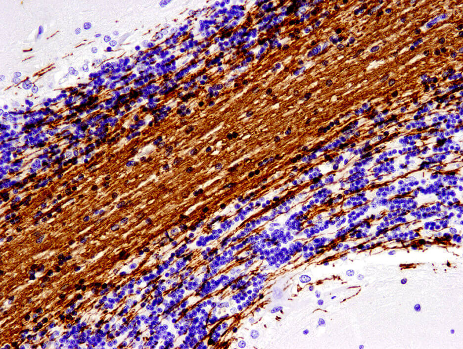
Immunohistochemical analysis of paraffin-embedded mouse brain using Neurofilament-L (C28E10) Rabbit mAb.
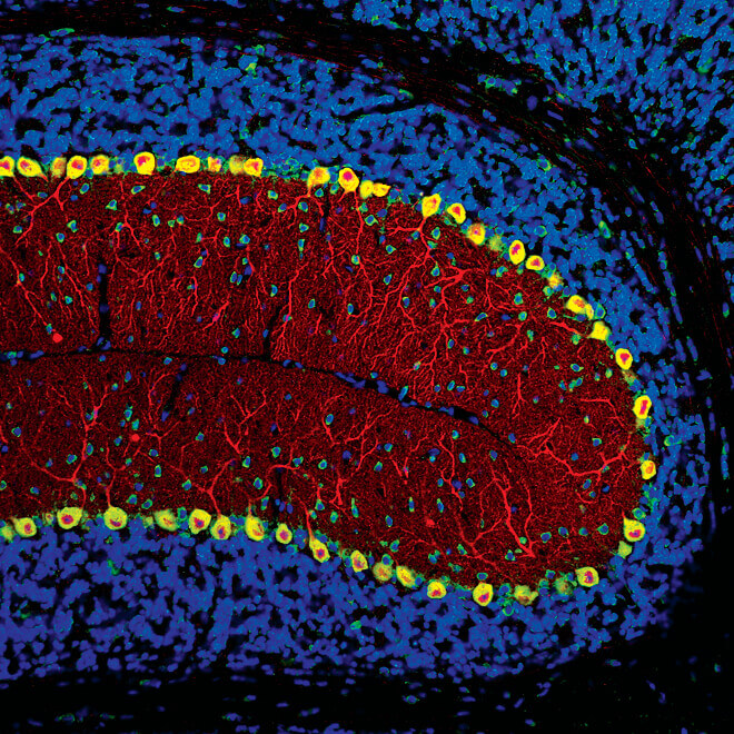
Confocal immunofluorescent image of mouse cerebellum labeled with Neurofilament-M (RMO 14.9) Mouse mAb (green) and Calbindin Antibody #2136 (red). Blue pseudocolor = DRAQ5 (fluorescent DNA dye).
Microglia are immune cells that are of myeloid origin and migrate to the CNS during development. The many functions of microglia include pruning of complex neural networks during embryogenesis, assisting in synaptic plasticity or preventing excitotoxicity by responding to neurotransmitter release, and mounting an inflammatory response to an acute infection or trauma to the brain. Specific markers for identification of these resident macrophages of the central nervous system (CNS) are integrin alpha M (ITGAM/CD11b), the surface glycoprotein F4/80, CD45, iba1, and TMEM119.
In disease states, distinct microglial populations can be identified by molecular signatures called disease-associated microglia (DAMs). DAMs mature in stages, both of which can be characterized by different biomarker expression: Stage 1 DAM microglia upregulate triggering receptor expressed on myeloid cells 2 (TREM2), ApoE, and Dap12, and downregulate P2ry12 and transmembrane protein 119 (TMEM119). Stage 2 DAMs upregulate osteopontin (Spp1) and cystatin 7 (CST7).
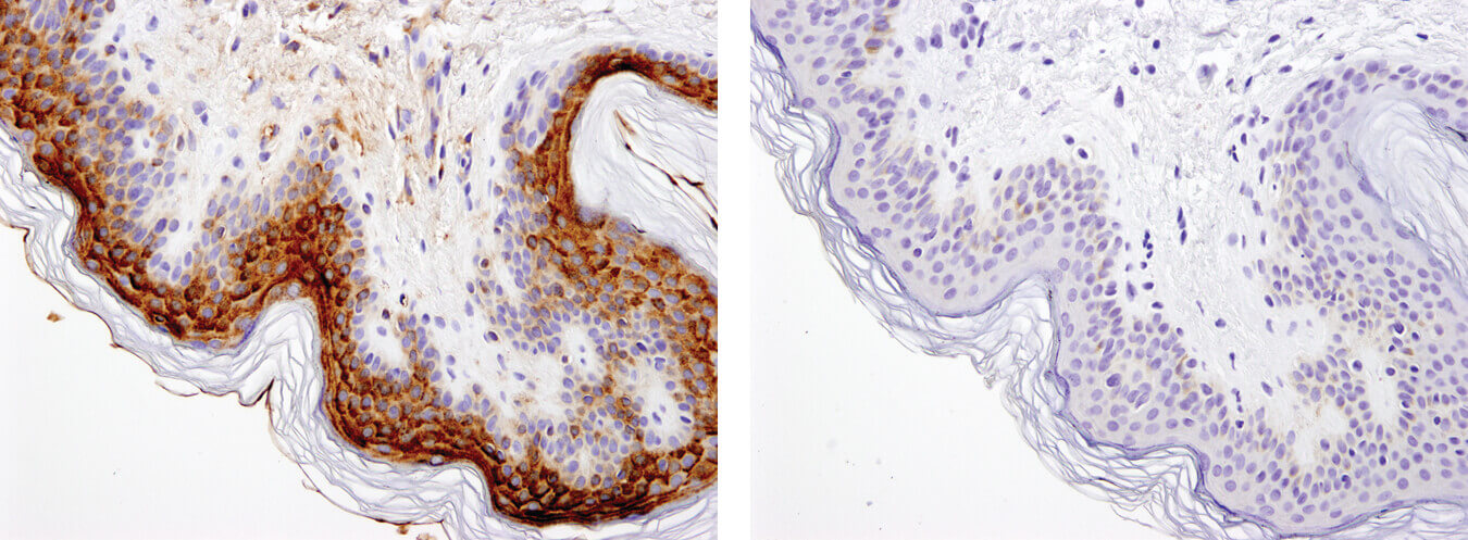
Immunohistochemical analysis of paraffin-embedded human skin using ApoE (pan) (D7I9N) Rabbit mAb in the presence of control peptide (left) and antigen-specific peptide (right).
Oligodendrocytes are a specialized glial cell type in CNS that produce myelin, which provides a protective sheath to the axons and improves the conduction velocity of signals between neurons. Oligodendrocytes are characterized by the expression of the myelin family proteins: myelin oligodendrocyte glycoprotein (MOG), myelin-associated glycoprotein (MAG), and Myelin Basic Protein (MBP).
Oligodendrocyte progenitor cells exist in the brain to facilitate regeneration of cells due to injury. However, breakdown of myelin and the inability to regenerate oligodendrocytes with insufficient or dysfunctional myelin have been correlated with the pathogenesis of several neurodegenerative diseases including AD, PD, ALS, and multiple sclerosis (MS).
Immunohistochemical analysis of paraffin-embedded human cerebellum using MOG (E5K6T) XP® Rabbit mAb.
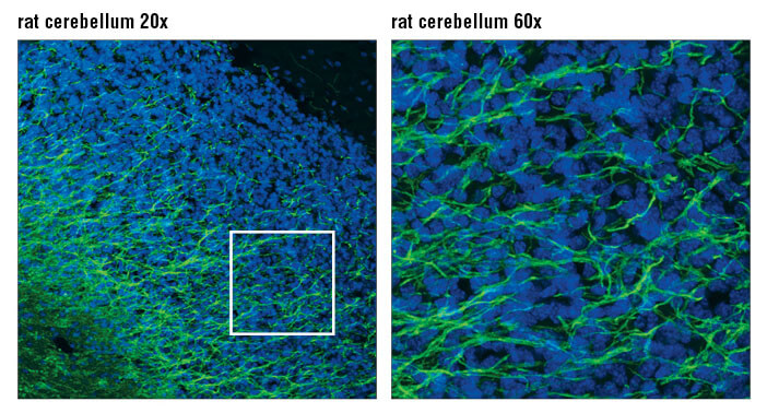
Confocal immunofluorescent analysis of rat cerebellum using MAG (D4G3) XP® Rabbit mAb (green). Blue pseudocolor = DRAQ5 #4084 (fluorescent DNA dye).
Astrocytes are another immunocompetent glial cell that arise within the CNS and are found within the brain. The most specific markers of astrocytes are GFAP and S100β.
In a healthy nervous system, astrocytes play essential roles in development, regulation of blood flow (by supporting endothelial cells in the blood brain barrier), synaptic transmission and function, and energy and metabolism (by providing nutrients to neurons and synthesizing certain neurotransmitters).
The loss or abnormal function of astrocytes has been implicated in a wide variety of neurodegenerative disease processes. Chronic activation of astrocytes results in the formation of lesions similar to those observed in AD and HD.
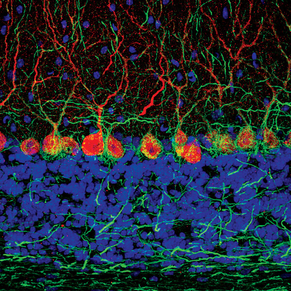
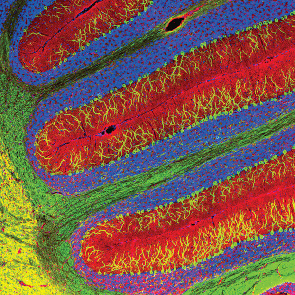
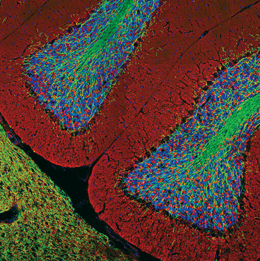
Confocal immunofluorescent analysis of rat cerebellum using GFAP (D1F4Q) XP® Rabbit mAb (green) and Neurofilament-H (RMdO 20) Mouse mAb #2836 (red). Blue pseudocolor = DRAQ5 #4084 (fluorescent DNA dye).
Confocal immunofluorescent analysis of mouse hippocampus (left), cortex (middle), and cerebellum (right) using NeuN (D4G4O) XP® Rabbit mAb (green). Actin filaments were labeled with DyLight 554 Phalloidin #13054 (red). Blue pseudocolor = DRAQ5 #4084 (fluorescent DNA dye).
Confocal immunofluorescent analysis of mouse cerebellum using β3-Tubulin (D71G9) XP® Rabbit mAb (green) and Tau (Tau46) Mouse mAb #4019 (red). Blue pseudocolor = DRAQ5 #4084 (fluorescent DNA dye).
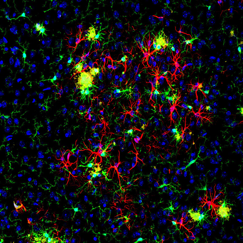
Confocal immunofluorescent analysis of mouse brain. using ASC/TMS1 (D2W8U) Rabbit mAb (Mouse Specific) #67824 (green) and APP/β-Amyloid (NAB228) Mouse mAb #2450 (yellow), GFAP (GA5) Mouse mAb (Alexa Fluor® 647 Conjugate) #3657 (red), and Hoechst 33342 #4082 (blue).
Confocal immunofluorescent analysis of rat brain using Myelin Basic Protein (D8X4Q) XP® Rabbit mAb (green) and Synaptophysin (7H12) Mouse mAb (IF Formulated) #9020 (red). Blue pseudocolor = DRAQ5 #4084 (fluorescent DNA dye).