Detect endogenous levels of B7-H3 and B7-H4 protein expression in human tissue, respectively.
 <
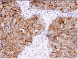
< B7-H3 (D9M2L) XP® Rabbit mAb #14058 IHC Analysis of paraffin-embedded ovarian carcinoma using #14058
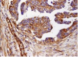
B7-H4 (D1M8I) XP® Rabbit mAb #14572 IHC analysis of paraffin-embedded human granulosa cell tumor of the ovary using #14572
Recognize B7-H3 and B7-H4 protein, respectively, and do not cross react with other B7 family members 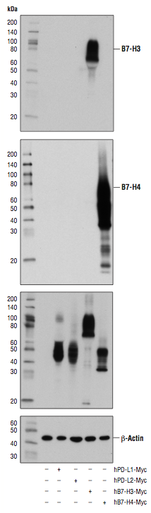
WB analysis of extracts from COS-7 cells, mock transfected (-) or transfected with constructs expressing Myc-tagged full-length human PD-L1, PD-L2, B7-H3, or B7-H4 protein (as indicated), using B7-H3 (D9M2L) XP® Rabbit mAb #14058 (upper), B7-H4 (D1M8I) XP® Rabbit mAb #14572 (upper middle), Myc-tag (71D10) Rabbit mAb #2278 (lower middle), and β-Actin (D6A8) Rabbit mAb #8457 (lower).
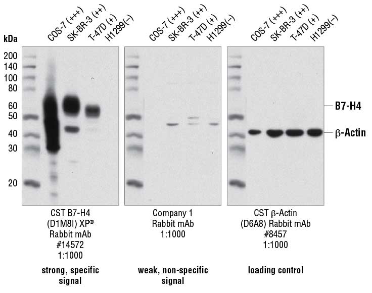
WB analysis of extracts from various cell lines using B7-H4 (D1M8I) XP® Rabbit mAb #14572 (left), Company 1 Rabbit mAb (middle), and β-Actin (D6A8) Rabbit mAb #8457 (right). The CST antibody #14572 (left) shows a strong signal proportional to the relative cellular expression levels. Company 1 rabbit mAb (middle) shows non-specific bands in the B7-H4-negative cell line, H1299.